Loading
PDBj
MenuPDBj@FacebookPDBj@TwitterPDBj@YouTubewwPDB FoundationwwPDB
RCSB PDBPDBeBMRBAdv. SearchSearch help

5QLI
DownloadVisualize
BU of 5qli by Molmil
Group deposition of library data - Crystal Structure of EcDsbA after initial refinement with no ligand modelled (structure C5_1)
Descriptor: Thiol:disulfide interchange protein
Authors:Ilyichova, O.V, Bentley, M.R, Doak, B.C, Scanlon, M.J.
Deposit date:2019-01-27
Release date:2020-02-05
Last modified:2020-07-22
Method:X-RAY DIFFRACTION (2.043 Å)
Cite:Rapid Elaboration of Fragments into Leads by X-ray Crystallographic Screening of Parallel Chemical Libraries (REFiLX).
J.Med.Chem., 63, 2020
5QLW
DownloadVisualize
BU of 5qlw by Molmil
Group deposition of library data - Crystal Structure of EcDsbA after initial refinement with no ligand modelled (structure D3_1)
Descriptor: Thiol:disulfide interchange protein
Authors:Ilyichova, O.V, Bentley, M.R, Doak, B.C, Scanlon, M.J.
Deposit date:2019-01-27
Release date:2020-02-05
Last modified:2020-07-22
Method:X-RAY DIFFRACTION (1.959 Å)
Cite:Rapid Elaboration of Fragments into Leads by X-ray Crystallographic Screening of Parallel Chemical Libraries (REFiLX).
J.Med.Chem., 63, 2020
5QMC
DownloadVisualize
BU of 5qmc by Molmil
Group deposition of library data - Crystal Structure of EcDsbA after initial refinement with no ligand modelled (structure E8_1)
Descriptor: Thiol:disulfide interchange protein
Authors:Ilyichova, O.V, Bentley, M.R, Doak, B.C, Scanlon, M.J.
Deposit date:2019-01-27
Release date:2020-02-05
Last modified:2020-07-22
Method:X-RAY DIFFRACTION (1.887 Å)
Cite:Rapid Elaboration of Fragments into Leads by X-ray Crystallographic Screening of Parallel Chemical Libraries (REFiLX).
J.Med.Chem., 63, 2020
5QMU
DownloadVisualize
BU of 5qmu by Molmil
Group deposition of library data - Crystal Structure of EcDsbA after initial refinement with no ligand modelled (structure G11_1)
Descriptor: Thiol:disulfide interchange protein
Authors:Ilyichova, O.V, Bentley, M.R, Doak, B.C, Scanlon, M.J.
Deposit date:2019-01-27
Release date:2020-02-05
Last modified:2020-07-22
Method:X-RAY DIFFRACTION (2.043 Å)
Cite:Rapid Elaboration of Fragments into Leads by X-ray Crystallographic Screening of Parallel Chemical Libraries (REFiLX).
J.Med.Chem., 63, 2020
5QN5
DownloadVisualize
BU of 5qn5 by Molmil
Group deposition of library data - Crystal Structure of EcDsbA after initial refinement with no ligand modelled (structure G8_2)
Descriptor: Thiol:disulfide interchange protein
Authors:Ilyichova, O.V, Bentley, M.R, Doak, B.C, Scanlon, M.J.
Deposit date:2019-01-27
Release date:2020-02-05
Last modified:2020-07-22
Method:X-RAY DIFFRACTION (1.891 Å)
Cite:Rapid Elaboration of Fragments into Leads by X-ray Crystallographic Screening of Parallel Chemical Libraries (REFiLX).
J.Med.Chem., 63, 2020
5H30
DownloadVisualize
BU of 5h30 by Molmil
Cryo-EM structure of zika virus complexed with Fab C10 at pH 6.5
Descriptor: IgG C10 heavy chain, IgG C10 light chain, structural protein E, ...
Authors:Zhang, S, Kostyuchenko, V, Ng, T.-S, Lok, S.-M.
Deposit date:2016-10-19
Release date:2016-11-30
Last modified:2024-05-29
Method:ELECTRON MICROSCOPY (4.4 Å)
Cite:Neutralization mechanism of a highly potent antibody against Zika virus
Nat Commun, 7, 2016
4DYN
DownloadVisualize
BU of 4dyn by Molmil
Crystal Structure of WSN/A Influenza Nucleoprotein with BMS-885838 Ligand Bound
Descriptor: N-[4-chloranyl-5-[4-[[3-(2-methoxyphenyl)-5-methyl-1,2-oxazol-4-yl]carbonyl]piperazin-1-yl]-2-nitro-phenyl]pyridine-2-carboxamide, Nucleocapsid protein
Authors:Lewis, H.A, Baldwin, E.T, Steinbacher, S, Maskos, K, Mortl, M, Kiefersauer, R, Edavettal, S, McDonnell, P.A, Pearce, B.C, Langley, D.R.
Deposit date:2012-02-29
Release date:2013-03-06
Last modified:2024-02-28
Method:X-RAY DIFFRACTION (2.4 Å)
Cite:To be determined
To be Published
5QKG
DownloadVisualize
BU of 5qkg by Molmil
Group deposition of library data - Crystal Structure of EcDsbA after initial refinement with no ligand modelled (structure A2_3)
Descriptor: Thiol:disulfide interchange protein
Authors:Ilyichova, O.V, Bentley, M.R, Doak, B.C, Scanlon, M.J.
Deposit date:2019-01-27
Release date:2020-02-05
Last modified:2020-07-22
Method:X-RAY DIFFRACTION (1.897 Å)
Cite:Rapid Elaboration of Fragments into Leads by X-ray Crystallographic Screening of Parallel Chemical Libraries (REFiLX).
J.Med.Chem., 63, 2020
5QKW
DownloadVisualize
BU of 5qkw by Molmil
Group deposition of library data - Crystal Structure of EcDsbA after initial refinement with no ligand modelled (structure B10_2)
Descriptor: Thiol:disulfide interchange protein
Authors:Ilyichova, O.V, Bentley, M.R, Doak, B.C, Scanlon, M.J.
Deposit date:2019-01-27
Release date:2020-02-05
Last modified:2020-07-22
Method:X-RAY DIFFRACTION (1.969 Å)
Cite:Rapid Elaboration of Fragments into Leads by X-ray Crystallographic Screening of Parallel Chemical Libraries (REFiLX).
J.Med.Chem., 63, 2020
5QLF
DownloadVisualize
BU of 5qlf by Molmil
Group deposition of library data - Crystal Structure of EcDsbA after initial refinement with no ligand modelled (structure C3_2)
Descriptor: Thiol:disulfide interchange protein
Authors:Ilyichova, O.V, Bentley, M.R, Doak, B.C, Scanlon, M.J.
Deposit date:2019-01-27
Release date:2020-02-05
Last modified:2020-07-22
Method:X-RAY DIFFRACTION (2.003 Å)
Cite:Rapid Elaboration of Fragments into Leads by X-ray Crystallographic Screening of Parallel Chemical Libraries (REFiLX).
J.Med.Chem., 63, 2020
5QLP
DownloadVisualize
BU of 5qlp by Molmil
Group deposition of library data - Crystal Structure of EcDsbA after initial refinement with no ligand modelled (structure D1_1)
Descriptor: Thiol:disulfide interchange protein
Authors:Ilyichova, O.V, Bentley, M.R, Doak, B.C, Scanlon, M.J.
Deposit date:2019-01-27
Release date:2020-02-05
Last modified:2020-07-22
Method:X-RAY DIFFRACTION (2.521 Å)
Cite:Rapid Elaboration of Fragments into Leads by X-ray Crystallographic Screening of Parallel Chemical Libraries (REFiLX).
J.Med.Chem., 63, 2020
5QM4
DownloadVisualize
BU of 5qm4 by Molmil
Group deposition of library data - Crystal Structure of EcDsbA after initial refinement with no ligand modelled (structure E10_1)
Descriptor: Thiol:disulfide interchange protein
Authors:Ilyichova, O.V, Bentley, M.R, Doak, B.C, Scanlon, M.J.
Deposit date:2019-01-27
Release date:2020-02-05
Last modified:2020-07-22
Method:X-RAY DIFFRACTION (2.007 Å)
Cite:Rapid Elaboration of Fragments into Leads by X-ray Crystallographic Screening of Parallel Chemical Libraries (REFiLX).
J.Med.Chem., 63, 2020
5QMK
DownloadVisualize
BU of 5qmk by Molmil
Group deposition of library data - Crystal Structure of EcDsbA after initial refinement with no ligand modelled (structure F2_1)
Descriptor: Thiol:disulfide interchange protein
Authors:Ilyichova, O.V, Bentley, M.R, Doak, B.C, Scanlon, M.J.
Deposit date:2019-01-27
Release date:2020-02-05
Last modified:2020-07-22
Method:X-RAY DIFFRACTION (1.836 Å)
Cite:Rapid Elaboration of Fragments into Leads by X-ray Crystallographic Screening of Parallel Chemical Libraries (REFiLX).
J.Med.Chem., 63, 2020
5QMZ
DownloadVisualize
BU of 5qmz by Molmil
Group deposition of library data - Crystal Structure of EcDsbA after initial refinement with no ligand modelled (structure G5_1)
Descriptor: Thiol:disulfide interchange protein
Authors:Ilyichova, O.V, Bentley, M.R, Doak, B.C, Scanlon, M.J.
Deposit date:2019-01-27
Release date:2020-02-05
Last modified:2020-07-22
Method:X-RAY DIFFRACTION (2.084 Å)
Cite:Rapid Elaboration of Fragments into Leads by X-ray Crystallographic Screening of Parallel Chemical Libraries (REFiLX).
J.Med.Chem., 63, 2020
5QNE
DownloadVisualize
BU of 5qne by Molmil
Group deposition of library data - Crystal Structure of EcDsbA after initial refinement with no ligand modelled (structure H7_1)
Descriptor: Thiol:disulfide interchange protein
Authors:Ilyichova, O.V, Bentley, M.R, Doak, B.C, Scanlon, M.J.
Deposit date:2019-01-27
Release date:2020-02-05
Last modified:2020-07-22
Method:X-RAY DIFFRACTION (2.394 Å)
Cite:Rapid Elaboration of Fragments into Leads by X-ray Crystallographic Screening of Parallel Chemical Libraries (REFiLX).
J.Med.Chem., 63, 2020
5QKE
DownloadVisualize
BU of 5qke by Molmil
Group deposition of library data - Crystal Structure of EcDsbA after initial refinement with no ligand modelled (structure A11_1)
Descriptor: Thiol:disulfide interchange protein
Authors:Ilyichova, O.V, Bentley, M.R, Doak, B.C, Scanlon, M.J.
Deposit date:2019-01-27
Release date:2020-02-05
Last modified:2020-07-22
Method:X-RAY DIFFRACTION (2.117 Å)
Cite:Rapid Elaboration of Fragments into Leads by X-ray Crystallographic Screening of Parallel Chemical Libraries (REFiLX).
J.Med.Chem., 63, 2020
5QKV
DownloadVisualize
BU of 5qkv by Molmil
Group deposition of library data - Crystal Structure of EcDsbA after initial refinement with no ligand modelled (structure B1_2)
Descriptor: Thiol:disulfide interchange protein
Authors:Ilyichova, O.V, Bentley, M.R, Doak, B.C, Scanlon, M.J.
Deposit date:2019-01-27
Release date:2020-02-05
Last modified:2020-07-22
Method:X-RAY DIFFRACTION (2.14 Å)
Cite:Rapid Elaboration of Fragments into Leads by X-ray Crystallographic Screening of Parallel Chemical Libraries (REFiLX).
J.Med.Chem., 63, 2020
5QLB
DownloadVisualize
BU of 5qlb by Molmil
Group deposition of library data - Crystal Structure of EcDsbA after initial refinement with no ligand modelled (structure C10_1)
Descriptor: Thiol:disulfide interchange protein
Authors:Ilyichova, O.V, Bentley, M.R, Doak, B.C, Scanlon, M.J.
Deposit date:2019-01-27
Release date:2020-02-05
Last modified:2020-07-22
Method:X-RAY DIFFRACTION (1.959 Å)
Cite:Rapid Elaboration of Fragments into Leads by X-ray Crystallographic Screening of Parallel Chemical Libraries (REFiLX).
J.Med.Chem., 63, 2020
3LXG
DownloadVisualize
BU of 3lxg by Molmil
Crystal structure of rat phosphodiesterase 10A in complex with ligand WEB-3
Descriptor: 2-methoxy-6,7-dimethyl-9-propylimidazo[1,5-a]pyrido[3,2-e]pyrazine, MAGNESIUM ION, ZINC ION, ...
Authors:Mosbacher, T, Jestel, A, Steinbacher, S.
Deposit date:2010-02-25
Release date:2010-05-19
Last modified:2024-02-21
Method:X-RAY DIFFRACTION (2.3 Å)
Cite:Discovery of imidazo[1,5-a]pyrido[3,2-e]pyrazines as a new class of phosphodiesterase 10A inhibitiors.
J.Med.Chem., 53, 2010
4YLQ
DownloadVisualize
BU of 4ylq by Molmil
Crystal Structure of a FVIIa-Trypsin Chimera (FT) in Complex with Soluble Tissue Factor
Descriptor: CALCIUM ION, Coagulation factor VII, N-PROPANOL, ...
Authors:Sorensen, A.B, Svensson, L.A, Gandhi, P.S.
Deposit date:2015-03-05
Release date:2015-12-30
Last modified:2024-01-10
Method:X-RAY DIFFRACTION (1.4 Å)
Cite:Molecular Basis of Enhanced Activity in Factor VIIa-Trypsin Variants Conveys Insights into Tissue Factor-mediated Allosteric Regulation of Factor VIIa Activity.
J.Biol.Chem., 291, 2016
6UTD
DownloadVisualize
BU of 6utd by Molmil
CRYSTAL STRUCTURE OF UNLIGANDED HIV-1 LM/HS CLADE A/E CRF01 GP120 CORE
Descriptor: 2-acetamido-2-deoxy-beta-D-glucopyranose, 4-(2-HYDROXYETHYL)-1-PIPERAZINE ETHANESULFONIC ACID, HIV-1 LM/HS clade A/E CRF01 gp120 core
Authors:Tolbert, W.D, Sherburn, R, Pazgier, M.
Deposit date:2019-10-29
Release date:2020-10-07
Last modified:2023-10-11
Method:X-RAY DIFFRACTION (2.2 Å)
Cite:The HIV-1 Env gp120 Inner Domain Shapes the Phe43 Cavity and the CD4 Binding Site.
Mbio, 11, 2020
6B2W
DownloadVisualize
BU of 6b2w by Molmil
C. Jejuni C315S Agmatine Deiminase with Substrate Bound
Descriptor: AGMATINE, POTASSIUM ION, Putative peptidyl-arginine deiminase family protein
Authors:Shek, R, Hicks, K.A, French, J.B.
Deposit date:2017-09-20
Release date:2017-12-13
Last modified:2023-10-04
Method:X-RAY DIFFRACTION (2.5 Å)
Cite:Structural and Functional Basis for Targeting Campylobacter jejuni Agmatine Deiminase To Overcome Antibiotic Resistance.
Biochemistry, 56, 2017
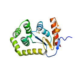
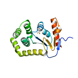
6ANK
DownloadVisualize
BU of 6ank by Molmil
Synaptotagmin-7, C2A- and C2B-domains
Descriptor: 1,2-ETHANEDIOL, CALCIUM ION, Synaptotagmin-7
Authors:Tomchick, D.R, Rizo, J, Voleti, R.
Deposit date:2017-08-13
Release date:2017-09-20
Last modified:2023-10-04
Method:X-RAY DIFFRACTION (2.254 Å)
Cite:Exceptionally tight membrane-binding may explain the key role of the synaptotagmin-7 C2A domain in asynchronous neurotransmitter release.
Proc. Natl. Acad. Sci. U.S.A., 114, 2017
4RJT
DownloadVisualize
BU of 4rjt by Molmil
Crystal Structure of Unliganded, Full Length hUGDH at pH 7.0
Descriptor: CHLORIDE ION, UDP-glucose 6-dehydrogenase
Authors:Sidlo, A.M, Wood, Z.A.
Deposit date:2014-10-09
Release date:2014-12-31
Last modified:2023-09-20
Method:X-RAY DIFFRACTION (2.7 Å)
Cite:Hysteresis in Human UDP-Glucose Dehydrogenase Is Due to a Restrained Hexameric Structure That Favors Feedback Inhibition.
Biochemistry, 53, 2014
5OIN
DownloadVisualize
BU of 5oin by Molmil
InhA (T2A mutant) complexed with N-(1-(pyrimidin-2-yl)piperidin-4-yl)acetamide
Descriptor: Enoyl-[acyl-carrier-protein] reductase [NADH], NICOTINAMIDE-ADENINE-DINUCLEOTIDE, ~{N}-(1-pyrimidin-2-ylpiperidin-4-yl)ethanamide
Authors:Convery, M.A.
Deposit date:2017-07-19
Release date:2018-02-14
Last modified:2024-05-08
Method:X-RAY DIFFRACTION (2.82 Å)
Cite:Screening of a Novel Fragment Library with Functional Complexity against Mycobacterium tuberculosis InhA.
ChemMedChem, 13, 2018

224931

數據於2024-09-11公開中

PDB statisticsPDBj update infoContact PDBjnumon