Loading
PDBj
MenuPDBj@FacebookPDBj@TwitterPDBj@YouTubewwPDB FoundationwwPDB
RCSB PDBPDBeBMRBAdv. SearchSearch help
PDB: 567 results

3B32
DownloadVisualize
BU of 3b32 by Molmil
Crystal Structure of Calcium-Saturated Calmodulin N-Terminal Domain Fragment, Residues 1-75
Descriptor: CALCIUM ION, Calmodulin
Authors:Newman, R.A, Shea, M.A.
Deposit date:2007-10-19
Release date:2007-11-13
Last modified:2023-08-30
Method:X-RAY DIFFRACTION (1.6 Å)
Cite:Thermodynamics and conformational change governing domain-domain interactions of calmodulin.
Methods Enzymol., 466, 2009
6T39
DownloadVisualize
BU of 6t39 by Molmil
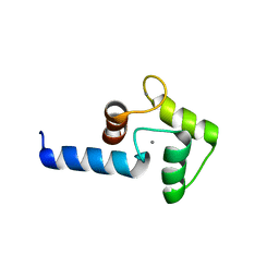
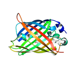
Crystal structure of rsEGFP2 in its off-state determined by SFX
Descriptor: Green fluorescent protein
Authors:Woodhouse, J, Coquelle, N, Adam, V, Barends, T.R.M, De La Mora, E, Bourgeois, D, Colletier, J.P, Schlichting, I, Weik, M.
Deposit date:2019-10-10
Release date:2020-02-19
Last modified:2024-01-24
Method:X-RAY DIFFRACTION (1.6 Å)
Cite:Photoswitching mechanism of a fluorescent protein revealed by time-resolved crystallography and transient absorption spectroscopy.
Nat Commun, 11, 2020
4KEX
DownloadVisualize
BU of 4kex by Molmil
Crystal structure analysis of a single amino acid deletion mutation in EGFP
Descriptor: Green fluorescent protein
Authors:Arpino, J.A.J, Rizkallah, P.J.
Deposit date:2013-04-26
Release date:2014-08-06
Last modified:2023-12-06
Method:X-RAY DIFFRACTION (1.6 Å)
Cite:Structural and dynamic changes associated with beneficial engineered single-amino-acid deletion mutations in enhanced green fluorescent protein.
Acta Crystallogr.,Sect.D, 70, 2014
1G4Y
DownloadVisualize
BU of 1g4y by Molmil
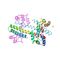
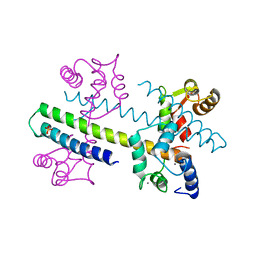
1.60 A CRYSTAL STRUCTURE OF THE GATING DOMAIN FROM SMALL CONDUCTANCE POTASSIUM CHANNEL COMPLEXED WITH CALCIUM-CALMODULIN
Descriptor: CALCIUM ION, CALCIUM-ACTIVATED POTASSIUM CHANNEL RSK2, CALMODULIN, ...
Authors:Schumacher, M.A, Rivard, A, Bachinger, H.P, Adelman, J.P.
Deposit date:2001-01-07
Release date:2001-05-09
Last modified:2024-04-03
Method:X-RAY DIFFRACTION (1.6 Å)
Cite:Structure of the gating domain of a Ca2+-activated K+ channel complexed with Ca2+/calmodulin.
Nature, 410, 2001
2AWJ
DownloadVisualize
BU of 2awj by Molmil
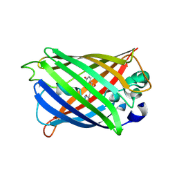
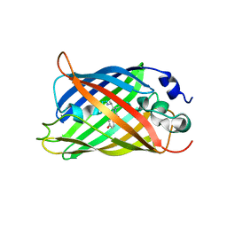
GFP R96M pre-cyclized intermediate in chromophore formation
Descriptor: MAGNESIUM ION, green-fluorescent protein
Authors:Wood, T.I, Barondeau, D.P, Hitomi, C, Kassmann, C.J, Tainer, J.A, Getzoff, E.D.
Deposit date:2005-09-01
Release date:2006-04-18
Last modified:2023-08-23
Method:X-RAY DIFFRACTION (1.6 Å)
Cite:Defining the role of arginine 96 in green fluorescent protein fluorophore biosynthesis.
Biochemistry, 44, 2005
1Q73
DownloadVisualize
BU of 1q73 by Molmil
S65T Q80R Y145C T203C Green Fluorescent Protein (GFP) pH 8.5
Descriptor: Green Fluorescent Protein
Authors:Jain, R.K, Ranganathan, R.
Deposit date:2003-08-15
Release date:2004-02-03
Last modified:2023-11-15
Method:X-RAY DIFFRACTION (1.6 Å)
Cite:Local complexity of amino acid interactions in a protein core.
Proc.Natl.Acad.Sci.USA, 101, 2004
3GEX
DownloadVisualize
BU of 3gex by Molmil
1.6 angstrom crystal structure of fluorescent protein Cypet
Descriptor: Green fluorescent protein
Authors:Hu, X, Liu, R.
Deposit date:2009-02-26
Release date:2010-03-09
Last modified:2023-11-22
Method:X-RAY DIFFRACTION (1.6 Å)
Cite:1.6 angstrom crystal structure of fluorescent protein Cypet
To be Published
3AI4
DownloadVisualize
BU of 3ai4 by Molmil
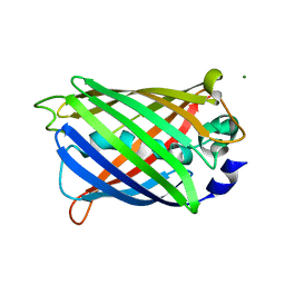
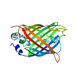
Crystal structure of yeast enhanced green fluorescent protein - mouse polymerase iota ubiquitin binding motif fusion protein
Descriptor: SULFATE ION, yeast enhanced green fluorescent protein,DNA polymerase iota
Authors:Suzuki, N, Wakatsuki, S, Kawasaki, M.
Deposit date:2010-05-10
Release date:2010-09-29
Last modified:2023-11-15
Method:X-RAY DIFFRACTION (1.6 Å)
Cite:Crystallization of small proteins assisted by green fluorescent protein
Acta Crystallogr.,Sect.D, 66, 2010
3G9A
DownloadVisualize
BU of 3g9a by Molmil
Green fluorescent protein bound to minimizer nanobody
Descriptor: Green fluorescent protein, Minimizer
Authors:Kirchhofer, A, Helma, J, Schmidthals, K, Frauer, C, Cui, S, Karcher, A, Pellis, M, Muyldermans, S, Delucci, C.C, Cardoso, M.C, Leonhardt, H, Hopfner, K.-P, Rothbauer, U.
Deposit date:2009-02-13
Release date:2009-12-08
Last modified:2023-11-15
Method:X-RAY DIFFRACTION (1.614 Å)
Cite:Modulation of protein properties in living cells using nanobodies
Nat.Struct.Mol.Biol., 17, 2010
6PFU
DownloadVisualize
BU of 6pfu by Molmil
rsEGFP2 with a chlorinated chromophore in the non-fluorescent off-state in a contracted unit cell
Descriptor: Green fluorescent protein, SULFATE ION
Authors:Chang, J, Romei, M.G, Boxer, S.G.
Deposit date:2019-06-22
Release date:2019-08-07
Last modified:2023-11-15
Method:X-RAY DIFFRACTION (1.619 Å)
Cite:Structural Evidence of Photoisomerization Pathways in Fluorescent Proteins.
J.Am.Chem.Soc., 141, 2019
4EN1
DownloadVisualize
BU of 4en1 by Molmil
The 1.62A structure of a FRET-optimized Cerulean Fluorescent Protein
Descriptor: ACETATE ION, DI(HYDROXYETHYL)ETHER, GLYCEROL, ...
Authors:Watkins, J.L.
Deposit date:2012-04-12
Release date:2013-04-24
Last modified:2023-12-06
Method:X-RAY DIFFRACTION (1.62 Å)
Cite:The 1.6 A resolution structure of a FRET-optimized Cerulean fluorescent protein.
Acta Crystallogr.,Sect.D, 69, 2013
3SSP
DownloadVisualize
BU of 3ssp by Molmil
Engineered low-affinity halide-binding protein derived from YFP: halide-free
Descriptor: Green fluorescent protein
Authors:Wang, W, Grimley, J.S, Beese, L.S, Hellinga, H.W.
Deposit date:2011-07-08
Release date:2012-07-11
Last modified:2023-11-15
Method:X-RAY DIFFRACTION (1.628 Å)
Cite:Determination of engineered chloride-binding site structures in fluorescent proteins reveals principles of halide recognition
To be Published
2YE1
DownloadVisualize
BU of 2ye1 by Molmil
X-ray structure of the cyan fluorescent proteinmTurquoise-GL (K206A mutant)
Descriptor: GREEN FLUORESCENT PROTEIN, MAGNESIUM ION
Authors:von Stetten, D, Noirclerc-Savoye, M, Goedhart, J, Gadella, T.W.J, Royant, A.
Deposit date:2011-03-25
Release date:2012-04-11
Last modified:2023-12-20
Method:X-RAY DIFFRACTION (1.63 Å)
Cite:Structural Characterization of the Cyan Fluorescent Protein Mturquoise-Gl
To be Published
4G28
DownloadVisualize
BU of 4g28 by Molmil
Calcium-calmodulin complexed with the calmodulin binding domain from a small conductance potassium channel splice variant and EBIO-1
Descriptor: 1-ethyl-1,3-dihydro-2H-benzimidazol-2-one, CALCIUM ION, Calmodulin, ...
Authors:Zhang, M, Pascal, J.M, Zhang, J.-F.
Deposit date:2012-07-11
Release date:2012-09-12
Last modified:2024-02-28
Method:X-RAY DIFFRACTION (1.63 Å)
Cite:Identification of the functional binding pocket for compounds targeting small-conductance Ca(2+)-activated potassium channels.
Nat Commun, 3, 2012
7AMF
DownloadVisualize
BU of 7amf by Molmil
Crystal structure of rsFolder2 in its non-fluorescent off-state
Descriptor: GLYCEROL, Green fluorescent protein
Authors:Moreno-Chicano, T, El Khatib, M, Colletier, J.-P.
Deposit date:2020-10-08
Release date:2022-08-24
Last modified:2024-01-31
Method:X-RAY DIFFRACTION (1.63 Å)
Cite:Rational Control of Off-State Heterogeneity in a Photoswitchable Fluorescent Protein Provides Switching Contrast Enhancement.
Chemphyschem, 23, 2022
7AMB
DownloadVisualize
BU of 7amb by Molmil
Crystal structure of rsFolder2 in its fluorescent on-state
Descriptor: GLYCEROL, Green fluorescent protein
Authors:Moreno-Chicano, T, El Khatib, M, Colletier, J.-P.
Deposit date:2020-10-08
Release date:2022-08-24
Last modified:2024-01-31
Method:X-RAY DIFFRACTION (1.63 Å)
Cite:Rational Control of Off-State Heterogeneity in a Photoswitchable Fluorescent Protein Provides Switching Contrast Enhancement.
Chemphyschem, 23, 2022
8A6O
DownloadVisualize
BU of 8a6o by Molmil
Room temperature rsEGFP2 with a chlorinated chromophore 600 fs after Photoexcitation
Descriptor: Green fluorescent protein
Authors:Fadini, A, van Thor, J.
Deposit date:2022-06-18
Release date:2023-07-19
Last modified:2024-02-07
Method:X-RAY DIFFRACTION (1.63 Å)
Cite:Serial Femtosecond Crystallography Reveals that Photoactivation in a Fluorescent Protein Proceeds via the Hula Twist Mechanism.
J.Am.Chem.Soc., 145, 2023
8A6S
DownloadVisualize
BU of 8a6s by Molmil
Room temperature rsEGFP2 with a chlorinated chromophore 1 microsecond after Photoexcitation
Descriptor: Green fluorescent protein
Authors:Fadini, A, van Thor, J.
Deposit date:2022-06-19
Release date:2023-07-19
Last modified:2024-02-07
Method:X-RAY DIFFRACTION (1.63 Å)
Cite:Serial Femtosecond Crystallography Reveals that Photoactivation in a Fluorescent Protein Proceeds via the Hula Twist Mechanism.
J.Am.Chem.Soc., 145, 2023
8A6N
DownloadVisualize
BU of 8a6n by Molmil
Room temperature rsEGFP2 with a chlorinated chromophore 300 fs after Photoexcitation
Descriptor: Green fluorescent protein
Authors:Fadini, A, van Thor, J.
Deposit date:2022-06-18
Release date:2023-07-19
Last modified:2024-02-07
Method:X-RAY DIFFRACTION (1.63 Å)
Cite:Serial Femtosecond Crystallography Reveals that Photoactivation in a Fluorescent Protein Proceeds via the Hula Twist Mechanism.
J.Am.Chem.Soc., 145, 2023
8A6R
DownloadVisualize
BU of 8a6r by Molmil
Room temperature rsEGFP2 with a chlorinated chromophore 100 ps after Photoexcitation
Descriptor: Green fluorescent protein
Authors:Fadini, A, van Thor, J.
Deposit date:2022-06-18
Release date:2023-07-19
Last modified:2024-02-07
Method:X-RAY DIFFRACTION (1.63 Å)
Cite:Serial Femtosecond Crystallography Reveals that Photoactivation in a Fluorescent Protein Proceeds via the Hula Twist Mechanism.
J.Am.Chem.Soc., 145, 2023
8A6G
DownloadVisualize
BU of 8a6g by Molmil
Room temperature rsEGFP2 with a chlorinated chromophore in the non-fluorescent OFF-state
Descriptor: Green fluorescent protein
Authors:Fadini, A, van Thor, J.
Deposit date:2022-06-17
Release date:2023-07-19
Last modified:2024-02-07
Method:X-RAY DIFFRACTION (1.63 Å)
Cite:Serial Femtosecond Crystallography Reveals that Photoactivation in a Fluorescent Protein Proceeds via the Hula Twist Mechanism.
J.Am.Chem.Soc., 145, 2023
8A6P
DownloadVisualize
BU of 8a6p by Molmil
Room temperature rsEGFP2 with a chlorinated chromophore 900 fs after photoexcitation
Descriptor: Green fluorescent protein
Authors:Fadini, A, van Thor, J.
Deposit date:2022-06-18
Release date:2023-07-19
Last modified:2024-02-07
Method:X-RAY DIFFRACTION (1.63 Å)
Cite:Serial Femtosecond Crystallography Reveals that Photoactivation in a Fluorescent Protein Proceeds via the Hula Twist Mechanism.
J.Am.Chem.Soc., 145, 2023
8A6Q
DownloadVisualize
BU of 8a6q by Molmil
Room temperature rsEGFP2 with a chlorinated chromophore 5 ps after photoexcitation
Descriptor: Green fluorescent protein
Authors:Fadini, A, van Thor, J.
Deposit date:2022-06-18
Release date:2023-07-19
Last modified:2024-02-07
Method:X-RAY DIFFRACTION (1.63 Å)
Cite:Serial Femtosecond Crystallography Reveals that Photoactivation in a Fluorescent Protein Proceeds via the Hula Twist Mechanism.
J.Am.Chem.Soc., 145, 2023
5WJ4
DownloadVisualize
BU of 5wj4 by Molmil
Crystal structure of redox-sensitive green fluorescent protein Clover mutant roClover1
Descriptor: Green fluorescent protein
Authors:Liu, C, Campbell, B.C, Petsko, G.A.
Deposit date:2017-07-21
Release date:2018-01-17
Last modified:2023-11-15
Method:X-RAY DIFFRACTION (1.631 Å)
Cite:Crystal Structure of Green Fluorescent Protein Clover and Design of Clover-Based Redox Sensors.
Structure, 26, 2018
7AMU
DownloadVisualize
BU of 7amu by Molmil
Crystal structure of rsEGFP2 T204A in its fluorescent on-state
Descriptor: GLYCEROL, Green fluorescent protein, SULFATE ION
Authors:Moreno-Chicano, T, Schlichting, I, Hartmann, E, Zala, N, Colletier, J.-P, Weik, M.
Deposit date:2020-10-09
Release date:2022-04-20
Last modified:2024-01-31
Method:X-RAY DIFFRACTION (1.64 Å)
Cite:Crystal structure of rsEGFP2 in its fluorescent on-state at pH 8.0
To Be Published

222415

數據於2024-07-10公開中

PDB statisticsPDBj update infoContact PDBjnumon