Loading
PDBj
MenuPDBj@FacebookPDBj@TwitterPDBj@YouTubewwPDB FoundationwwPDB
RCSB PDBPDBeBMRBAdv. SearchSearch help

2M0Y
DownloadVisualize
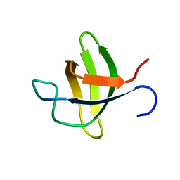
BU of 2m0y by Molmil
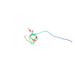
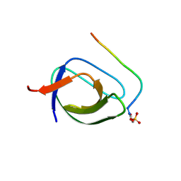
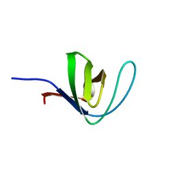
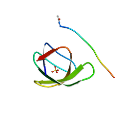
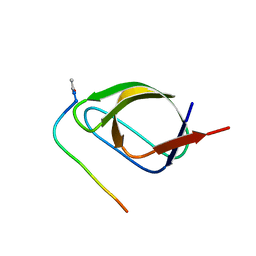
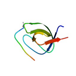
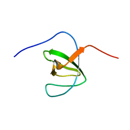
Solution structure of the SH3 domain of DOCK180
Descriptor: Dedicator of cytokinesis protein 1
Authors:Liu, X, Wen, W.
Deposit date:2012-11-08
Release date:2012-12-19
Last modified:2024-05-15
Method:SOLUTION NMR
Cite:Solution structure of the SH3 domain of DOCK180.
Proteins, 81, 2013
2O2W
DownloadVisualize
BU of 2o2w by Molmil
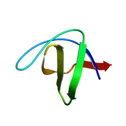
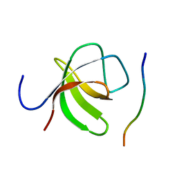
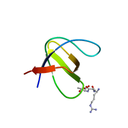
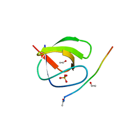
Extending powder diffraction to proteins: structure solution of the second SH3 domain from ponsin
Descriptor: Ponsin
Authors:Pinotsis, N, Margiolaki, I.
Deposit date:2006-11-30
Release date:2007-10-23
Last modified:2023-12-27
Method:POWDER DIFFRACTION
Cite:Second SH3 domain of ponsin solved from powder diffraction
J.Am.Chem.Soc., 129, 2007
2O31
DownloadVisualize
BU of 2o31 by Molmil
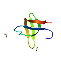
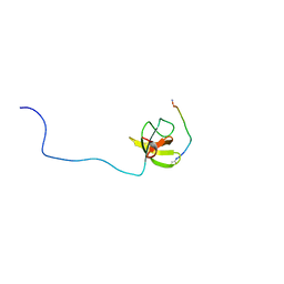
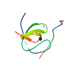
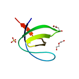
Crystal structure of the second SH3 domain from ponsin
Descriptor: FORMIC ACID, Ponsin
Authors:Pinotsis, N, Wilmanns, M, Margiolaki, I.
Deposit date:2006-11-30
Release date:2007-10-23
Last modified:2023-10-25
Method:X-RAY DIFFRACTION (1.5 Å)
Cite:Second SH3 domain of ponsin solved from powder diffraction
J.Am.Chem.Soc., 129, 2007
2O9S
DownloadVisualize
BU of 2o9s by Molmil
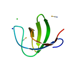
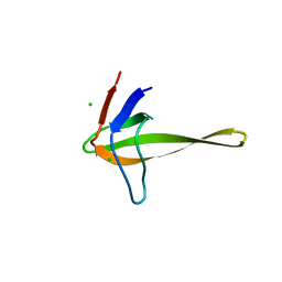
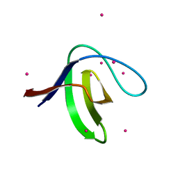
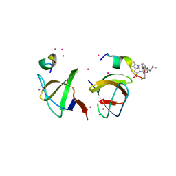
The second SH3 domain from ponsin
Descriptor: CHLORIDE ION, Ponsin, SODIUM ION, ...
Authors:Pinotsis, N, Wilmanns, M.
Deposit date:2006-12-14
Release date:2007-10-30
Last modified:2023-10-25
Method:X-RAY DIFFRACTION (0.83 Å)
Cite:Paxillin and ponsin interact in nascent costameres of muscle cells
J.Mol.Biol., 369, 2007
2OJ2
DownloadVisualize
BU of 2oj2 by Molmil
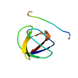
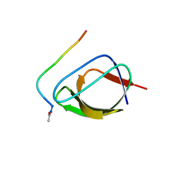
NMR Structure Analysis of the Hematopoetic Cell Kinase SH3 Domain complexed with an artificial high affinity ligand (PD1)
Descriptor: Hematopoetic Cell Kinase, SH3 domain, artificial peptide PD1
Authors:Schmidt, H, Hoffmann, S, Tran, T, Stoldt, M, Stangler, T, Wiesehan, K, Willbold, D.
Deposit date:2007-01-12
Release date:2007-01-30
Last modified:2023-12-27
Method:SOLUTION NMR
Cite:Solution Structure of a Hck SH3 Domain Ligand Complex Reveals Novel Interaction Modes
J.Mol.Biol., 365, 2007
2NUZ
DownloadVisualize
BU of 2nuz by Molmil
crystal structure of alpha spectrin SH3 domain measured at room temperature
Descriptor: Spectrin alpha chain, brain
Authors:Agarwal, V, Faelber, K, Hologne, M, Chevelkov, V, Oschkinat, H, Diehl, A, Reif, B.
Deposit date:2006-11-10
Release date:2007-05-15
Last modified:2023-10-25
Method:X-RAY DIFFRACTION (1.85 Å)
Cite:Comparison of MAS solid-state NMR and X-ray crystallographic data in the analysis of protein dynamics in the solid state
To be Published
2O88
DownloadVisualize
BU of 2o88 by Molmil
Crystal structure of the N114A mutant of ABL-SH3 domain complexed with a designed high-affinity peptide ligand: implications for SH3-ligand interactions
Descriptor: P41 peptide, Proto-oncogene tyrosine-protein kinase ABL1, SULFATE ION
Authors:Camara-Artigas, A.
Deposit date:2006-12-12
Release date:2007-05-01
Last modified:2023-08-30
Method:X-RAY DIFFRACTION (1.75 Å)
Cite:Crystallization by capillary counter-diffusion and structure determination of the N114A mutant of the SH3 domain of Abl tyrosine kinase complexed with a high-affinity peptide ligand.
Acta Crystallogr.,Sect.D, 63, 2007
2O9V
DownloadVisualize
BU of 2o9v by Molmil
The second SH3 domain from Ponsin in complex with the paxillin proline rich region
Descriptor: Paxillin, Ponsin
Authors:Pinotsis, N, Wilmanns, M.
Deposit date:2006-12-14
Release date:2007-10-30
Last modified:2023-10-25
Method:X-RAY DIFFRACTION (1.63 Å)
Cite:Paxillin and ponsin interact in nascent costameres of muscle cells
J.Mol.Biol., 369, 2007
2OI3
DownloadVisualize
BU of 2oi3 by Molmil
NMR Structure Analysis of the Hematopoetic Cell Kinase SH3 Domain complexed with an artificial high affinity ligand (PD1)
Descriptor: Tyrosine-protein kinase HCK, artificial peptide PD1
Authors:Schmidt, H, Stoldt, M, Hoffmann, S, Tran, T, Willbold, D.
Deposit date:2007-01-10
Release date:2007-02-20
Last modified:2023-12-27
Method:SOLUTION NMR
Cite:Solution Structure of a Hck SH3 Domain Ligand Complex Reveals Novel Interaction Modes
J.Mol.Biol., 365, 2007
2OAW
DownloadVisualize
BU of 2oaw by Molmil
Structure of SHH variant of "Bergerac" chimera of spectrin SH3
Descriptor: CHLORIDE ION, Spectrin alpha chain, brain
Authors:Gabdoulkhakov, A.G, Gushchina, L.V, Nikulin, A.D, Nikonov, S.V, Viguera, A.R, Serrano, L, Filimonov, V.V.
Deposit date:2006-12-18
Release date:2008-04-08
Last modified:2023-08-30
Method:X-RAY DIFFRACTION (1.9 Å)
Cite:Structural and thermodynamic studies of Bergerac-SH3 chimeras.
Biophys.Chem., 139, 2009
4FSS
DownloadVisualize
BU of 4fss by Molmil
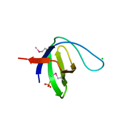
Crystal structure of a RAS p21 protein activator (RASA1) from Homo sapiens at 2.25 A resolution
Descriptor: CHLORIDE ION, GLYCEROL, PHOSPHATE ION, ...
Authors:Joint Center for Structural Genomics (JCSG), Partnership for T-Cell Biology (TCELL)
Deposit date:2012-06-27
Release date:2012-07-18
Last modified:2023-02-01
Method:X-RAY DIFFRACTION (2.25 Å)
Cite:Crystal structure of a RAS p21 protein activator (RASA1) from Homo sapiens at 2.25 A resolution
To be published
4HCK
DownloadVisualize
BU of 4hck by Molmil
HUMAN HCK SH3 DOMAIN, NMR, 25 STRUCTURES
Descriptor: HEMATOPOIETIC CELL KINASE
Authors:Horita, D.A, Baldisseri, D.M, Zhang, W, Altieri, A.S, Smithgall, T.E, Gmeiner, W.H, Byrd, R.A.
Deposit date:1998-03-09
Release date:1998-06-17
Last modified:2024-05-01
Method:SOLUTION NMR
Cite:Solution structure of the human Hck SH3 domain and identification of its ligand binding site.
J.Mol.Biol., 278, 1998
4HXJ
DownloadVisualize
BU of 4hxj by Molmil
Crystal structure of SH3:RGT complex
Descriptor: C-terminal 3-mer peptide from Integrin beta-3, Proto-oncogene tyrosine-protein kinase Src
Authors:Xiao, R, Meng, G.
Deposit date:2012-11-11
Release date:2012-11-28
Last modified:2023-11-08
Method:X-RAY DIFFRACTION (2 Å)
Cite:Structural framework of c-Src activation by integrin beta 3
Blood, 121, 2013
4HVW
DownloadVisualize
BU of 4hvw by Molmil
Crystal structure of the T98E c-Src-SH3 domain mutant in complex with the high affinity peptide VSL12
Descriptor: ACETYL GROUP, Proto-oncogene tyrosine-protein kinase Src, SULFATE ION, ...
Authors:Camara-Artigas, A.
Deposit date:2012-11-07
Release date:2013-05-01
Last modified:2023-09-20
Method:X-RAY DIFFRACTION (0.98 Å)
Cite:Atomic resolution structures of the c-Src SH3 domain in complex with two high-affinity peptides from classes I and II.
Acta Crystallogr.,Sect.D, 69, 2013
4HVV
DownloadVisualize
BU of 4hvv by Molmil
Crystal structure of the T98E c-Src-SH3 domain mutant in complex with the high affinity peptide APP12
Descriptor: Proto-oncogene tyrosine-protein kinase Src, SULFATE ION, SYNTHETIC PEPTIDE Acetyl-APPLPPRNRP
Authors:Camara-Artigas, A.
Deposit date:2012-11-07
Release date:2013-05-01
Last modified:2023-09-20
Method:X-RAY DIFFRACTION (1.1 Å)
Cite:Atomic resolution structures of the c-Src SH3 domain in complex with two high-affinity peptides from classes I and II.
Acta Crystallogr.,Sect.D, 69, 2013
4HVU
DownloadVisualize
BU of 4hvu by Molmil
Crystal structure of the T98D c-Src-SH3 domain mutant in complex with the high affinity peptide APP12
Descriptor: ACETYL GROUP, Proto-oncogene tyrosine-protein kinase Src, SULFATE ION, ...
Authors:Camara-Artigas, A.
Deposit date:2012-11-07
Release date:2013-05-01
Last modified:2023-09-20
Method:X-RAY DIFFRACTION (0.98 Å)
Cite:Atomic resolution structures of the c-Src SH3 domain in complex with two high-affinity peptides from classes I and II.
Acta Crystallogr.,Sect.D, 69, 2013
4GLM
DownloadVisualize
BU of 4glm by Molmil
Crystal structure of the SH3 Domain of DNMBP protein [Homo sapiens]
Descriptor: Dynamin-binding protein, UNKNOWN ATOM OR ION
Authors:Dong, A, Guan, X, Huang, H, Tempel, W, Gu, J, Sidhu, S, Bountra, C, Arrowsmith, C.H, Edwards, A.M, Tong, Y, Structural Genomics Consortium (SGC)
Deposit date:2012-08-14
Release date:2012-11-21
Last modified:2023-09-13
Method:X-RAY DIFFRACTION (1.9 Å)
Cite:Crystal structure of the SH3 Domain of DNMBP protein [Homo sapiens]
to be published
4GBQ
DownloadVisualize
BU of 4gbq by Molmil
SOLUTION NMR STRUCTURE OF THE GRB2 N-TERMINAL SH3 DOMAIN COMPLEXED WITH A TEN-RESIDUE PEPTIDE DERIVED FROM SOS DIRECT REFINEMENT AGAINST NOES, J-COUPLINGS, AND 1H AND 13C CHEMICAL SHIFTS, 15 STRUCTURES
Descriptor: GRB2, SOS-1
Authors:Wittekind, M, Mapelli, C, Lee, V, Goldfarb, V, Friedrichs, M.S, Meyers, C.A, Mueller, L.
Deposit date:1996-12-23
Release date:1997-09-04
Last modified:2022-03-16
Method:SOLUTION NMR
Cite:Solution structure of the Grb2 N-terminal SH3 domain complexed with a ten-residue peptide derived from SOS: direct refinement against NOEs, J-couplings and 1H and 13C chemical shifts.
J.Mol.Biol., 267, 1997
4J9E
DownloadVisualize
BU of 4j9e by Molmil
Crystal structure of the N114A mutant of the Abl-SH3 domain complexed with the high affinity peptide P17
Descriptor: P17, SULFATE ION, Tyrosine-protein kinase ABL1
Authors:Camara-Artigas, A.
Deposit date:2013-02-16
Release date:2014-01-29
Last modified:2023-09-20
Method:X-RAY DIFFRACTION (1.4 Å)
Cite:Crystal structure of the N114A mutant of the Abl-SH3 domain complexed with the high affinity peptide P17
To be Published
4IIM
DownloadVisualize
BU of 4iim by Molmil
Crystal structure of the Second SH3 Domain of ITSN1 bound with a synthetic peptide
Descriptor: Intersectin-1, UNKNOWN ATOM OR ION, peptide ligand
Authors:Dong, A, Guan, X, Huang, H, Wernimont, A, Gu, J, Sidhu, S, Bountra, C, Arrowsmith, C.H, Edwards, A.M, Tong, Y, Structural Genomics Consortium (SGC)
Deposit date:2012-12-20
Release date:2013-01-23
Last modified:2023-09-20
Method:X-RAY DIFFRACTION (1.8 Å)
Cite:Crystal structure of the Second SH3 Domain of ITSN1 bound with a synthetic peptide
To be Published
4J9B
DownloadVisualize
BU of 4j9b by Molmil
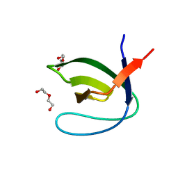
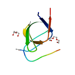
Crystal structure of the Abl-SH3 domain H59Q-N96T mutant
Descriptor: DI(HYDROXYETHYL)ETHER, Tyrosine-protein kinase ABL1
Authors:Camara-Artigas, A.
Deposit date:2013-02-16
Release date:2014-01-29
Last modified:2023-09-20
Method:X-RAY DIFFRACTION (1.702 Å)
Cite:Crystal structure of the Abl-SH3 domain H59Q-N96T mutant
To be Published
4J9D
DownloadVisualize
BU of 4j9d by Molmil
Crystal structure of the N114A mutant of the Abl-SH3 domain complexed with the high affinity peptide P0
Descriptor: P0, SULFATE ION, Tyrosine-protein kinase ABL1
Authors:Camara-Artigas, A.
Deposit date:2013-02-16
Release date:2014-01-29
Last modified:2023-09-20
Method:X-RAY DIFFRACTION (1.5 Å)
Cite:Crystal structure of the N114A mutant of the Abl-SH3 domain complexed with the high affinity peptide P0
To be Published
4JJB
DownloadVisualize
BU of 4jjb by Molmil
Crystal structure of the Abl-SH3 domain at pH3
Descriptor: DI(HYDROXYETHYL)ETHER, SULFATE ION, Tyrosine-protein kinase ABL1
Authors:Camara-Artigas, A, Martin-Garcia, J.M.
Deposit date:2013-03-07
Release date:2014-03-12
Last modified:2023-11-08
Method:X-RAY DIFFRACTION (1.65 Å)
Cite:Crystal structure of the Abl-SH3 domain at pH3
To be Published
4J9H
DownloadVisualize
BU of 4j9h by Molmil
Crystal structure of the ABL-SH3 domain complexed with the designed high-affinity peptide ligand P7 at pH 8
Descriptor: P7, SULFATE ION, Tyrosine-protein kinase ABL1
Authors:Camara-Artigas, A.
Deposit date:2013-02-16
Release date:2014-01-29
Last modified:2023-09-20
Method:X-RAY DIFFRACTION (1.7 Å)
Cite:Crystal structure of the ABL-SH3 domain complexed with the designed high-affinity peptide ligand P7
To be Published
4J9C
DownloadVisualize
BU of 4j9c by Molmil
Crystal structure of the Abl-SH3 domain H59Q-N96T mutant complexed with the designed high-affinity peptide ligand P17
Descriptor: DI(HYDROXYETHYL)ETHER, P17, TRIETHYLENE GLYCOL, ...
Authors:Camara-Artigas, A.
Deposit date:2013-02-16
Release date:2014-01-29
Last modified:2023-09-20
Method:X-RAY DIFFRACTION (1.051 Å)
Cite:Crystal structure of the Abl-SH3 domain H59Q-N96T mutant complexed with the designed high-affinity peptide ligand P17
To be Published

222415

数据于2024-07-10公开中

PDB statisticsPDBj update infoContact PDBjnumon