Loading
PDBj
MenuPDBj@FacebookPDBj@TwitterPDBj@YouTubewwPDB FoundationwwPDB
RCSB PDBPDBeBMRBAdv. SearchSearch help

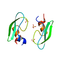
2ZVX
DownloadVisualize
BU of 2zvx by Molmil
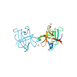
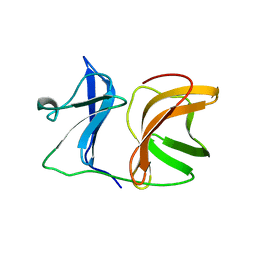
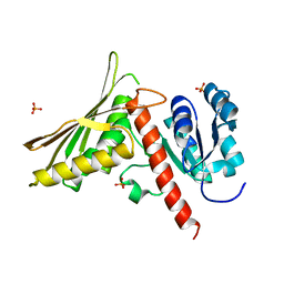
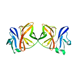
Structure of a BPTI-[5,55] variant containing Gly/Val at the 14/38th positions
Descriptor: Pancreatic trypsin inhibitor, SULFATE ION
Authors:Islam, M.M, Sohya, S, Noguchi, K, Yohda, M, Kuroda, Y.
Deposit date:2008-11-24
Release date:2009-10-13
Last modified:2023-11-01
Method:X-RAY DIFFRACTION (1.09 Å)
Cite:Thermodynamic and structural analysis of highly stabilized BPTIs by single and double mutations
Proteins, 77, 2009
4II0
DownloadVisualize
BU of 4ii0 by Molmil
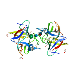
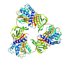
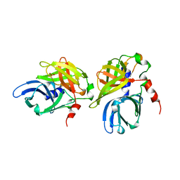
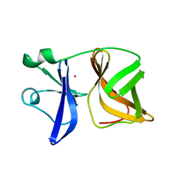
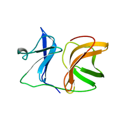
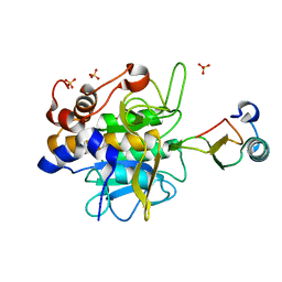
Crystal structure of CrataBL, a trypsin inhibitor from Crataeva tapia
Descriptor: 2-acetamido-2-deoxy-beta-D-glucopyranose, CrataBL, GLYCEROL, ...
Authors:Zhou, D, Wlodawer, A.
Deposit date:2012-12-19
Release date:2013-07-24
Last modified:2020-07-29
Method:X-RAY DIFFRACTION (1.75 Å)
Cite:Crystal Structure of Crataeva tapia Bark Protein (CrataBL) and Its Effect in Human Prostate Cancer Cell Lines.
Plos One, 8, 2013
4IHZ
DownloadVisualize
BU of 4ihz by Molmil
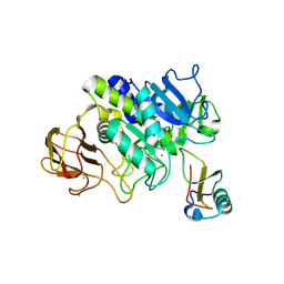
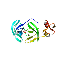
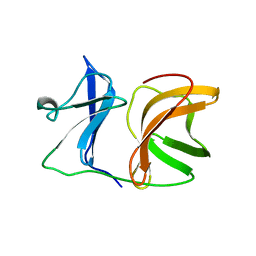
Crystal structure of CrataBL, a trypsin inhibitor from Crataeva tapia
Descriptor: 2-acetamido-2-deoxy-beta-D-glucopyranose, CHLORIDE ION, CrataBL, ...
Authors:Zhou, D, Wlodawer, A.
Deposit date:2012-12-19
Release date:2013-07-24
Last modified:2020-07-29
Method:X-RAY DIFFRACTION (1.5 Å)
Cite:Crystal Structure of Crataeva tapia Bark Protein (CrataBL) and Its Effect in Human Prostate Cancer Cell Lines.
Plos One, 8, 2013
1DTK
DownloadVisualize
BU of 1dtk by Molmil
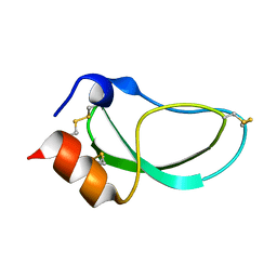
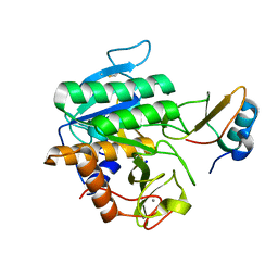
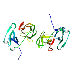
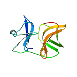
THE NMR SOLUTION STRUCTURE OF DENDROTOXIN K FROM THE VENOM OF DENDROASPIS POLYLEPIS POLYLEPIS
Descriptor: DENDROTOXIN K
Authors:Berndt, K, Guntert, P, Wuthrich, K.
Deposit date:1993-04-02
Release date:1994-01-31
Last modified:2024-10-16
Method:SOLUTION NMR
Cite:Nuclear magnetic resonance solution structure of dendrotoxin K from the venom of Dendroaspis polylepis polylepis.
J.Mol.Biol., 234, 1993
1GM2
DownloadVisualize
BU of 1gm2 by Molmil
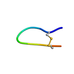
The independent structure of the antitryptic reactive site loop of Bowman-Birk inhibitor and sunflower trypsin inhibitor-1
Descriptor: BOWMAN-BIRK INHIBITOR DERIVED PEPTIDE
Authors:Brauer, A.B.E, Kelly, G, Matthews, S.J, Leatherbarrow, R.J.
Deposit date:2001-09-08
Release date:2002-08-29
Last modified:2024-10-16
Method:SOLUTION NMR
Cite:The (1)H-NMR solution structure of the antitryptic core peptide of Bowman-Birk inhibitor proteins: a minimal canonical loop.
J.Biomol.Struct.Dyn., 20, 2002
1KXA
DownloadVisualize
BU of 1kxa by Molmil
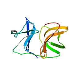
SINDBIS VIRUS CAPSID, (WILD-TYPE) RESIDUES 106-264, TETRAGONAL CRYSTAL FORM
Descriptor: SINDBIS VIRUS CAPSID PROTEIN
Authors:Choi, H.-K, Rossmann, M.G.
Deposit date:1996-05-05
Release date:1996-11-08
Last modified:2024-02-14
Method:X-RAY DIFFRACTION (3.1 Å)
Cite:Structural analysis of Sindbis virus capsid mutants involving assembly and catalysis.
J.Mol.Biol., 262, 1996
6KGA
DownloadVisualize
BU of 6kga by Molmil
Structure of Ovalbumin from Emu (Dromaius novaehollandiae)
Descriptor: Ovalbumin
Authors:Yasutake, Y, Maehashi, K, Matano, M, Takeuchi, J.
Deposit date:2019-07-11
Release date:2019-07-24
Last modified:2024-10-30
Method:X-RAY DIFFRACTION (3.3 Å)
Cite:Structure of Emu (Dromaius novaehollandiae) ovalbumin revealed alpha-1-antitrypsin-like domain-swapped trimer.
To Be Published
5FBZ
DownloadVisualize
BU of 5fbz by Molmil
Structure of subtilase SubHal from Bacillus halmapalus - complex with chymotrypsin inhibitor CI2A
Descriptor: Autoproteolytic fragment of enzyme subtilase SubHal, CALCIUM ION, Enzyme subtilase SubHal from Bacillus halmapalus, ...
Authors:Dohnalek, J, Brzozowski, A.M, Svendsen, A, Wilson, K.S.
Deposit date:2015-12-14
Release date:2016-05-18
Last modified:2024-07-10
Method:X-RAY DIFFRACTION (1.9 Å)
Cite:Stabilization of Enzymes by Metal Binding: Structures of Two Alkalophilic Bacillus Subtilases and Analysis of the Second Metal-Binding Site of the Subtilase Family
Book, 2016
5FFN
DownloadVisualize
BU of 5ffn by Molmil
Complex of subtilase SubTY from Bacillus sp. TY145 with chymotrypsin inhibitor CI2A
Descriptor: CALCIUM ION, Enzyme subtilase SubTY from Bacillus sp. TY145, SODIUM ION, ...
Authors:McAuley, K.E, Svendsen, A, Oestergaard, P.R, Dohnalek, J, Wilson, K.S.
Deposit date:2015-12-18
Release date:2016-05-18
Last modified:2024-01-10
Method:X-RAY DIFFRACTION (1.8 Å)
Cite:Stabilization of Enzymes by Metal Binding: Structures of Two Alkalophilic Bacillus Subtilases and Analysis of the Second Metal-Binding Site of the Subtilase Family
Book, 2016
7Z3S
DownloadVisualize
BU of 7z3s by Molmil
CRYSTAL STRUCTURE of PheF from Geobacillus stearothermophilus
Descriptor: Acetaldehyde dehydrogenase, PHOSPHATE ION
Authors:Driller, R, Haack, M, Brueck, T, Loll, B.
Deposit date:2022-03-02
Release date:2023-01-11
Last modified:2024-01-31
Method:X-RAY DIFFRACTION (1.99 Å)
Cite:CRYSTAL STRUCTURE OF THE P1 trifluoroethylglycine (TfeGly) BPTI MUTANT- BOVINE CHYMOTRYPSIN COMPLEX
To Be Published
1HAV
DownloadVisualize
BU of 1hav by Molmil
HEPATITIS A VIRUS 3C PROTEINASE
Descriptor: CHLORIDE ION, HEPATITIS A VIRUS 3C PROTEINASE
Authors:Bergmann, E.M, James, M.N.G.
Deposit date:1996-10-23
Release date:1996-12-23
Last modified:2024-04-03
Method:X-RAY DIFFRACTION (2 Å)
Cite:The refined crystal structure of the 3C gene product from hepatitis A virus: specific proteinase activity and RNA recognition.
J.Virol., 71, 1997
3FAO
DownloadVisualize
BU of 3fao by Molmil
Crystal structure of S118A mutant 3CLSP of PRRSV
Descriptor: Non-structural protein, PHOSPHATE ION
Authors:Gao, F, Peng, H, Tian, X, Lu, G, Liu, Y, Gao, G.F.
Deposit date:2008-11-17
Release date:2009-10-20
Last modified:2023-12-27
Method:X-RAY DIFFRACTION (2.01 Å)
Cite:Structure and cleavage specificity of the chymotrypsin-like serine protease (3CLSP/nsp4) of Porcine Reproductive and Respiratory Syndrome Virus (PRRSV).
J.Mol.Biol., 392, 2009
2SNW
DownloadVisualize
BU of 2snw by Molmil
SINDBIS VIRUS CAPSID PROTEIN, TYPE3 CRYSTAL FORM
Descriptor: COAT PROTEIN C
Authors:Choi, H.-K, Lee, S, Zhang, Y.-P, Mckinney, B.R, Wengler, G, Rossmann, M.G, Kuhn, R.J.
Deposit date:1998-02-17
Release date:1998-04-08
Last modified:2024-02-21
Method:X-RAY DIFFRACTION (2.7 Å)
Cite:Structural analysis of Sindbis virus capsid mutants involving assembly and catalysis.
J.Mol.Biol., 262, 1996
1VCQ
DownloadVisualize
BU of 1vcq by Molmil
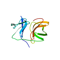
SEMLIKI FOREST VIRUS CAPSID PROTEIN (CRYSTAL FORM II)
Descriptor: SEMLIKI FOREST VIRUS CAPSID PROTEIN
Authors:Lu, G, Choi, H.-K, Rossmann, M.G.
Deposit date:1996-03-04
Release date:1996-12-07
Last modified:2024-11-06
Method:X-RAY DIFFRACTION (3.1 Å)
Cite:Structure of Semliki Forest virus core protein.
Proteins, 27, 1997
1VCP
DownloadVisualize
BU of 1vcp by Molmil
SEMLIKI FOREST VIRUS CAPSID PROTEIN (CRYSTAL FORM I)
Descriptor: MERCURY (II) ION, SEMLIKI FOREST VIRUS CAPSID PROTEIN
Authors:Lu, G, Choi, H.-K, Rossmann, M.G.
Deposit date:1996-03-04
Release date:1996-12-07
Last modified:2024-02-14
Method:X-RAY DIFFRACTION (3 Å)
Cite:Structure of Semliki Forest virus core protein.
Proteins, 27, 1997
1KXC
DownloadVisualize
BU of 1kxc by Molmil
SINDBIS VIRUS CAPSID (N190K MUTANT), TETRAGONAL CRYSTAL FORM
Descriptor: SINDBIS VIRUS CAPSID PROTEIN
Authors:Choi, H.-K, Rossmann, M.G.
Deposit date:1996-05-05
Release date:1996-11-08
Last modified:2024-02-14
Method:X-RAY DIFFRACTION (3.1 Å)
Cite:Structural analysis of Sindbis virus capsid mutants involving assembly and catalysis.
J.Mol.Biol., 262, 1996
1KXD
DownloadVisualize
BU of 1kxd by Molmil
SINDBIS VIRUS CAPSID (N222L MUTANT), TETRAGONAL CRYSTAL FORM
Descriptor: SINDBIS VIRUS CAPSID PROTEIN
Authors:Choi, H.-K, Rossmann, M.G.
Deposit date:1996-05-05
Release date:1996-11-08
Last modified:2024-02-14
Method:X-RAY DIFFRACTION (3 Å)
Cite:Structural analysis of Sindbis virus capsid mutants involving assembly and catalysis.
J.Mol.Biol., 262, 1996
1KXE
DownloadVisualize
BU of 1kxe by Molmil
SINDBIS VIRUS CAPSID (Y180S, E183G DOUBLE MUTANT), TETRAGONAL CRYSTAL FORM
Descriptor: SINDBIS VIRUS CAPSID PROTEIN
Authors:Choi, H.-K, Rossmann, M.G.
Deposit date:1996-05-05
Release date:1996-11-08
Last modified:2024-02-14
Method:X-RAY DIFFRACTION (3.2 Å)
Cite:Structural analysis of Sindbis virus capsid mutants involving assembly and catalysis.
J.Mol.Biol., 262, 1996
1KXB
DownloadVisualize
BU of 1kxb by Molmil
SINDBIS VIRUS CAPSID (S215A MUTANT), TETRAGONAL CRYSTAL FORM
Descriptor: SINDBIS VIRUS CAPSID PROTEIN
Authors:Choi, H.-K, Rossmann, M.G.
Deposit date:1996-05-05
Release date:1996-11-08
Last modified:2024-02-14
Method:X-RAY DIFFRACTION (2.9 Å)
Cite:Structural analysis of Sindbis virus capsid mutants involving assembly and catalysis.
J.Mol.Biol., 262, 1996
1KXF
DownloadVisualize
BU of 1kxf by Molmil
SINDBIS VIRUS CAPSID, (WILD-TYPE) RESIDUES 1-264, TETRAGONAL CRYSTAL FORM (FORM II)
Descriptor: SINDBIS VIRUS CAPSID PROTEIN
Authors:Choi, H.-K, Rossmann, M.G.
Deposit date:1996-05-05
Release date:1996-12-07
Last modified:2024-02-14
Method:X-RAY DIFFRACTION (2.38 Å)
Cite:Structural analysis of Sindbis virus capsid mutants involving assembly and catalysis.
J.Mol.Biol., 262, 1996
3OEU
DownloadVisualize
BU of 3oeu by Molmil
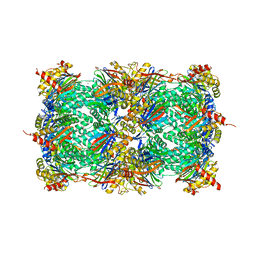
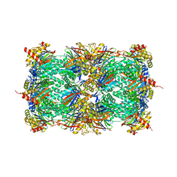
Structure of yeast 20S open-gate proteasome with Compound 24
Descriptor: 2-(N-MORPHOLINO)-ETHANESULFONIC ACID, MAGNESIUM ION, N-{(2S)-1-[(2-chlorobenzyl)amino]-1-oxo-4-phenylbutan-2-yl}-N~2~-[3-(2-methylphenyl)propanoyl]-L-threoninamide, ...
Authors:Sintchak, M.D.
Deposit date:2010-08-13
Release date:2011-07-20
Last modified:2024-02-21
Method:X-RAY DIFFRACTION (2.6 Å)
Cite:Optimization of a series of dipeptides with a P3 threonine residue as non-covalent inhibitors of the chymotrypsin-like activity of the human 20S proteasome.
Bioorg.Med.Chem.Lett., 20, 2010
3OEV
DownloadVisualize
BU of 3oev by Molmil
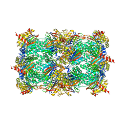
Structure of yeast 20S open-gate proteasome with Compound 25
Descriptor: 2-(N-MORPHOLINO)-ETHANESULFONIC ACID, 4-(benzyloxy)-N-[(2S,3R)-3-hydroxy-1-{[(2S)-1-{[(3-methylthiophen-2-yl)methyl]amino}-1-oxo-4-phenylbutan-2-yl]amino}-1-oxobutan-2-yl]benzamide, MAGNESIUM ION, ...
Authors:Sintchak, M.D.
Deposit date:2010-08-13
Release date:2011-07-20
Last modified:2024-02-21
Method:X-RAY DIFFRACTION (2.85 Å)
Cite:Optimization of a series of dipeptides with a P3 threonine residue as non-covalent inhibitors of the chymotrypsin-like activity of the human 20S proteasome.
Bioorg.Med.Chem.Lett., 20, 2010
3SDI
DownloadVisualize
BU of 3sdi by Molmil
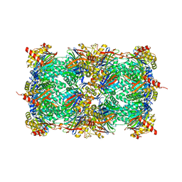
Structure of yeast 20S open-gate proteasome with Compound 20
Descriptor: 2-(N-MORPHOLINO)-ETHANESULFONIC ACID, MAGNESIUM ION, N~4~-(2,2-dimethylpropyl)-N~1~-{(2S)-1-[(4-methylbenzyl)amino]-1-oxo-4-phenylbutan-2-yl}-N~2~-[(5-methyl-1,2-oxazol-3-yl)carbonyl]-L-aspartamide, ...
Authors:Sintchak, M.D.
Deposit date:2011-06-09
Release date:2012-06-13
Last modified:2023-09-13
Method:X-RAY DIFFRACTION (2.65 Å)
Cite:Optimization of a series of dipeptides with a P3 threonine residue as non-covalent inhibitors of the chymotrypsin-like activity of the human 20S proteasome.
Bioorg.Med.Chem.Lett., 20, 2010
3SDK
DownloadVisualize
BU of 3sdk by Molmil
Structure of yeast 20S open-gate proteasome with Compound 34
Descriptor: 2-(N-MORPHOLINO)-ETHANESULFONIC ACID, MAGNESIUM ION, N-[(2S)-3-(3-tert-butyl-1,2,4-oxadiazol-5-yl)-1-({(2S)-1-[(4-methylbenzyl)amino]-1-oxo-4-phenylbutan-2-yl}amino)-1-oxopropan-2-yl]-5-methyl-1,2-oxazole-3-carboxamide, ...
Authors:Sintchak, M.D.
Deposit date:2011-06-09
Release date:2012-06-13
Last modified:2023-09-13
Method:X-RAY DIFFRACTION (2.7 Å)
Cite:Optimization of a series of dipeptides with a P3 threonine residue as non-covalent inhibitors of the chymotrypsin-like activity of the human 20S proteasome.
Bioorg.Med.Chem.Lett., 20, 2010
1LW6
DownloadVisualize
BU of 1lw6 by Molmil
Crystal Structure of the Complex of Subtilisin BPN' with Chymotrypsin Inhibitor 2 at 1.5 Angstrom Resolution
Descriptor: CALCIUM ION, SUBTILISIN BPN', SUBTILISIN-CHYMOTRYPSIN INHIBITOR-2A, ...
Authors:Radisky, E.S, Koshland JR, D.E.
Deposit date:2002-05-30
Release date:2002-08-21
Last modified:2024-02-14
Method:X-RAY DIFFRACTION (1.5 Å)
Cite:A clogged gutter mechanism for protease inhibitors.
Proc.Natl.Acad.Sci.USA, 99, 2002

227111

数据于2024-11-06公开中

PDB statisticsPDBj update infoContact PDBjnumon