Loading
PDBj
MenuPDBj@FacebookPDBj@TwitterPDBj@YouTubewwPDB FoundationwwPDB
RCSB PDBPDBeBMRBAdv. SearchSearch help

3FIQ
DownloadVisualize
BU of 3fiq by Molmil
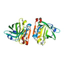
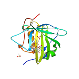
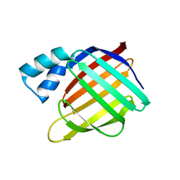
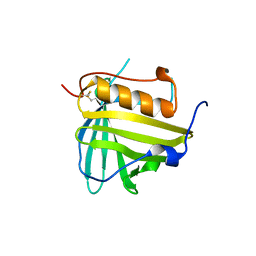
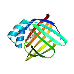
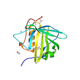
Odorant Binding Protein OBP1
Descriptor: 1,2-ETHANEDIOL, Odorant-binding protein 1F
Authors:White, S.A.
Deposit date:2008-12-12
Release date:2009-05-19
Last modified:2023-11-01
Method:X-RAY DIFFRACTION (1.599 Å)
Cite:Structure of rat odorant-binding protein OBP1 at 1.6 A resolution
Acta Crystallogr.,Sect.D, 65, 2009
6MLB
DownloadVisualize
BU of 6mlb by Molmil
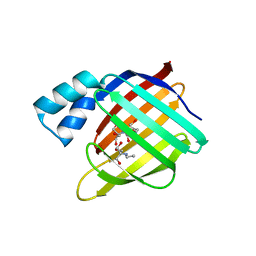
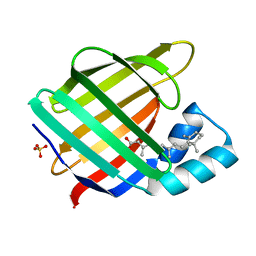
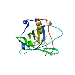
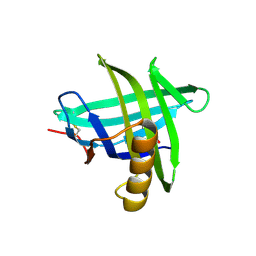
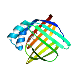
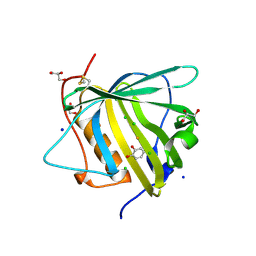
Crystal structure of the holo retinal-bound domain-swapped dimer Q108K:K40L:T51F mutant of human cellular retinol binding protein II
Descriptor: ACETATE ION, GLYCEROL, RETINAL, ...
Authors:Ghanbarpour, A, Geiger, J.
Deposit date:2018-09-27
Release date:2019-10-16
Last modified:2023-10-11
Method:X-RAY DIFFRACTION (2.15 Å)
Cite:Engineering the hCRBPII Domain-Swapped Dimer into a New Class of Protein Switches.
J.Am.Chem.Soc., 141, 2019
4QGW
DownloadVisualize
BU of 4qgw by Molmil
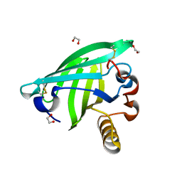
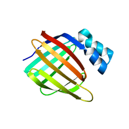
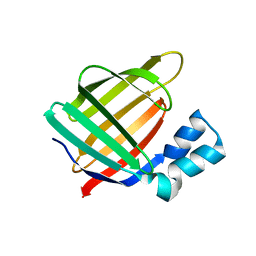
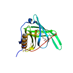
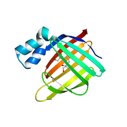
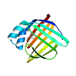
Crystal sturcture of the R132K:R111L:L121D mutant of Cellular Retinoic Acid Binding ProteinII complexed with a synthetic ligand (Merocyanine) at 1.77 angstrom resolution
Descriptor: (2E,4E,6E)-3-methyl-6-(1,3,3-trimethyl-1,3-dihydro-2H-indol-2-ylidene)hexa-2,4-dienal, 2-[3-(2-HYDROXY-1,1-DIHYDROXYMETHYL-ETHYLAMINO)-PROPYLAMINO]-2-HYDROXYMETHYL-PROPANE-1,3-DIOL, Cellular retinoic acid-binding protein 2
Authors:Nosrati, M, Yapici, I, Geiger, J.H.
Deposit date:2014-05-26
Release date:2015-01-28
Last modified:2015-02-11
Method:X-RAY DIFFRACTION (1.77 Å)
Cite:"Turn-on" protein fluorescence: in situ formation of cyanine dyes.
J.Am.Chem.Soc., 137, 2015
6MR0
DownloadVisualize
BU of 6mr0 by Molmil
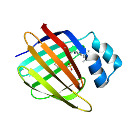
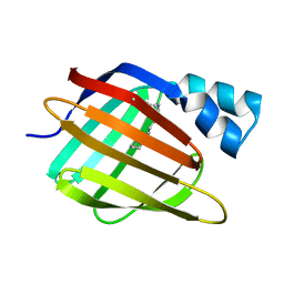
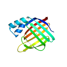
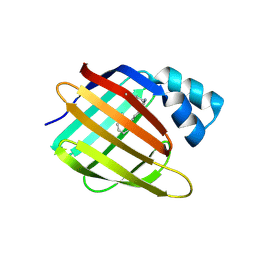
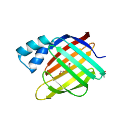
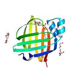
Crystal Structure of the All-trans Retinal Bound R111K:Y134F:T54V:R132Q:P39Q:R59Y:L121E Human Cellular Retinoic Acid Binding Protein II Mutant After 5 Minutes UV irradiation at 2.6 Angstrom Resolution
Descriptor: Cellular retinoic acid-binding protein 2, RETINAL
Authors:Ghanbarpour, A, Geiger, J.
Deposit date:2018-10-11
Release date:2019-01-09
Last modified:2023-10-11
Method:X-RAY DIFFRACTION (2.649 Å)
Cite:Mimicking Microbial Rhodopsin Isomerization in a Single Crystal.
J. Am. Chem. Soc., 141, 2019
6MOX
DownloadVisualize
BU of 6mox by Molmil
Crystal Structure of the All-trans Retinal-Bound R111K:Y134F:T54V:R132Q:P39Q:R59Y:L121E Human Cellular Retinoic Acid Binding Protein II Mutant in the Dark at 2.18 Angstrom Resolution
Descriptor: Cellular retinoic acid-binding protein 2, RETINAL
Authors:Ghanbarpour, A, Geiger, J.
Deposit date:2018-10-04
Release date:2019-01-09
Last modified:2023-10-11
Method:X-RAY DIFFRACTION (2.18 Å)
Cite:Mimicking Microbial Rhodopsin Isomerization in a Single Crystal.
J. Am. Chem. Soc., 141, 2019
6MKV
DownloadVisualize
BU of 6mkv by Molmil
Crystal structure of Retinal-bound holo Q108K:K40L:T51W domain-swapped dimer of human cellular retinol binding protein 2
Descriptor: ACETATE ION, GLYCEROL, RETINAL, ...
Authors:Ghanbarpour, A, Geiger, J.
Deposit date:2018-09-26
Release date:2019-10-16
Last modified:2023-10-11
Method:X-RAY DIFFRACTION (2.107 Å)
Cite:Engineering the hCRBPII Domain-Swapped Dimer into a New Class of Protein Switches.
J.Am.Chem.Soc., 141, 2019
6MQX
DownloadVisualize
BU of 6mqx by Molmil
Crystal Structure of All-trans Retinal-Bound R111K:Y134F:T54V:R132Q:P39Y:R59Y:L121E Human Cellular Retinoic Acid Binding Protein II Irradiated with 400 nm Laser (30 seconds) and Subsequently Dark Adapted (25 minutes) at 2.0 Angstrom Resolution
Descriptor: Cellular retinoic acid-binding protein 2, GLYCEROL, RETINAL
Authors:Ghanbarpour, A, Geiger, J.
Deposit date:2018-10-11
Release date:2019-01-09
Last modified:2023-10-11
Method:X-RAY DIFFRACTION (2.01 Å)
Cite:Mimicking Microbial Rhodopsin Isomerization in a Single Crystal.
J. Am. Chem. Soc., 141, 2019
6MQI
DownloadVisualize
BU of 6mqi by Molmil
Crystal Structure of the All-trans Retinal bound R111K:Y134F:T54V:R132Q:P39Y:R59Y:L121Q mutant of Human Cellular Retinoic Acid Binding Protein II Irradiated with 400 nm Laser for 5 minutes at 1.87 Angstrom Resolution
Descriptor: Cellular retinoic acid-binding protein 2, RETINAL
Authors:Ghanbarpour, A, Geiger, J.
Deposit date:2018-10-09
Release date:2019-01-09
Last modified:2023-10-11
Method:X-RAY DIFFRACTION (1.87 Å)
Cite:Mimicking Microbial Rhodopsin Isomerization in a Single Crystal.
J. Am. Chem. Soc., 141, 2019
6MQZ
DownloadVisualize
BU of 6mqz by Molmil
Crystal Structure of the 13-cis Product of the All-trans Retinal-Bound R111K:Y134F:T54V:R132Q:P39Y:R59Y:L121E Mutant of Human Cellular Retinoic Acid Binding Protein II Irradiated with 400 nm Laser (5 minutes) at 2.07 Angstrom
Descriptor: Cellular retinoic acid-binding protein 2, GLYCEROL, RETINAL
Authors:Ghanbarpour, A, Geiger, J.
Deposit date:2018-10-11
Release date:2019-01-09
Last modified:2023-10-11
Method:X-RAY DIFFRACTION (2.07 Å)
Cite:Mimicking Microbial Rhodopsin Isomerization in a Single Crystal.
J. Am. Chem. Soc., 141, 2019
3FEP
DownloadVisualize
BU of 3fep by Molmil
Crystal structure of the R132K:R111L:L121E:R59W-CRABPII mutant complexed with a synthetic ligand (merocyanin) at 2.60 angstrom resolution.
Descriptor: (2E,4E,6E)-3-methyl-6-(1,3,3-trimethyl-1,3-dihydro-2H-indol-2-ylidene)hexa-2,4-dienal, 2-(N-MORPHOLINO)-ETHANESULFONIC ACID, Cellular retinoic acid-binding protein 2
Authors:Jia, X, Geiger, J.H.
Deposit date:2008-11-30
Release date:2009-11-10
Last modified:2023-09-06
Method:X-RAY DIFFRACTION (2.6 Å)
Cite:"Turn-on" protein fluorescence: in situ formation of cyanine dyes.
J.Am.Chem.Soc., 137, 2015
6NKQ
DownloadVisualize
BU of 6nkq by Molmil
The structure of bovine beta-lactoglobulin in novel crystals grown at pH 3.8
Descriptor: Beta-lactoglobulin, CALCIUM ION
Authors:McPherson, A.
Deposit date:2019-01-07
Release date:2019-01-23
Last modified:2023-10-11
Method:X-RAY DIFFRACTION (2.3 Å)
Cite:The structure of bovine beta-lactoglobulin in crystals grown at pH 3.8 exhibiting novel threefold twinning.
Acta Crystallogr.,Sect.F, 75, 2019
3FA9
DownloadVisualize
BU of 3fa9 by Molmil
Crystal structure of the apo R132K:Y134F:R111L:L121D mutant of cellular retinoic acid-binding protein II at 1.94 angstrom resolution
Descriptor: ACETATE ION, Cellular retinoic acid-binding protein 2
Authors:Jia, X, Geiger, J.H.
Deposit date:2008-11-16
Release date:2009-10-27
Last modified:2023-12-27
Method:X-RAY DIFFRACTION (1.94 Å)
Cite:Crystal structures of apo cellular retinoic acid-binding protein II mutants: Structural integrity investigated through multiple site mutations
To be Published
3DSZ
DownloadVisualize
BU of 3dsz by Molmil
Engineered human lipocalin 2 in complex with Y-DTPA
Descriptor: N-{(1S,2S)-2-[bis(carboxymethyl)amino]cyclohexyl}-N-{(2R)-2-[bis(carboxymethyl)amino]-3-[4-({[2-hydroxy-1,1-bis(hydroxymethyl)ethyl]carbamothioyl}amino)phenyl]propyl}glycine, YTTRIUM (III) ION, engineered human lipocalin 2
Authors:Eichinger, A, Skerra, A.
Deposit date:2008-07-14
Release date:2009-05-19
Last modified:2011-07-13
Method:X-RAY DIFFRACTION (2 Å)
Cite:High-affinity recognition of lanthanide(III) chelate complexes by a reprogrammed human lipocalin 2
J.Am.Chem.Soc., 131, 2009
6NNX
DownloadVisualize
BU of 6nnx by Molmil
Crystal Structure of the All-Trans Retinal-Bound R111K:Y134F:T54V:R132Q:P39Y:R59Y:L121M Mutant of Human Cellular Retinoic Acid Binding Protein II in the Dark at 1.87 Angstrom Resolution
Descriptor: Cellular retinoic acid-binding protein 2, RETINAL
Authors:Ghanbarpour, A, Geiger, J.
Deposit date:2019-01-15
Release date:2019-01-30
Last modified:2023-10-11
Method:X-RAY DIFFRACTION (1.87 Å)
Cite:Crystal Structure of the All-Trans Retinal-Bound R111K:Y134F:T54V:R132Q:P39Y:R59Y:L121M Mutant of Human Cellular Retionic Acid Binding Protein II in the Dark at 1.87 Angstrom Resolution
To Be Published
3CMP
DownloadVisualize
BU of 3cmp by Molmil
Crystal structure of Siderocalin (NGAL, Lipocalin 2) K125A mutant complexed with Ferric Enterobactin
Descriptor: 2,3-DIHYDROXY-BENZOIC ACID, FE (III) ION, GLYCEROL, ...
Authors:Clifton, M.C, Strong, R.K.
Deposit date:2008-03-24
Release date:2009-05-12
Last modified:2023-08-30
Method:X-RAY DIFFRACTION (2.8 Å)
Cite:Parsing the functional specificity of Siderocalin / Lipocalin 2 / NGAL for siderophores and related small-molecule ligands
To be Published
6NNY
DownloadVisualize
BU of 6nny by Molmil
Crystal Structure of the All-Trans Retinal-Bound R111K:Y134F:T54V:R132Q:P39E:R59Y:L121E Mutant of Human Cellular Retinoic Acid Binding Protein II in the Dark at 1.67 Angstrom Resolution
Descriptor: Cellular retinoic acid-binding protein 2, RETINAL
Authors:Ghanbarpour, A, Geiger, J.
Deposit date:2019-01-15
Release date:2019-01-30
Last modified:2023-10-11
Method:X-RAY DIFFRACTION (1.668 Å)
Cite:Crystal Structure of the All-Trans Retinal-Bound R111K:Y134F:T54V:R132Q:P39E:R59Y:L121E Mutant of Human Cellular Retinoic Acid Binding Protein II in the Dark at 1.68 Angstrom Resolution
To Be Published
4ORR
DownloadVisualize
BU of 4orr by Molmil
Threedimensional structure of the C65A mutant of Human lipocalin-type Prostaglandin D Synthase olo-form
Descriptor: 3,6,9,12,15,18,21,24,27,30,33,36,39-TRIDECAOXAHENTETRACONTANE-1,41-DIOL, Prostaglandin-H2 D-isomerase, SULFATE ION
Authors:Perduca, M, Bovi, M, Bertinelli, M, Bertini, E, Destefanis, L, Carrizo, M.E, Capaldi, S, Monaco, H.L.
Deposit date:2014-02-12
Release date:2014-08-06
Last modified:2023-09-20
Method:X-RAY DIFFRACTION (1.4 Å)
Cite:High-resolution structures of mutants of residues that affect access to the ligand-binding cavity of human lipocalin-type prostaglandin D synthase.
Acta Crystallogr.,Sect.D, 70, 2014
4OS0
DownloadVisualize
BU of 4os0 by Molmil
Three-dimensional structure of the C65A-W54F double mutant of Human lipocalin-type Prostaglandin D Synthase apo-form
Descriptor: Prostaglandin-H2 D-isomerase
Authors:Perduca, M, Bovi, M, Bertinelli, M, Bertini, E, Destefanis, L, Carrizo, M.E, Capaldi, S, Monaco, H.L.
Deposit date:2014-02-12
Release date:2014-08-06
Last modified:2014-12-17
Method:X-RAY DIFFRACTION (1.75 Å)
Cite:High-resolution structures of mutants of residues that affect access to the ligand-binding cavity of human lipocalin-type prostaglandin D synthase.
Acta Crystallogr.,Sect.D, 70, 2014
3D96
DownloadVisualize
BU of 3d96 by Molmil
Crystal Structure of the R132K:Y134F Mutant of Apo-Cellular Retinoic Acid Binding Protein Type II at 1.71 Angstroms Resolution
Descriptor: ACETATE ION, Cellular retinoic acid-binding protein 2
Authors:Vaezeslami, S, Geiger, J.H.
Deposit date:2008-05-26
Release date:2008-07-22
Last modified:2024-02-21
Method:X-RAY DIFFRACTION (1.71 Å)
Cite:Structural analysis of site-directed mutants of cellular retinoic acid-binding protein II addresses the relationship between structural integrity and ligand binding.
Acta Crystallogr.,Sect.D, 64, 2008
6MQW
DownloadVisualize
BU of 6mqw by Molmil
Crystal Structure of All-trans Retinal-Bound R111K:Y134F:T54V:R132Q:P39Y:R59Y:L121E Human Cellular Retinoic Acid Binding Protein II Irradiated with 400 nm laser (30 seconds) and subsequently dark adapted (10 minutes) at 2.1 Angstrom Resolution
Descriptor: Cellular retinoic acid-binding protein 2, RETINAL
Authors:Ghanbarpour, A, Geiger, J.
Deposit date:2018-10-11
Release date:2019-01-09
Last modified:2023-10-11
Method:X-RAY DIFFRACTION (2.09 Å)
Cite:Mimicking Microbial Rhodopsin Isomerization in a Single Crystal.
J. Am. Chem. Soc., 141, 2019
4NLI
DownloadVisualize
BU of 4nli by Molmil
Crystal structure of sheep beta-lactoglobulin (space group P3121)
Descriptor: Beta-lactoglobulin-1/B, SULFATE ION
Authors:Loch, J.I, Molenda, M, Kopec, M, Swiatek, S, Lewinski, K.
Deposit date:2013-11-14
Release date:2014-03-12
Last modified:2023-11-08
Method:X-RAY DIFFRACTION (1.9 Å)
Cite:Structure of two crystal forms of sheep beta-lactoglobulin with EF-loop in closed conformation
Biopolymers, 101, 2014
3FA8
DownloadVisualize
BU of 3fa8 by Molmil
Crystal structure of the apo R132K:Y134F:R111L:L121E mutant of cellular retinoic acid-binding protein II at 1.78 angstrom resolution
Descriptor: Cellular retinoic acid-binding protein 2
Authors:Jia, X, Geiger, J.H.
Deposit date:2008-11-16
Release date:2009-10-27
Last modified:2023-09-06
Method:X-RAY DIFFRACTION (1.78 Å)
Cite:Crystal structures of apo cellular retinoic acid-binding protein II mutants: Structural integrity investigated through multiple site mutations
To be Published
3FEN
DownloadVisualize
BU of 3fen by Molmil
Crystal structure of the R132K:R111L:A32E mutant of cellular retinoic acid-binding protein II at 1.56 angstrom resolution
Descriptor: CHLORIDE ION, Cellular retinoic acid-binding protein 2
Authors:Jia, X, Geiger, J.H.
Deposit date:2008-11-30
Release date:2009-11-10
Last modified:2023-09-06
Method:X-RAY DIFFRACTION (1.56 Å)
Cite:Crystal structures of apo cellular retinoic acid-binding protein II mutants: Structural integrity investigated through multiple site mutations
To be Published
3EYC
DownloadVisualize
BU of 3eyc by Molmil
New crystal structure of human tear lipocalin in complex with 1,4-butanediol in space group P21
Descriptor: 1,4-BUTANEDIOL, Lipocalin-1
Authors:Breustedt, D.A, Keil, L, Skerra, A.
Deposit date:2008-10-20
Release date:2009-10-06
Last modified:2023-11-01
Method:X-RAY DIFFRACTION (2.6 Å)
Cite:A new crystal form of human tear lipocalin reveals high flexibility in the loop region and induced fit in the ligand cavity
Acta Crystallogr.,Sect.D, 65, 2009
6NOE
DownloadVisualize
BU of 6noe by Molmil
Crystal Structure of the All-Trans Retinal-Bound R111K:Y134F:T54V:R132Q:P39Y:R59Y:L121E:I63D Mutant of Human Cellular Retinoic Acid Binding Protein II in the Dark at 1.97 Angstrom Resolution
Descriptor: Cellular retinoic acid-binding protein 2, RETINAL
Authors:Ghanbarpour, A, Geiger, J.
Deposit date:2019-01-16
Release date:2019-01-30
Last modified:2023-10-11
Method:X-RAY DIFFRACTION (1.969 Å)
Cite:Crystal Structure of the All-Trans Retinal-Bound R111K:Y134F:T54V:R132Q:P39Y:R59Y:L121E:I63D Mutant of Human Cellular Retinoic Acid Binding Protein II in the Dark at 1.97 Angstrom Resolution
To Be Published

221051

数据于2024-06-12公开中

PDB statisticsPDBj update infoContact PDBjnumon