Loading
PDBj
MenuPDBj@FacebookPDBj@TwitterPDBj@YouTubewwPDB FoundationwwPDB
RCSB PDBPDBeBMRBAdv. SearchSearch help

1ENZ
DownloadVisualize
BU of 1enz by Molmil
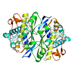
CRYSTAL STRUCTURE AND FUNCTION OF THE ISONIAZID TARGET OF MYCOBACTERIUM TUBERCULOSIS
Descriptor: ENOYL-ACYL CARRIER PROTEIN (ACP) REDUCTASE, NICOTINAMIDE-ADENINE-DINUCLEOTIDE
Authors:Dessen, A, Quemard, A, Blanchard, J.S, Jacobs Jr, W.R, Sacchettini, J.C, TB Structural Genomics Consortium (TBSGC)
Deposit date:1995-01-27
Release date:1996-01-29
Last modified:2024-02-07
Method:X-RAY DIFFRACTION (2.7 Å)
Cite:Crystal structure and function of the isoniazid target of Mycobacterium tuberculosis.
Science, 267, 1995
1YRN
DownloadVisualize
BU of 1yrn by Molmil
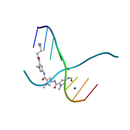
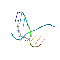
CRYSTAL STRUCTURE OF THE MATA1/MATALPHA2 HOMEODOMAIN HETERODIMER BOUND TO DNA
Descriptor: DNA (5'-D(*TP*AP*CP*AP*TP*GP*TP*AP*AP*TP*TP*TP*AP*TP*TP*AP*C P*AP*TP*CP*A)-3'), DNA (5'-D(*TP*AP*TP*GP*AP*TP*GP*TP*AP*AP*TP*AP*AP*AP*TP*TP*A P*CP*AP*TP*G)-3'), PROTEIN (MAT A1 HOMEODOMAIN), ...
Authors:Li, T, Stark, M.R, Johnson, A.D, Wolberger, C.
Deposit date:1995-11-02
Release date:1996-01-29
Last modified:2024-02-14
Method:X-RAY DIFFRACTION (2.5 Å)
Cite:Crystal structure of the MATa1/MAT alpha 2 homeodomain heterodimer bound to DNA.
Science, 270, 1995
1ETN
DownloadVisualize
BU of 1etn by Molmil
MOLECULAR STRUCTURE OF THE TOXIC DOMAIN OF HEAT-STABLE ENTEROTOXIN PRODUCED BY A PATHOGENIC STRAIN OF ESCHERICHIA COLI
Descriptor: 5-BETA-MERCAPTOPROPIONATE HEAT-STABLE ENTEROTOXIN
Authors:Sato, T, Shimonishi, Y.
Deposit date:1994-03-15
Release date:1996-01-29
Last modified:2017-11-29
Method:X-RAY DIFFRACTION (0.89 Å)
Cite:Molecular structure of the toxin domain of heat-stable enterotoxin produced by a pathogenic strain of Escherichia coli. A putative binding site for a binding protein on rat intestinal epithelial cell membranes.
J.Biol.Chem., 266, 1991
2ANH
DownloadVisualize
BU of 2anh by Molmil
ALKALINE PHOSPHATASE (D153H)
Descriptor: ALKALINE PHOSPHATASE, PHOSPHATE ION, ZINC ION
Authors:Murphy, J.E, Tibbitts, T.T, Kantrowitz, E.R.
Deposit date:1995-09-06
Release date:1996-01-29
Last modified:2021-11-03
Method:X-RAY DIFFRACTION (2.4 Å)
Cite:Mutations at positions 153 and 328 in Escherichia coli alkaline phosphatase provide insight towards the structure and function of mammalian and yeast alkaline phosphatases.
J.Mol.Biol., 253, 1995
1ZAQ
DownloadVisualize
BU of 1zaq by Molmil
FOURTH EGF-LIKE DOMAIN OF THROMBOMODULIN, NMR, 12 STRUCTURES
Descriptor: THROMBOMODULIN
Authors:Meininger, D.P, Komives, E.A.
Deposit date:1995-09-09
Release date:1996-01-29
Last modified:2022-03-02
Method:SOLUTION NMR
Cite:Synthesis, Activity, and Preliminary Structure of the Fourth Egf-Like Domain of Thrombomodulin
Protein Sci., 4, 1995
1GBK
DownloadVisualize
BU of 1gbk by Molmil
ALPHA-LYTIC PROTEASE WITH MET 190 REPLACED BY ALA COMPLEX WITH METHOXYSUCCINYL-ALA-ALA-PRO-ALANINE BORONIC ACID
Descriptor: ALPHA-LYTIC PROTEASE, METHOXYSUCCINYL-ALA-ALA-PRO-ALANINE BORONIC ACID INHIBITOR, SULFATE ION
Authors:Mace, J.E, Agard, D.A.
Deposit date:1995-09-06
Release date:1996-01-29
Last modified:2024-06-05
Method:X-RAY DIFFRACTION (2.13 Å)
Cite:Kinetic and structural characterization of mutations of glycine 216 in alpha-lytic protease: a new target for engineering substrate specificity.
J.Mol.Biol., 254, 1995
1GBB
DownloadVisualize
BU of 1gbb by Molmil
Alpha-lytic protease with met 190 replaced by ALA AND GLY 216 replaced by ALA complex with METHOXYSUCCINYL-ALA-ALA-PRO-ALANINE BORONIC ACID
Descriptor: ALPHA-LYTIC PROTEASE, METHOXYSUCCINYL-ALA-ALA-PRO-ALANINE BORONIC ACID INHIBITOR, SULFATE ION
Authors:Mace, J.E, Agard, D.A.
Deposit date:1995-09-06
Release date:1996-01-29
Last modified:2024-06-05
Method:X-RAY DIFFRACTION (2.15 Å)
Cite:Kinetic and structural characterization of mutations of glycine 216 in alpha-lytic protease: a new target for engineering substrate specificity.
J.Mol.Biol., 254, 1995
1ARX
DownloadVisualize
BU of 1arx by Molmil
CRYSTAL STRUCTURES OF CYANIDE-AND TRIIODIDE-BOUND FORMS OF ARTHROMYCES RAMOSUS PEROXIDASE AT DIFFERENT PH VALUES. PERTURBATIONS OF ACTIVE SITE RESIDUES AND THEIR IMPLICATION IN ENZYME CATALYSIS
Descriptor: 2-acetamido-2-deoxy-beta-D-glucopyranose-(1-4)-2-acetamido-2-deoxy-beta-D-glucopyranose, CALCIUM ION, IODIDE ION, ...
Authors:Fukuyama, K, Kunishima, N, Amada, F.
Deposit date:1995-04-25
Release date:1996-01-29
Last modified:2020-07-29
Method:X-RAY DIFFRACTION (1.9 Å)
Cite:Crystal structures of cyanide- and triiodide-bound forms of Arthromyces ramosus peroxidase at different pH values. Perturbations of active site residues and their implication in enzyme catalysis.
J.Biol.Chem., 270, 1995
1ARY
DownloadVisualize
BU of 1ary by Molmil
CRYSTAL STRUCTURES OF CYANIDE-AND TRIIODIDE-BOUND FORMS OF ARTHROMYCES RAMOSUS PEROXIDASE AT DIFFERENT PH VALUES. PERTURBATIONS OF ACTIVE SITE RESIDUES AND THEIR IMPLICATION IN ENZYME CATALYSIS
Descriptor: 2-acetamido-2-deoxy-beta-D-glucopyranose-(1-4)-2-acetamido-2-deoxy-beta-D-glucopyranose, CALCIUM ION, IODIDE ION, ...
Authors:Fukuyama, K, Kunishima, N, Amada, F.
Deposit date:1995-04-25
Release date:1996-01-29
Last modified:2020-07-29
Method:X-RAY DIFFRACTION (1.9 Å)
Cite:Crystal structures of cyanide- and triiodide-bound forms of Arthromyces ramosus peroxidase at different pH values. Perturbations of active site residues and their implication in enzyme catalysis.
J.Biol.Chem., 270, 1995
1TSD
DownloadVisualize
BU of 1tsd by Molmil
THYMIDYLATE SYNTHASE COMPLEX WITH 2'-DEOXYURIDINE 5'-MONOPHOSPHATE (DUMP) AND FOLATE ANALOG 1843U89
Descriptor: 2'-DEOXYURIDINE 5'-MONOPHOSPHATE, BETA-MERCAPTOETHANOL, S)-2-(5(((1,2-DIHYDRO-3-METHYL-1-OXOBENZO(F)QUINAZOLIN-9-YL)METHYL)AMINO)1-OXO-2-ISOINDOLINYL)GLUTARIC ACID, ...
Authors:Weichsel, A, Montfort, W.R.
Deposit date:1995-08-15
Release date:1996-01-29
Last modified:2018-04-18
Method:X-RAY DIFFRACTION (1.95 Å)
Cite:Ligand-induced distortion of an active site in thymidylate synthase upon binding anticancer drug 1843U89.
Nat.Struct.Biol., 2, 1995
217D
DownloadVisualize
BU of 217d by Molmil
CRYSTAL STRUCTURES OF THE B-FORM DNA-RNA CHIMER (5'-D(*IP*)-R(*CP*)-D(*IP*)-R(*CP*)-D(*IP*CP*IP*C)-3') COMPLEXED WITH DISTAMYCIN
Descriptor: DISTAMYCIN A, DNA/RNA (5'-D(*IP*)-R(*CP*)-D(*IP*)-R(*CP*)-D(*IP*CP*IP*C)-3'), MAGNESIUM ION
Authors:Chen, X, Ramakrishnan, B, Sundaralingam, M.
Deposit date:1995-07-18
Release date:1996-01-29
Last modified:2024-02-14
Method:X-RAY DIFFRACTION (1.7 Å)
Cite:Crystal structures of B-form DNA-RNA chimers complexed with distamycin.
Nat.Struct.Biol., 2, 1995
216D
DownloadVisualize
BU of 216d by Molmil
CRYSTAL STRUCTURES OF THE B-FORM DNA-RNA CHIMER (5'-D(*IP*)-R(*CP*)-D(*IP*CP*IP*CP*IP*C)-3') COMPLEXED WITH DISTAMYCIN
Descriptor: DISTAMYCIN A, DNA/RNA (5'-D(*IP*)-R(*CP*)-D(*IP*CP*IP*CP*IP*C)-3'), MAGNESIUM ION
Authors:Chen, X, Ramakrishnan, B, Sundaralingam, M.
Deposit date:1995-07-18
Release date:1996-01-29
Last modified:2024-02-14
Method:X-RAY DIFFRACTION (1.73 Å)
Cite:Crystal structures of B-form DNA-RNA chimers complexed with distamycin.
Nat.Struct.Biol., 2, 1995
1GBM
DownloadVisualize
BU of 1gbm by Molmil
ALPHA-LYTIC PROTEASE WITH MET 190 REPLACED BY ALA COMPLEX WITH METHOXYSUCCINYL-ALA-ALA-PRO-PHENYLALANINE BORONIC ACID
Descriptor: ALPHA-LYTIC PROTEASE, METHOXYSUCCINYL-ALA-ALA-PRO-PHENYLALANINE BORONIC ACID INHIBITOR, SULFATE ION
Authors:Mace, J.E, Agard, D.A.
Deposit date:1995-09-06
Release date:1996-01-29
Last modified:2021-11-03
Method:X-RAY DIFFRACTION (2.28 Å)
Cite:Kinetic and structural characterization of mutations of glycine 216 in alpha-lytic protease: a new target for engineering substrate specificity.
J.Mol.Biol., 254, 1995
1CYN
DownloadVisualize
BU of 1cyn by Molmil
CYCLOPHILIN B COMPLEXED WITH [D-(CHOLINYLESTER)SER8]-CYCLOSPORIN
Descriptor: CYCLOSPORIN A, PEPTIDYL-PROLYL CIS-TRANS ISOMERASE B
Authors:Mikol, V, Kallen, J, Walkinshaw, M.D.
Deposit date:1995-05-22
Release date:1996-01-29
Last modified:2018-09-12
Method:X-RAY DIFFRACTION (1.85 Å)
Cite:X-Ray Structure of a Cyclophilin B/Cyclosporin Complex: Comparison with Cyclophilin a and Delineation of its Calcineurin-Binding Domain.
Proc.Natl.Acad.Sci.USA, 91, 1994
1CWB
DownloadVisualize
BU of 1cwb by Molmil
THE X-RAY STRUCTURE OF (MEBM2T)1-CYCLOSPORIN COMPLEXED WITH CYCLOPHILIN A PROVIDES AN EXPLANATION FOR ITS ANOMALOUSLY HIGH IMMUNOSUPPRESSIVE ACTIVITY
Descriptor: CYCLOSPORIN A, PEPTIDYL-PROLYL CIS-TRANS ISOMERASE A
Authors:Mikol, V, Kallen, J, Walkinshaw, M.D.
Deposit date:1995-09-06
Release date:1996-01-29
Last modified:2024-06-05
Method:X-RAY DIFFRACTION (2.2 Å)
Cite:The X-Ray Structure of (Mebm2T)1-Cyclosporin Complexed with Cyclophilin a Provides an Explanation for its Anomalously High Immunosuppressive Activity.
Protein Eng., 7, 1994
1GPM
DownloadVisualize
BU of 1gpm by Molmil
ESCHERICHIA COLI GMP SYNTHETASE COMPLEXED WITH AMP AND PYROPHOSPHATE
Descriptor: ADENOSINE MONOPHOSPHATE, CITRIC ACID, GMP SYNTHETASE, ...
Authors:Tesmer, J.J.G.
Deposit date:1995-04-04
Release date:1996-01-29
Last modified:2024-02-07
Method:X-RAY DIFFRACTION (2.2 Å)
Cite:The crystal structure of GMP synthetase reveals a novel catalytic triad and is a structural paradigm for two enzyme families.
Nat.Struct.Biol., 3, 1996
2HFT
DownloadVisualize
BU of 2hft by Molmil
THE CRYSTAL STRUCTURE OF THE EXTRACELLULAR DOMAIN OF HUMAN TISSUE FACTOR AT 1.7 ANGSTROMS RESOLUTION
Descriptor: HUMAN TISSUE FACTOR, SULFATE ION
Authors:Muller, Y.A, De Vos, A.M.
Deposit date:1995-09-08
Release date:1996-01-29
Last modified:2011-07-13
Method:X-RAY DIFFRACTION (1.69 Å)
Cite:The crystal structure of the extracellular domain of human tissue factor refined to 1.7 A resolution.
J.Mol.Biol., 256, 1996
1CLF
DownloadVisualize
BU of 1clf by Molmil
CLOSTRIDIUM PASTEURIANUM FERREDOXIN
Descriptor: FERREDOXIN, IRON/SULFUR CLUSTER
Authors:Bertini, I, Donaire, A, Feinberg, B.A, Luchinat, C, Piccioli, M, Yuan, H.
Deposit date:1995-06-21
Release date:1996-01-29
Last modified:2024-05-22
Method:SOLUTION NMR
Cite:Solution structure of the oxidized 2[4Fe-4S] ferredoxin from Clostridium pasteurianum.
Eur.J.Biochem., 232, 1995
1UDG
DownloadVisualize
BU of 1udg by Molmil
THE STRUCTURAL BASIS OF SPECIFIC BASE EXCISION REPAIR BY URACIL-DNA GLYCOSYLASE
Descriptor: SULFATE ION, URACIL-DNA GLYCOSYLASE
Authors:Pearl, L.H, Savva, R.
Deposit date:1995-06-23
Release date:1996-01-29
Last modified:2024-02-14
Method:X-RAY DIFFRACTION (1.75 Å)
Cite:The structural basis of specific base-excision repair by uracil-DNA glycosylase.
Nature, 373, 1995
1HOR
DownloadVisualize
BU of 1hor by Molmil
STRUCTURE AND CATALYTIC MECHANISM OF GLUCOSAMINE 6-PHOSPHATE DEAMINASE FROM ESCHERICHIA COLI AT 2.1 ANGSTROMS RESOLUTION
Descriptor: 2-DEOXY-2-AMINO GLUCITOL-6-PHOSPHATE, GLUCOSAMINE 6-PHOSPHATE DEAMINASE, PHOSPHATE ION
Authors:Oliva, G, Fontes, M.R.M, Garratt, R.C, Altamirano, M.M, Calcagno, M.L, Horjales, E.
Deposit date:1995-09-13
Release date:1996-01-29
Last modified:2024-02-07
Method:X-RAY DIFFRACTION (2.4 Å)
Cite:Structure and catalytic mechanism of glucosamine 6-phosphate deaminase from Escherichia coli at 2.1 A resolution.
Structure, 3, 1995
1HIK
DownloadVisualize
BU of 1hik by Molmil
INTERLEUKIN-4 (WILD-TYPE)
Descriptor: INTERLEUKIN-4
Authors:Mueller, T, Buehner, M.
Deposit date:1995-06-01
Release date:1996-01-29
Last modified:2024-06-05
Method:X-RAY DIFFRACTION (2.6 Å)
Cite:Human interleukin-4 and variant R88Q: phasing X-ray diffraction data by molecular replacement using X-ray and nuclear magnetic resonance models.
J.Mol.Biol., 247, 1995
1CIR
DownloadVisualize
BU of 1cir by Molmil
COMPLEX OF TWO FRAGMENTS OF CI2 [(1-40)(DOT)(41-64)]
Descriptor: CHYMOTRYPSIN INHIBITOR 2
Authors:Davis, B.J, Fersht, A.R.
Deposit date:1995-10-02
Release date:1996-01-29
Last modified:2024-05-22
Method:SOLUTION NMR
Cite:Towards the complete structural characterization of a protein folding pathway: the structures of the denatured, transition and native states for the association/folding of two complementary fragments of cleaved chymotrypsin inhibitor 2. Direct evidence for a nucleation-condensation mechanism
Structure Fold.Des., 1, 1996
1XYZ
DownloadVisualize
BU of 1xyz by Molmil
A COMMON PROTEIN FOLD AND SIMILAR ACTIVE SITE IN TWO DISTINCT FAMILIES OF BETA-GLYCANASES
Descriptor: 1,4-BETA-D-XYLAN-XYLANOHYDROLASE
Authors:Alzari, P.M, Spinelli, S, Dominguez, R.
Deposit date:1995-06-07
Release date:1996-01-29
Last modified:2024-02-14
Method:X-RAY DIFFRACTION (1.4 Å)
Cite:A common protein fold and similar active site in two distinct families of beta-glycanases.
Nat.Struct.Biol., 2, 1995
1CHD
DownloadVisualize
BU of 1chd by Molmil
CHEB METHYLESTERASE DOMAIN
Descriptor: CHEB METHYLESTERASE
Authors:West, A.H, Martinez-Hackert, E, Stock, A.M.
Deposit date:1995-03-09
Release date:1996-01-29
Last modified:2024-02-07
Method:X-RAY DIFFRACTION (1.75 Å)
Cite:Crystal structure of the catalytic domain of the chemotaxis receptor methylesterase, CheB.
J.Mol.Biol., 250, 1995
1FNF
DownloadVisualize
BU of 1fnf by Molmil
FRAGMENT OF HUMAN FIBRONECTIN ENCOMPASSING TYPE-III REPEATS 7 THROUGH 10
Descriptor: FIBRONECTIN
Authors:Leahy, D.J, Aukhil, I, Erickson, H.P.
Deposit date:1995-09-30
Release date:1996-01-29
Last modified:2024-02-07
Method:X-RAY DIFFRACTION (2 Å)
Cite:2.0 A crystal structure of a four-domain segment of human fibronectin encompassing the RGD loop and synergy region.
Cell(Cambridge,Mass.), 84, 1996

222415

数据于2024-07-10公开中

PDB statisticsPDBj update infoContact PDBjnumon