7P01
 
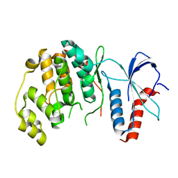
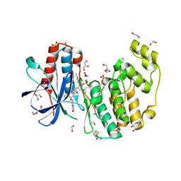
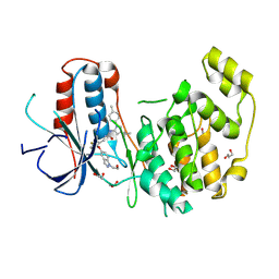
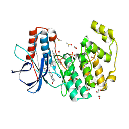
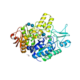 | | Structure of the maltase BaAG2 from Blastobotrys adeninivorans in complex with acarbose | | Descriptor: | 1-methylpyrrolidin-2-one, 4,6-dideoxy-4-{[(1S,4R,5S,6S)-4,5,6-trihydroxy-3-(hydroxymethyl)cyclohex-2-en-1-yl]amino}-alpha-D-glucopyranose-(1-4)-alpha-D-glucopyranose-(1-4)-alpha-D-glucopyranose, BaAG2, ... | | Authors: | Ernits, K, Visnapuu, T, Persson, K. | | Deposit date: | 2021-06-29 | | Release date: | 2021-10-06 | | Last modified: | 2024-01-31 | | Method: | X-RAY DIFFRACTION (2.12 Å) | | Cite: | Structural Insight into a Yeast Maltase-The Ba AG2 from Blastobotrys adeninivorans with Transglycosylating Activity.
J Fungi (Basel), 7, 2021
|
|
7P07
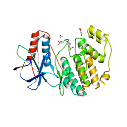
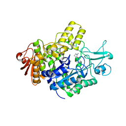 | | Structure of the maltase BaAG2 from Blastobotrys adeninivorans in complex with glucose | | Descriptor: | 1-methylpyrrolidin-2-one, BaAG2, CALCIUM ION, ... | | Authors: | Ernits, K, Visnapuu, T, Persson, K. | | Deposit date: | 2021-06-29 | | Release date: | 2021-10-06 | | Last modified: | 2024-01-31 | | Method: | X-RAY DIFFRACTION (2.13 Å) | | Cite: | Structural Insight into a Yeast Maltase-The Ba AG2 from Blastobotrys adeninivorans with Transglycosylating Activity.
J Fungi (Basel), 7, 2021
|
|
7OLY
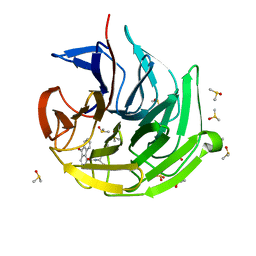
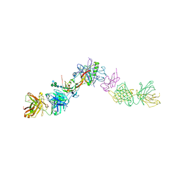 | | Structure of activin A in complex with an ActRIIB-Alk4 fusion reveal insight into activin receptor interactions | | Descriptor: | 2-acetamido-2-deoxy-beta-D-glucopyranose, 2-acetamido-2-deoxy-beta-D-glucopyranose-(1-6)-alpha-D-mannopyranose-(1-6)-[alpha-D-mannopyranose-(1-3)]alpha-D-mannopyranose-(1-4)-2-acetamido-2-deoxy-beta-D-glucopyranose-(1-4)-2-acetamido-2-deoxy-beta-D-glucopyranose, Activin receptor type-1B, ... | | Authors: | Hakansson, M, Rose, N.C, Castonguay, R, Logan, D.T, Krishnan, L. | | Deposit date: | 2021-05-20 | | Release date: | 2022-02-23 | | Last modified: | 2024-11-06 | | Method: | X-RAY DIFFRACTION (3.265 Å) | | Cite: | Structures of activin ligand traps using natural sets of type I and type II TGF beta receptors.
Iscience, 25, 2022
|
|
7OKR
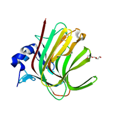
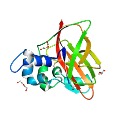 | | Catalytic domain from the Aliivibrio salmonicida lytic polysaccharide monooxygenase AsLPMO10B | | Descriptor: | COPPER (II) ION, Chitinase B, DI(HYDROXYETHYL)ETHER | | Authors: | Cordara, G, Leitl, K.D, Loose, J.S.M, Vaaje-Kolstad, G, Krengel, U. | | Deposit date: | 2021-05-18 | | Release date: | 2022-06-01 | | Last modified: | 2024-10-09 | | Method: | X-RAY DIFFRACTION (1.35 Å) | | Cite: | Chitinolytic enzymes contribute to the pathogenicity of Aliivibrio salmonicida LFI1238 in the invasive phase of cold-water vibriosis.
Bmc Microbiol., 22, 2022
|
|
6RFO
 | | ERK2 MAP kinase with the activation loop of p38alpha | | Descriptor: | Mitogen-activated protein kinase 1,Mitogen-activated protein kinase 14,Mitogen-activated protein kinase 1 | | Authors: | Livnah, O, Eitan-Wexler, M. | | Deposit date: | 2019-04-15 | | Release date: | 2020-05-27 | | Last modified: | 2024-01-24 | | Method: | X-RAY DIFFRACTION (1.7 Å) | | Cite: | The bacterial metalloprotease NleD selectively cleaves mitogen-activated protein kinases that have high flexibility in their activation loop.
J.Biol.Chem., 295, 2020
|
|
7OOF
 | | Crystal structure of Paradendryphiella salina PL7A alginate lyase in complex with tri-mannuronic acid | | Descriptor: | Alginate lyase (PL7), TETRAETHYLENE GLYCOL, alpha-D-mannopyranuronic acid, ... | | Authors: | Fredslund, F, Welner, D.H, Wilkens, C. | | Deposit date: | 2021-05-27 | | Release date: | 2022-06-08 | | Last modified: | 2025-07-02 | | Method: | X-RAY DIFFRACTION (1.3 Å) | | Cite: | Unraveling the molecular mechanism of polysaccharide lyases for efficient alginate degradation.
Nat Commun, 16, 2025
|
|
7ORY
 | |
7OWL
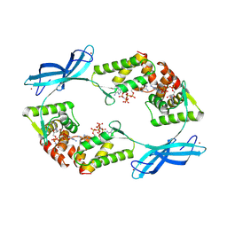
 | | Odinarchaeota Adenylate kinase (OdinAK) in complex with CTP | | Descriptor: | Adenylate kinase, CYTIDINE-5'-TRIPHOSPHATE | | Authors: | Aberg-Zingmark, E, Grundstrom, C, Verma, A, Wolf-Watz, M, Sauer, U.H, Sauer-Eriksson, A.E. | | Deposit date: | 2021-06-18 | | Release date: | 2022-09-28 | | Last modified: | 2024-01-31 | | Method: | X-RAY DIFFRACTION (2.9 Å) | | Cite: | Insights into the evolution of enzymatic specificity and catalysis: From Asgard archaea to human adenylate kinases.
Sci Adv, 8, 2022
|
|
7OWH
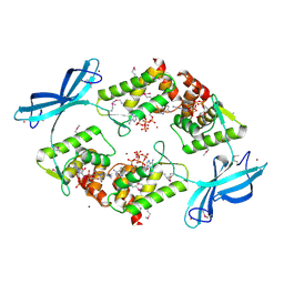
 | | Odinarchaeota Adenylate kinase (OdinAK) native structure | | Descriptor: | Adenylate kinase, CHLORIDE ION | | Authors: | Aberg-Zingmark, E, Grundstrom, C, Verma, A, Wolf-Watz, M, Sauer, U.H, Sauer-Eriksson, A.E. | | Deposit date: | 2021-06-18 | | Release date: | 2022-09-28 | | Last modified: | 2024-01-31 | | Method: | X-RAY DIFFRACTION (1.85 Å) | | Cite: | Insights into the evolution of enzymatic specificity and catalysis: From Asgard archaea to human adenylate kinases.
Sci Adv, 8, 2022
|
|
7OY1
 | | DnrK mutant RTCR | | Descriptor: | Carminomycin 4-O-methyltransferase DnrK,Methyltransferase domain-containing protein,Aclacinomycin 10-hydroxylase RdmB, S-ADENOSYL-L-HOMOCYSTEINE, methyl (1R,2R,4S)-2-ethyl-2,5,7-trihydroxy-6,11-dioxo-4-{[2,3,6-trideoxy-3-(dimethylamino)-alpha-L-lyxo-hexopyranosyl]oxy}-1,2,3,4,6,11-hexahydrotetracene-1-carboxylate | | Authors: | Dinis, P, MetsaKetela, M. | | Deposit date: | 2021-06-23 | | Release date: | 2022-07-13 | | Last modified: | 2024-11-13 | | Method: | X-RAY DIFFRACTION (2.39 Å) | | Cite: | Evolution-inspired engineering of anthracycline methyltransferases.
Pnas Nexus, 2, 2023
|
|
7OWK
 | | Odinarchaeota Adenylate kinase (OdinAK) in complex with dTTP | | Descriptor: | Adenylate kinase, THYMIDINE-5'-TRIPHOSPHATE | | Authors: | Aberg-Zingmark, E, Grundstrom, C, Verma, A, Wolf-Watz, M, Sauer, U.H, Sauer-Eriksson, A.E. | | Deposit date: | 2021-06-18 | | Release date: | 2022-09-28 | | Last modified: | 2024-01-31 | | Method: | X-RAY DIFFRACTION (3.1 Å) | | Cite: | Insights into the evolution of enzymatic specificity and catalysis: From Asgard archaea to human adenylate kinases.
Sci Adv, 8, 2022
|
|
7OWJ
 | | Odinarchaeota Adenylate kinase (OdinAK) in complex with GTP | | Descriptor: | Adenylate kinase, GUANOSINE-5'-TRIPHOSPHATE | | Authors: | Aberg-Zingmark, E, Grundstrom, C, Verma, A, Wolf-Watz, M, Sauer, U.H, Sauer-Eriksson, A.E. | | Deposit date: | 2021-06-18 | | Release date: | 2022-09-28 | | Last modified: | 2024-01-31 | | Method: | X-RAY DIFFRACTION (2.5 Å) | | Cite: | Insights into the evolution of enzymatic specificity and catalysis: From Asgard archaea to human adenylate kinases.
Sci Adv, 8, 2022
|
|
9CJ4
 | | Dual phosphorylated human p38 alpha bound to BIRB796 | | Descriptor: | 1,2-ETHANEDIOL, 1-(5-TERT-BUTYL-2-P-TOLYL-2H-PYRAZOL-3-YL)-3-[4-(2-MORPHOLIN-4-YL-ETHOXY)-NAPHTHALEN-1-YL]-UREA, DI(HYDROXYETHYL)ETHER, ... | | Authors: | Stadnicki, E.J, Ludewig, H, Prem Kumar, R, Kern, D, Bradshaw, N. | | Deposit date: | 2024-07-05 | | Release date: | 2024-11-13 | | Method: | X-RAY DIFFRACTION (1.8 Å) | | Cite: | Dual-Action Kinase Inhibitors Influence p38 alpha MAP Kinase Dephosphorylation.
Biorxiv, 2024
|
|
9CJ1
 | | Dual phosphorylated human p38 alpha bound to nilotinib | | Descriptor: | 1,2-ETHANEDIOL, Mitogen-activated protein kinase 14, Nilotinib | | Authors: | Stadnicki, E.J, Ludewig, H, Prem Kumar, R, Kern, D, Bradshaw, N. | | Deposit date: | 2024-07-05 | | Release date: | 2024-11-13 | | Method: | X-RAY DIFFRACTION (1.8 Å) | | Cite: | Dual-Action Kinase Inhibitors Influence p38 alpha MAP Kinase Dephosphorylation.
Biorxiv, 2024
|
|
9CJ2
 | | Dual phosphorylated human p38 alpha | | Descriptor: | 1,2-ETHANEDIOL, DI(HYDROXYETHYL)ETHER, Mitogen-activated protein kinase 14 | | Authors: | Stadnicki, E.J, Ludewig, H, Prem Kumar, R, Kern, D, Bradshaw, N. | | Deposit date: | 2024-07-05 | | Release date: | 2024-11-13 | | Method: | X-RAY DIFFRACTION (2.83 Å) | | Cite: | Dual-Action Kinase Inhibitors Influence p38 alpha MAP Kinase Dephosphorylation.
Biorxiv, 2024
|
|
7OFF
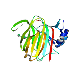
 | | Keap1 kelch domain bound to a small molecule inhibitor of the Keap1-Nrf2 protein-protein interaction | | Descriptor: | (2~{R})-2-[(9-oxidanylidenefluoren-4-yl)carbonylamino]-2-phenyl-ethanoic acid, DIMETHYL SULFOXIDE, Kelch-like ECH-associated protein 1, ... | | Authors: | Narayanan, D, Bach, A, Gajhede, M. | | Deposit date: | 2021-05-04 | | Release date: | 2022-11-02 | | Last modified: | 2024-01-31 | | Method: | X-RAY DIFFRACTION (1.37 Å) | | Cite: | Development of Noncovalent Small-Molecule Keap1-Nrf2 Inhibitors by Fragment-Based Drug Discovery.
J.Med.Chem., 65, 2022
|
|
9CJ5
 | | Unphosphorylated human p38 alpha bound to pexmetinib | | Descriptor: | 1,2-ETHANEDIOL, 1,3-PROPANDIOL, Mitogen-activated protein kinase 14, ... | | Authors: | Stadnicki, E.J, Ludewig, H, Prem Kumar, R, Kern, D, Bradshaw, N. | | Deposit date: | 2024-07-05 | | Release date: | 2024-11-13 | | Method: | X-RAY DIFFRACTION (3.3 Å) | | Cite: | Dual-Action Kinase Inhibitors Influence p38 alpha MAP Kinase Dephosphorylation.
Biorxiv, 2024
|
|
9GUB
 | | SARS-CoV-2 Mac1 in complex with MCD-628 | | Descriptor: | (2~{S})-3-(1~{H}-indol-3-yl)-2-(7~{H}-pyrrolo[2,3-d]pyrimidin-4-ylamino)propanoic acid, Papain-like protease nsp3 | | Authors: | Duong, M, Paakkonen, J, Lehtio, L. | | Deposit date: | 2024-09-19 | | Release date: | 2025-06-11 | | Last modified: | 2025-06-25 | | Method: | X-RAY DIFFRACTION (1.1 Å) | | Cite: | Identification of a series of pyrrolo-pyrimidine-based SARS-CoV-2 Mac1 inhibitors that repress coronavirus replication.
Mbio, 16, 2025
|
|
9CJ3
 | | Dual phosphorylated human p38 alpha bound to pexmetinib | | Descriptor: | 1,2-ETHANEDIOL, 1,3-PROPANDIOL, DIMETHYL SULFOXIDE, ... | | Authors: | Stadnicki, E.J, Ludewig, H, Prem Kumar, R, Kern, D, Bradshaw, N. | | Deposit date: | 2024-07-05 | | Release date: | 2024-11-13 | | Method: | X-RAY DIFFRACTION (1.95 Å) | | Cite: | Dual-Action Kinase Inhibitors Influence p38 alpha MAP Kinase Dephosphorylation.
Biorxiv, 2024
|
|
9FXK
 | |
9FVR
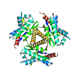
 | | Transcription repressor NrdR from E. coli, ATP/dATP-bound state, SeMet protein | | Descriptor: | 2'-DEOXYADENOSINE 5'-TRIPHOSPHATE, ADENOSINE-5'-TRIPHOSPHATE, Transcriptional repressor NrdR, ... | | Authors: | Bimai, O, Sjoberg, B.M, Rozman Grinberg, I, Logan, D.T. | | Deposit date: | 2024-06-28 | | Release date: | 2025-03-19 | | Last modified: | 2025-07-02 | | Method: | X-RAY DIFFRACTION (3.099 Å) | | Cite: | Bacterial transcriptional repressor NrdR - a flexible multifactorial nucleotide sensor.
Febs J., 292, 2025
|
|
5HL3
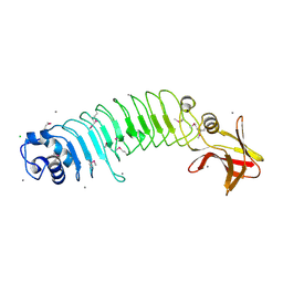 | | Crystal structure of Listeria monocytogenes InlP | | Descriptor: | CALCIUM ION, CHLORIDE ION, Lmo2470 protein | | Authors: | Nocadello, S, Light, S.H, Minasov, G, Kiryukhina, O, Kwon, K, Faralla, C, Bakardjiev, A, Anderson, W.F, Center for Structural Genomics of Infectious Diseases (CSGID) | | Deposit date: | 2016-01-14 | | Release date: | 2017-01-18 | | Last modified: | 2024-10-23 | | Method: | X-RAY DIFFRACTION (1.4 Å) | | Cite: | Listeria monocytogenes InlP interacts with afadin and facilitates basement membrane crossing.
Plos Pathog., 14, 2018
|
|
9FYJ
 | | N-terminal domain of human galectin-8 in complex with an alpha-galactoside ligand | | Descriptor: | 2-[[(2~{R},3~{R},4~{S},5~{S},6~{R})-2-(3,4-dichlorophenyl)sulfanyl-6-(hydroxymethyl)-5-oxidanyl-3-prop-2-ynoxy-oxan-4-yl]oxymethyl]-3-methyl-benzimidazole-5-carboxylic acid, CHLORIDE ION, Isoform 1 of Galectin-8 | | Authors: | Adrover Forteza, J, Puric, E, Nilsson, U.J, Anderluh, M, Logan, D.T. | | Deposit date: | 2024-07-03 | | Release date: | 2025-03-12 | | Method: | X-RAY DIFFRACTION (1.08 Å) | | Cite: | Nanomolar inhibitor of the galectin-8 N-terminal domain binds via a non-canonical cation-pi interaction.
Commun Chem, 8, 2025
|
|
9FHF
 | |
9FKC
 | |


