Loading
PDBj
MenuPDBj@FacebookPDBj@TwitterPDBj@YouTubewwPDB FoundationwwPDB
RCSB PDBPDBeBMRBAdv. SearchSearch help

4LHI
DownloadVisualize
BU of 4lhi by Molmil
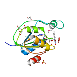
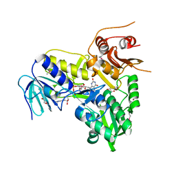
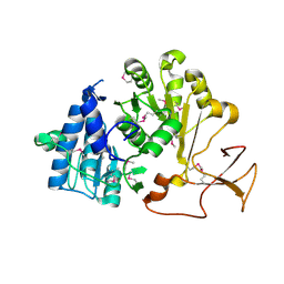
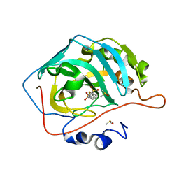
Crystal structure of human carbonic anhydrase isozyme II with 5-(phenylsulfonyl)thiophene-2-sulfonamide
Descriptor: 5-(phenylsulfonyl)thiophene-2-sulfonamide, Carbonic anhydrase 2, DIMETHYL SULFOXIDE, ...
Authors:Smirnov, A, Manakova, E, Grazulis, S.
Deposit date:2013-07-01
Release date:2014-07-02
Last modified:2023-11-08
Method:X-RAY DIFFRACTION (1.6 Å)
Cite:Crystal structure of human carbonic anhydrase isozyme II with 5-(phenylsulfonyl)thiophene-2-sulfonamide
To be Published
4LGQ
DownloadVisualize
BU of 4lgq by Molmil
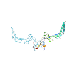
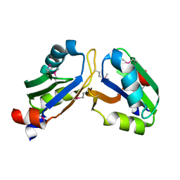
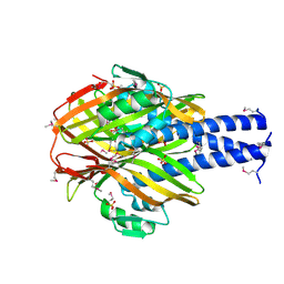
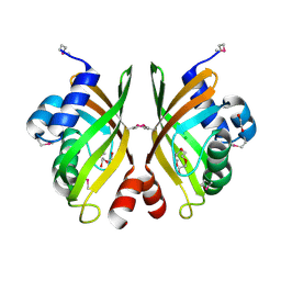
Crystal structure of a putative polyketide cyclase (CV_0247) from Chromobacterium violaceum ATCC 12472 at 2.72 A resolution
Descriptor: 1,2-ETHANEDIOL, CHLORIDE ION, DI(HYDROXYETHYL)ETHER, ...
Authors:Joint Center for Structural Genomics (JCSG)
Deposit date:2013-06-28
Release date:2013-07-24
Last modified:2023-02-01
Method:X-RAY DIFFRACTION (2.72 Å)
Cite:Crystal structure of a putative polyketide cyclase (CV_0247) from Chromobacterium violaceum ATCC 12472 at 2.72 A resolution
To be published
4LJX
DownloadVisualize
BU of 4ljx by Molmil
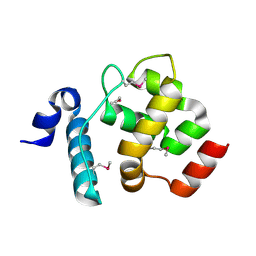
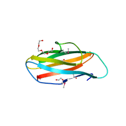
Crystal structure of an AT-rich interactive domain-containing protein 3A (ARID3A) from Homo sapiens at 2.21 A resolution
Descriptor: AT-rich interactive domain-containing protein 3A
Authors:Joint Center for Structural Genomics (JCSG), Partnership for Stem Cell Biology (STEMCELL)
Deposit date:2013-07-05
Release date:2013-08-28
Last modified:2023-12-06
Method:X-RAY DIFFRACTION (2.21 Å)
Cite:Crystal structure of an AT-rich interactive domain-containing protein 3A (ARID3A) from Homo sapiens at 2.21 A resolution
To be published
4JD2
DownloadVisualize
BU of 4jd2 by Molmil
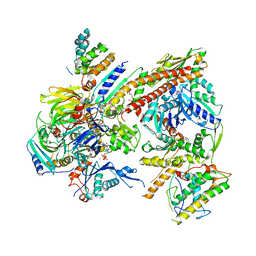
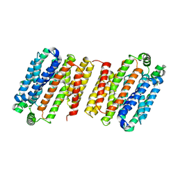
Crystal structure of Bos taurus Arp2/3 complex binding with Mus musculus GMF
Descriptor: ADENOSINE-5'-TRIPHOSPHATE, Actin-related protein 2, Actin-related protein 2/3 complex subunit 1B, ...
Authors:Nolen, B.J, Luan, Q.
Deposit date:2013-02-22
Release date:2013-07-17
Last modified:2024-02-28
Method:X-RAY DIFFRACTION (3.08 Å)
Cite:Structural basis for regulation of Arp2/3 complex by GMF.
Nat.Struct.Mol.Biol., 20, 2013
4JHY
DownloadVisualize
BU of 4jhy by Molmil
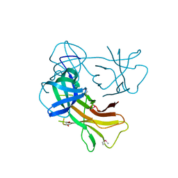
Crystal structure of a TBP-like protein (BDI_3606) from Parabacteroides distasonis ATCC 8503 at 1.90 A resolution
Descriptor: hypothetical protein
Authors:Joint Center for Structural Genomics (JCSG)
Deposit date:2013-03-05
Release date:2013-04-03
Last modified:2023-02-01
Method:X-RAY DIFFRACTION (1.9 Å)
Cite:Crystal structure of a hypothetical protein (BDI_3606) from Parabacteroides distasonis ATCC 8503 at 1.90 A resolution
To be published
4JQT
DownloadVisualize
BU of 4jqt by Molmil
Crystal structure of a putative glycosyl hydrolase (BT3469) from Bacteroides thetaiotaomicron VPI-5482 at 2.49 A resolution
Descriptor: 1,2-ETHANEDIOL, SODIUM ION, putative glycosyl hydrolase
Authors:Joint Center for Structural Genomics (JCSG)
Deposit date:2013-03-20
Release date:2013-04-17
Last modified:2023-02-01
Method:X-RAY DIFFRACTION (2.49 Å)
Cite:Crystal structure of a putative glycosyl hydrolase (BT3469) from Bacteroides thetaiotaomicron VPI-5482 at 2.49 A resolution
To be published
4J8P
DownloadVisualize
BU of 4j8p by Molmil
Crystal structure of a Putative flavoprotein (BACUNI_04544) from Bacteroides uniformis ATCC 8492 at 1.50 A resolution
Descriptor: CHLORIDE ION, FLAVIN MONONUCLEOTIDE, Flavodoxin, ...
Authors:Joint Center for Structural Genomics (JCSG)
Deposit date:2013-02-14
Release date:2013-03-13
Last modified:2024-10-09
Method:X-RAY DIFFRACTION (1.5 Å)
Cite:Crystal structure of a Putative flavoprotein (BACUNI_04544) from Bacteroides uniformis ATCC 8492 at 1.50 A resolution
To be published
4L8J
DownloadVisualize
BU of 4l8j by Molmil
Crystal structure of a Putative efflux transporter (BACEGG_01895) from Bacteroides eggerthii DSM 20697 at 2.06 A resolution
Descriptor: DI(HYDROXYETHYL)ETHER, GLYCEROL, Putative efflux transporter
Authors:Joint Center for Structural Genomics (JCSG)
Deposit date:2013-06-17
Release date:2013-07-03
Last modified:2023-02-01
Method:X-RAY DIFFRACTION (2.06 Å)
Cite:Crystal structure of a Putative efflux transporter (BACEGG_01895) from Bacteroides eggerthii DSM 20697 at 2.06 A resolution
To be published
4LBA
DownloadVisualize
BU of 4lba by Molmil
Crystal structure of a conjugative transposon lipoprotein (BACEGG_03088) from Bacteroides eggerthii DSM 20697 at 1.70 A resolution
Descriptor: 1,2-ETHANEDIOL, PENTAETHYLENE GLYCOL, conjugative transposon lipoprotein
Authors:Joint Center for Structural Genomics (JCSG)
Deposit date:2013-06-20
Release date:2013-08-07
Last modified:2023-02-01
Method:X-RAY DIFFRACTION (1.7 Å)
Cite:Crystal structure of a conjugative transposon lipoprotein (BACEGG_03088) from Bacteroides eggerthii DSM 20697 at 1.70 A resolution
To be published
4LDS
DownloadVisualize
BU of 4lds by Molmil
The inward-facing structure of the glucose transporter from Staphylococcus epidermidis
Descriptor: Glucose transporter GlcP
Authors:Choe, J, Aleshin, A, Iancu, C.V.
Deposit date:2013-06-25
Release date:2013-10-16
Last modified:2024-02-28
Method:X-RAY DIFFRACTION (3.2 Å)
Cite:Crystal structure of a glucose/H+ symporter and its mechanism of action.
Proc.Natl.Acad.Sci.USA, 110, 2013
4LII
DownloadVisualize
BU of 4lii by Molmil
Crystal structure of an apoptosis-inducing factor, mitochondrion-associated, 1 (AIFM1) from Homo sapiens at 1.88 A resolution
Descriptor: 1,2-ETHANEDIOL, Apoptosis-inducing factor 1, mitochondrial, ...
Authors:Joint Center for Structural Genomics (JCSG), Partnership for T-Cell Biology (TCELL)
Deposit date:2013-07-02
Release date:2013-09-04
Last modified:2024-10-30
Method:X-RAY DIFFRACTION (1.88 Å)
Cite:Crystal structure of an apoptosis-inducing factor, mitochondrion-associated, 1 (AIFM1) from Homo sapiens at 1.88 A resolution
To be published
4LIR
DownloadVisualize
BU of 4lir by Molmil
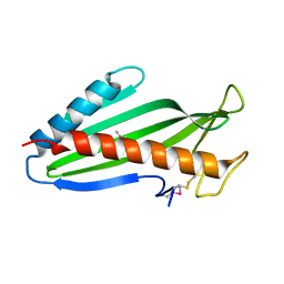
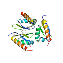
Crystal structure of a nucleoporin 35kDa (NUP35) from Homo sapiens at 2.46 A resolution
Descriptor: Nucleoporin NUP53
Authors:Joint Center for Structural Genomics (JCSG), Partnership for T-Cell Biology (TCELL)
Deposit date:2013-07-03
Release date:2013-09-11
Last modified:2023-02-01
Method:X-RAY DIFFRACTION (2.46 Å)
Cite:Crystal structure of a nucleoporin 35kDa (NUP35) from Homo sapiens at 2.46 A resolution
To be published
4LG9
DownloadVisualize
BU of 4lg9 by Molmil
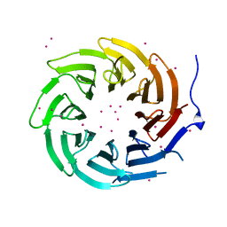
Crystal structure of TBL1XR1 WD40 repeats
Descriptor: F-box-like/WD repeat-containing protein TBL1XR1, UNKNOWN ATOM OR ION
Authors:Xu, C, Tempel, W, He, H, Wu, X, Seitova, A, Bountra, C, Arrowsmith, C.H, Edwards, A.M, Min, J, Structural Genomics Consortium (SGC)
Deposit date:2013-06-27
Release date:2014-04-16
Last modified:2023-09-20
Method:X-RAY DIFFRACTION (2.28 Å)
Cite:Crystal structure of TBL1XR1 WD40 repeats
TO BE PUBLISHED
4J8Q
DownloadVisualize
BU of 4j8q by Molmil
Crystal structure of a NigD-like protein (BF0700) from Bacteroides fragilis NCTC 9343 at 2.50 A resolution
Descriptor: 1,2-ETHANEDIOL, Putative uncharacterized protein
Authors:Joint Center for Structural Genomics (JCSG)
Deposit date:2013-02-14
Release date:2013-03-06
Last modified:2024-10-16
Method:X-RAY DIFFRACTION (2.5 Å)
Cite:Crystal structure of a hypothetical protein (BF0700) from Bacteroides fragilis NCTC 9343 at 2.50 A resolution
To be published
4JH0
DownloadVisualize
BU of 4jh0 by Molmil
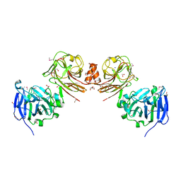
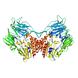
Crystal structure of dipeptidyl-peptidase 4 (CD26, adenosine deaminase complexing protein 2) (DPP-IV-WT) complex with bms-767778 AKA 2-(3-(aminomethyl)-4-(2,4- dichlorophenyl)-2-methyl-5-oxo-5,7-dihydro-6h-pyrrolo[3,4- b]pyridin-6-yl)-n,n-dimethylacetamide
Descriptor: 2-[3-(aminomethyl)-4-(2,4-dichlorophenyl)-2-methyl-5-oxo-5,7-dihydro-6H-pyrrolo[3,4-b]pyridin-6-yl]-N,N-dimethylacetamide, Dipeptidyl peptidase 4
Authors:Klei, H.E.
Deposit date:2013-03-04
Release date:2013-09-04
Last modified:2024-10-09
Method:X-RAY DIFFRACTION (2.35 Å)
Cite:Optimization of Activity, Selectivity, and Liability Profiles in 5-Oxopyrrolopyridine DPP4 Inhibitors Leading to Clinical Candidate (Sa)-2-(3-(Aminomethyl)-4-(2,4-dichlorophenyl)-2-methyl-5-oxo-5H-pyrrolo[3,4-b]pyridin-6(7H)-yl)-N,N-dimethylacetamide (BMS-767778).
J.Med.Chem., 56, 2013
4JX0
DownloadVisualize
BU of 4jx0 by Molmil
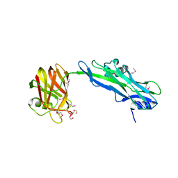
Crystal structure of a two domain protein with unknown function (BF3416) from Bacteroides fragilis NCTC 9343 at 2.90 A resolution
Descriptor: MAGNESIUM ION, TETRAETHYLENE GLYCOL, hypothetical protein
Authors:Joint Center for Structural Genomics (JCSG)
Deposit date:2013-03-27
Release date:2013-05-15
Last modified:2024-10-09
Method:X-RAY DIFFRACTION (2.9 Å)
Cite:Crystal structure of a hypothetical protein (BF3416) from Bacteroides fragilis NCTC 9343 at 2.90 A resolution
To be published
4LIJ
DownloadVisualize
BU of 4lij by Molmil
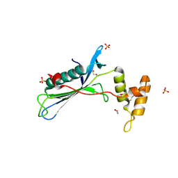
Crystal structure of a far upstream element (FUSE) binding protein 1 (FUBP1) from Homo sapiens at 1.95 A resolution
Descriptor: Far upstream element-binding protein 1, PHOSPHATE ION
Authors:Joint Center for Structural Genomics (JCSG), Partnership for T-Cell Biology, Partnership for T-Cell Biology (TCELL)
Deposit date:2013-07-02
Release date:2013-11-27
Last modified:2024-10-09
Method:X-RAY DIFFRACTION (1.8 Å)
Cite:Crystal structure of a far upstream element (FUSE) binding protein 1 (FUBP1) from Homo sapiens at 1.95 A resolution
To be published
4LG3
DownloadVisualize
BU of 4lg3 by Molmil
Crystal structure of a DUF487 family protein (DESPIG_00776) from Desulfovibrio piger ATCC 29098 at 2.49 A resolution
Descriptor: 1,2-ETHANEDIOL, SULFATE ION, Uncharacterized protein
Authors:Joint Center for Structural Genomics (JCSG)
Deposit date:2013-06-27
Release date:2013-08-21
Last modified:2024-10-30
Method:X-RAY DIFFRACTION (2.4 Å)
Cite:Crystal structure of a hypothetical protein (DESPIG_00776) from Desulfovibrio piger ATCC 29098 at 2.49 A resolution
To be published
4LG8
DownloadVisualize
BU of 4lg8 by Molmil
Crystal structure of PRPF19 WD40 repeats
Descriptor: Pre-mRNA-processing factor 19, SODIUM ION, UNKNOWN ATOM OR ION
Authors:Xu, C, Tempel, W, He, H, Dobrovetsky, E, Seitova, A, Bountra, C, Arrowsmith, C.H, Edwards, A.M, Min, J, Structural Genomics Consortium (SGC)
Deposit date:2013-06-27
Release date:2013-08-07
Last modified:2023-09-20
Method:X-RAY DIFFRACTION (1.89 Å)
Cite:Crystal structure of the WD40 domain of human PRPF19.
Biochem. Biophys. Res. Commun., 493, 2017
4LGC
DownloadVisualize
BU of 4lgc by Molmil
Crystal structure of a bile acid-coenzyme A ligase (baiB) from Clostridium scindens (VPI 12708) at 2.19 A resolution
Descriptor: Bile acid-coenzyme A ligase
Authors:Joint Center for Structural Genomics (JCSG)
Deposit date:2013-06-27
Release date:2013-11-13
Last modified:2017-11-15
Method:X-RAY DIFFRACTION (2.19 Å)
Cite:Crystal structure of a bile acid-coenzyme A ligase (baiB) from Clostridium scindens (VPI 12708) at 2.19 A resolution
To be published
4LEH
DownloadVisualize
BU of 4leh by Molmil
Crystal structure of a bile-acid 7-alpha dehydratase (CLOSCI_03134) from Clostridium scindens ATCC 35704 at 2.90 A resolution
Descriptor: Bile acid 7-alpha dehydratase, BaiE, SULFATE ION, ...
Authors:Joint Center for Structural Genomics (JCSG)
Deposit date:2013-06-25
Release date:2013-08-21
Last modified:2023-02-01
Method:X-RAY DIFFRACTION (2.9 Å)
Cite:Crystal structure of a bile-acid 7-alpha dehydratase (CLOSCI_03134) from Clostridium scindens ATCC 35704 at 2.90 A resolution
To be published
4LG7
DownloadVisualize
BU of 4lg7 by Molmil
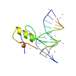
Crystal structure MBD4 MBD domain in complex with methylated CpG DNA
Descriptor: DNA (5'-D(*GP*CP*CP*AP*AP*(5CM)P*GP*TP*TP*GP*GP*C)-3'), Methyl-CpG-binding domain protein 4, UNKNOWN ATOM OR ION
Authors:Xu, C, Tempel, W, Wernimont, A.K, Bountra, C, Arrowsmith, C.H, Edwards, A.M, Min, J, Structural Genomics Consortium (SGC)
Deposit date:2013-06-27
Release date:2013-07-17
Last modified:2024-10-30
Method:X-RAY DIFFRACTION (2.5 Å)
Cite:Crystal structure MBD4 MBD domain in complex with methylated CpG DNA
To be Published
4LGJ
DownloadVisualize
BU of 4lgj by Molmil
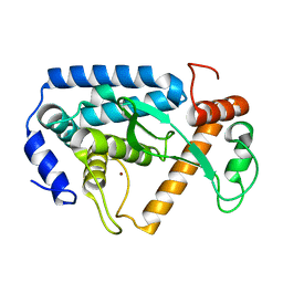
Crystal structure and mechanism of a type III secretion protease
Descriptor: Uncharacterized protein, ZINC ION
Authors:Li, W.Q, Liu, Y.X, Sheng, X.L, Yan, C.Y, Wang, J.W.
Deposit date:2013-06-28
Release date:2014-01-15
Last modified:2023-11-08
Method:X-RAY DIFFRACTION (1.55 Å)
Cite:Structure and mechanism of a type III secretion protease, NleC
Acta Crystallogr.,Sect.D, 70, 2014
4K70
DownloadVisualize
BU of 4k70 by Molmil
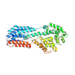
Crystal Structure of N-terminal half of Pseudorabiesvirus UL37 protein
Descriptor: CALCIUM ION, CHLORIDE ION, GLYCEROL, ...
Authors:Pitts, J.D, Heldwein, E.E.
Deposit date:2013-04-16
Release date:2014-03-19
Last modified:2024-02-28
Method:X-RAY DIFFRACTION (2 Å)
Cite:Crystal Structure of the Herpesvirus Inner Tegument Protein UL37 Supports Its Essential Role in Control of Viral Trafficking.
J.Virol., 88, 2014
4K24
DownloadVisualize
BU of 4k24 by Molmil
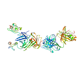
Structure of anti-uPAR Fab ATN-658 in complex with uPAR
Descriptor: 2-acetamido-2-deoxy-beta-D-glucopyranose, 2-acetamido-2-deoxy-beta-D-glucopyranose-(1-4)-2-acetamido-2-deoxy-beta-D-glucopyranose, Urokinase plasminogen activator surface receptor, ...
Authors:Huang, M.D, Xu, X, Yuan, C.
Deposit date:2013-04-08
Release date:2014-02-26
Last modified:2023-11-08
Method:X-RAY DIFFRACTION (4.5 Å)
Cite:Identification of a New Epitope in uPAR as a Target for the Cancer Therapeutic Monoclonal Antibody ATN-658, a Structural Homolog of the uPAR Binding Integrin CD11b ( alpha M)
Plos One, 9, 2014

226707

건을2024-10-30부터공개중

PDB statisticsPDBj update infoContact PDBjnumon