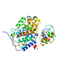
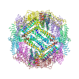
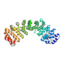
1V9Y
 
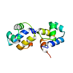
 | | Crystal Structure of the heme PAS sensor domain of Ec DOS (ferric form) | | 分子名称: | Heme pas sensor protein, PROTOPORPHYRIN IX CONTAINING FE | | 著者 | Kurokawa, H, Lee, D.S, Watanabe, M, Sagami, I, Mikami, B, Raman, C.S, Shimizu, T. | | 登録日 | 2004-02-04 | | 公開日 | 2004-05-25 | | 最終更新日 | 2023-12-27 | | 実験手法 | X-RAY DIFFRACTION (1.32 Å) | | 主引用文献 | A redox-controlled molecular switch revealed by the crystal structure of a bacterial heme PAS sensor.
J.Biol.Chem., 279, 2004
|
|
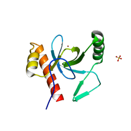
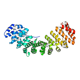
1V9Z
 | | Crystal Structure of the heme PAS sensor domain of Ec DOS (Ferrous Form) | | 分子名称: | Heme pas sensor protein, PROTOPORPHYRIN IX CONTAINING FE | | 著者 | Kurokawa, H, Lee, D.S, Watanabe, M, Sagami, I, Mikami, B, Raman, C.S, Shimizu, T. | | 登録日 | 2004-02-04 | | 公開日 | 2004-05-25 | | 最終更新日 | 2023-12-27 | | 実験手法 | X-RAY DIFFRACTION (1.9 Å) | | 主引用文献 | A redox-controlled molecular switch revealed by the crystal structure of a bacterial heme PAS sensor.
J.Biol.Chem., 279, 2004
|
|
1TKQ
 | | SOLUTION STRUCTURE OF A LINKED UNSYMMETRIC GRAMICIDIN IN A MEMBRANE-ISOELECTRICAL SOLVENTS MIXTURE IN THE PRESENCE OF CsCl | | 分子名称: | GRAMICIDIN A, MINI-GRAMICIDIN A, SUCCINIC ACID | | 著者 | Xie, X, Al-Momani, L, Bockelmann, D, Griesinger, C, Koert, U. | | 登録日 | 2004-06-09 | | 公開日 | 2004-07-13 | | 最終更新日 | 2019-09-25 | | 実験手法 | SOLUTION NMR | | 主引用文献 | An Asymmetric Ion Channel Derived from Gramicidin A. Synthesis, Function and NMR Structure.
FEBS J., 272, 2005
|
|
1ZY6
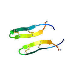 | | Membrane-bound dimer structure of Protegrin-1 (PG-1), a beta-Hairpin Antimicrobial Peptide in Lipid Bilayers from Rotational-Echo Double-Resonance Solid-State NMR | | 分子名称: | Protegrin 1 | | 著者 | Wu, X, Mani, R, Tang, M, Buffy, J.J, Waring, A.J, Sherman, M.A, Hong, M. | | 登録日 | 2005-06-09 | | 公開日 | 2006-06-13 | | 最終更新日 | 2022-03-02 | | 実験手法 | SOLID-STATE NMR | | 主引用文献 | Membrane-Bound Dimer Structure of a beta-Hairpin Antimicrobial Peptide from Rotational-Echo Double-Resonance Solid-State NMR.
Biochemistry, 45, 2006
|
|
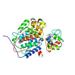
2FOT
 | | Crystal structure of the complex between calmodulin and alphaII-spectrin | | 分子名称: | CALCIUM ION, Calmodulin, alpha-II spectrin Spectrin | | 著者 | Simonovic, M, Zhang, Z, Cianci, C.D, Steitz, T.A, Morrow, J.S. | | 登録日 | 2006-01-13 | | 公開日 | 2006-09-05 | | 最終更新日 | 2023-08-30 | | 実験手法 | X-RAY DIFFRACTION (2.45 Å) | | 主引用文献 | Structure of the calmodulin alphaII-spectrin complex provides insight into the regulation of cell plasticity.
J.Biol.Chem., 281, 2006
|
|
2GB8
 | | Solution structure of the complex between yeast iso-1-cytochrome c and yeast cytochrome c peroxidase | | 分子名称: | Cytochrome c iso-1, Cytochrome c peroxidase, HEME C, ... | | 著者 | Volkov, A.N, Worrall, J.A.R, Ubbink, M. | | 登録日 | 2006-03-10 | | 公開日 | 2006-11-21 | | 最終更新日 | 2022-03-09 | | 実験手法 | SOLUTION NMR | | 主引用文献 | Solution structure and dynamics of the complex between cytochrome c and cytochrome c peroxidase determined by paramagnetic NMR.
Proc.Natl.Acad.Sci.Usa, 103, 2006
|
|
2JTI
 | |
2VX9
 | | H. salinarum dodecin E45A mutant | | 分子名称: | CHLORIDE ION, DODECIN, RIBOFLAVIN, ... | | 著者 | Grininger, M, Staudt, H, Johansson, P, Wachtveitl, J, Oesterhelt, D. | | 登録日 | 2008-07-01 | | 公開日 | 2009-02-17 | | 最終更新日 | 2023-12-13 | | 実験手法 | X-RAY DIFFRACTION (1.65 Å) | | 主引用文献 | Dodecin is the Key Player in Flavin Homeostasis of Archaea.
J.Biol.Chem., 284, 2009
|
|
2VXA
 | | H. halophila dodecin in complex with riboflavin | | 分子名称: | CHLORIDE ION, DODECIN, RIBOFLAVIN | | 著者 | Grininger, M, Staudt, H, Johansson, P, Wachtveitl, J, Oesterhelt, D. | | 登録日 | 2008-07-01 | | 公開日 | 2009-02-17 | | 最終更新日 | 2023-12-13 | | 実験手法 | X-RAY DIFFRACTION (2.6 Å) | | 主引用文献 | Dodecin is the Key Player in Flavin Homeostasis of Archaea.
J.Biol.Chem., 284, 2009
|
|
3E6S
 | |
3E6R
 | |
3NAR
 | | Crystal structure of ZHX1 HD4 (zinc-fingers and homeoboxes protein 1, homeodomain 4) | | 分子名称: | SULFATE ION, Zinc fingers and homeoboxes protein 1 | | 著者 | Ren, J, Bird, L.E, Owens, R.J, Stammers, D.K, Oxford Protein Production Facility (OPPF) | | 登録日 | 2010-06-02 | | 公開日 | 2010-07-07 | | 最終更新日 | 2023-11-01 | | 実験手法 | X-RAY DIFFRACTION (2.6 Å) | | 主引用文献 | Novel structural features in two ZHX homeodomains derived from a systematic study of single and multiple domains
Bmc Struct.Biol., 10, 2010
|
|
3PFS
 | | PWWP Domain of Human Bromodomain and PHD finger-containing protein 3 | | 分子名称: | Bromodomain and PHD finger-containing protein 3, SULFATE ION, ZINC ION | | 著者 | Lam, R, Zeng, H, Kania, J, Bountra, C, Weigelt, J, Arrowsmith, C.H, Edwards, A.M, Min, J, Wu, H, Structural Genomics Consortium (SGC) | | 登録日 | 2010-10-29 | | 公開日 | 2010-11-10 | | 最終更新日 | 2023-09-06 | | 実験手法 | X-RAY DIFFRACTION (1.9 Å) | | 主引用文献 | Structural and histone binding ability characterizations of human PWWP domains.
Plos One, 6, 2011
|
|
3PMI
 | | PWWP Domain of Human Mutated Melanoma-Associated Antigen 1 | | 分子名称: | DI(HYDROXYETHYL)ETHER, PWWP domain-containing protein MUM1, SULFATE ION, ... | | 著者 | Lam, R, Zeng, H, Loppnau, P, Bountra, C, Weigelt, J, Arrowsmith, C.H, Edwards, A.M, Bochkarev, A, Min, J, Wu, H, Structural Genomics Consortium (SGC) | | 登録日 | 2010-11-17 | | 公開日 | 2010-12-15 | | 最終更新日 | 2017-11-08 | | 実験手法 | X-RAY DIFFRACTION (2.82 Å) | | 主引用文献 | Structural and histone binding ability characterizations of human PWWP domains.
Plos One, 6, 2011
|
|
2L9G
 | |
3QQ6
 | | The N-terminal DNA binding domain of SinR from Bacillus subtilis | | 分子名称: | HTH-type transcriptional regulator sinR | | 著者 | Colledge, V, Fogg, M.J, Levdikov, V.M, Dodson, E.J, Wilkinson, A.J. | | 登録日 | 2011-02-15 | | 公開日 | 2011-06-15 | | 最終更新日 | 2023-09-13 | | 実験手法 | X-RAY DIFFRACTION (1.9 Å) | | 主引用文献 | Structure and Organisation of SinR, the Master Regulator of Biofilm Formation in Bacillus subtilis.
J.Mol.Biol., 411, 2011
|
|
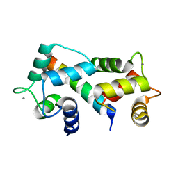
2YAL
 | | SinR, Master Regulator of biofilm formation in Bacillus subtilis | | 分子名称: | HTH-TYPE TRANSCRIPTIONAL REGULATOR SINR, NICKEL (II) ION | | 著者 | Colledge, V.L, Fogg, M.J, Levdikov, V.M, Leech, A, Dodson, E.J, Wilkinson, A.J. | | 登録日 | 2011-02-23 | | 公開日 | 2011-06-08 | | 最終更新日 | 2023-12-20 | | 実験手法 | X-RAY DIFFRACTION (2.27 Å) | | 主引用文献 | Structure and Organisation of Sinr, the Master Regulator of Biofilm Formation in Bacillus Subtilis.
J.Mol.Biol., 411, 2011
|
|
2YD6
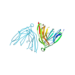 | | Crystal structure of the N-terminal Ig1-2 module of Human Receptor Protein Tyrosine Phosphatase Delta | | 分子名称: | CHLORIDE ION, CITRATE ANION, PTPRD PROTEIN | | 著者 | Coles, C.H, Shen, Y, Tenney, A.P, Siebold, C, Sutton, G.C, Lu, W, Gallagher, J.T, Jones, E.Y, Flanagan, J.G, Aricescu, A.R. | | 登録日 | 2011-03-17 | | 公開日 | 2011-04-13 | | 最終更新日 | 2023-12-20 | | 実験手法 | X-RAY DIFFRACTION (1.35 Å) | | 主引用文献 | Proteoglycan-Specific Molecular Switch for Rptp Sigma Clustering and Neuronal Extension.
Science, 332, 2011
|
|
3T9Q
 | |
3VGN
 | | Crystal Structure of Ketosteroid Isomerase D40N from Pseudomonas putida (pKSI) with bound 3-fluoro-4-nitrophenol | | 分子名称: | 3-fluoro-4-nitrophenol, Steroid Delta-isomerase | | 著者 | Caaveiro, J.M.M, Pybus, B, Ringe, D, Petsko, G.A, Sigala, P.A. | | 登録日 | 2011-08-16 | | 公開日 | 2012-08-22 | | 最終更新日 | 2023-11-08 | | 実験手法 | X-RAY DIFFRACTION (1.3 Å) | | 主引用文献 | Quantitative dissection of hydrogen bond-mediated proton transfer in the ketosteroid isomerase active site
Proc.Natl.Acad.Sci.USA, 110, 2013
|
|
3TPM
 | |
3TPO
 | |
3TPQ
 | |
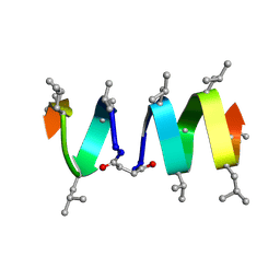
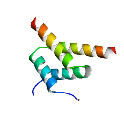
3TYY
 | | Crystal Structure of Human Lamin-B1 Coil 2 Segment | | 分子名称: | Lamin-B1 | | 著者 | Lam, R, Xu, C, Bian, C.B, Mackenzie, F, Walker, J.R, Bountra, C, Weigelt, J, Arrowsmith, C.H, Edwards, A.M, Bochkarev, A, Min, J, Structural Genomics Consortium (SGC) | | 登録日 | 2011-09-26 | | 公開日 | 2011-10-05 | | 最終更新日 | 2023-09-13 | | 実験手法 | X-RAY DIFFRACTION (2.399 Å) | | 主引用文献 | Crystal structures of the coil 2B fragment and the globular tail domain of human lamin B1.
Febs Lett., 586, 2012
|
|
3U43
 | |


