6HU5
 
 | |
7ZUG
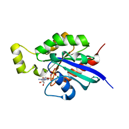
 | | Heterogeneous nuclear ribonucleoprotein H1, qRRM2 domain | | 分子名称: | CHLORIDE ION, Heterogeneous nuclear ribonucleoprotein H, N-terminally processed, ... | | 著者 | Winter, N, Kumar, M, Isupov, M.N, Wiener, R. | | 登録日 | 2022-05-12 | | 公開日 | 2023-05-24 | | 最終更新日 | 2024-06-19 | | 実験手法 | X-RAY DIFFRACTION (1.075 Å) | | 主引用文献 | Heterogeneous nuclear ribonucleoprotein H1, qRRM2 domain
To Be Published
|
|
5GNM
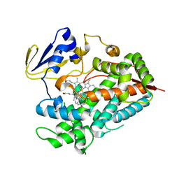
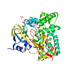
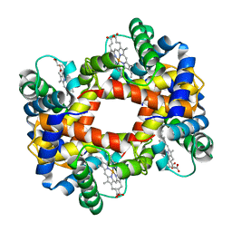
 | | Cytochrome P450 Vdh (CYP107BR1) L348M mutant | | 分子名称: | PROTOPORPHYRIN IX CONTAINING FE, Vitamin D(3) 25-hydroxylase | | 著者 | Yasutake, Y, Tamura, T. | | 登録日 | 2016-07-22 | | 公開日 | 2017-05-17 | | 最終更新日 | 2023-11-08 | | 実験手法 | X-RAY DIFFRACTION (2.7 Å) | | 主引用文献 | Structural insights into the mechanism of the drastic changes in enzymatic activity of the cytochrome P450 vitamin D3 hydroxylase (CYP107BR1) caused by a mutation distant from the active site
Acta Crystallogr F Struct Biol Commun, 73, 2017
|
|
7AA0
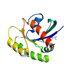
 | | Structural comparison of cellular retinoic acid binding protein I and II in the presence and absence of natural and synthetic ligands | | 分子名称: | (~{E})-3-[4-(4,4-dimethyl-1-propan-2-yl-2,3-dihydroquinolin-6-yl)phenyl]prop-2-enoic acid, Cellular retinoic acid-binding protein 2 | | 著者 | Tomlinson, C.W.E, Cornish, K.A.S, Pohl, E. | | 登録日 | 2020-09-02 | | 公開日 | 2021-02-17 | | 最終更新日 | 2024-01-31 | | 実験手法 | X-RAY DIFFRACTION (1.82 Å) | | 主引用文献 | Structure-functional relationship of cellular retinoic acid-binding proteins I and II interacting with natural and synthetic ligands.
Acta Crystallogr D Struct Biol, 77, 2021
|
|
8PN4
 | |
6HXU
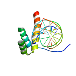
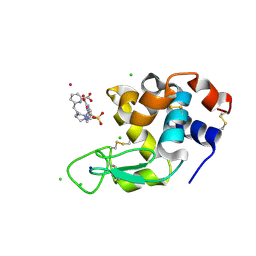
 | | Crystal structure of Human RHOB Q63L in complex with GTP | | 分子名称: | GUANOSINE-5'-TRIPHOSPHATE, MAGNESIUM ION, Rho-related GTP-binding protein RhoB | | 著者 | Soulie, S, Gence, R, Cabantous, S, Lajoie-Mazenc, I, Favre, G, Pedelacq, J.D. | | 登録日 | 2018-10-18 | | 公開日 | 2019-09-25 | | 最終更新日 | 2024-01-24 | | 実験手法 | X-RAY DIFFRACTION (1.19 Å) | | 主引用文献 | A Targeted Protein Degradation Cell-Based Screening for Nanobodies Selective toward the Cellular RHOB GTP-Bound Conformation.
Cell Chem Biol, 26, 2019
|
|
4R20
 | | Zebra fish cytochrome P450 17A2 with Abiraterone | | 分子名称: | Abiraterone, Cytochrome P450 family 17 polypeptide 2, MERCURY (II) ION, ... | | 著者 | Pallan, P.S, Egli, M. | | 登録日 | 2014-08-08 | | 公開日 | 2014-12-31 | | 最終更新日 | 2024-02-28 | | 実験手法 | X-RAY DIFFRACTION (2.86 Å) | | 主引用文献 | Structural and Kinetic Basis of Steroid 17 alpha, 20-Lyase Activity in Teleost Fish Cytochrome P450 17A1 and Its Absence in Cytochrome P450 17A2.
J.Biol.Chem., 290, 2015
|
|
8PIY
 | |
8PNA
 | |
4R2E
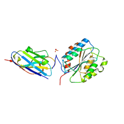
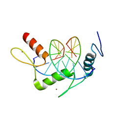 | | Wilms Tumor Protein (WT1) zinc fingers in complex with methylated DNA | | 分子名称: | DNA (5'-D(*AP*GP*CP*GP*TP*GP*GP*GP*(5CM)P*GP*T)-3'), DNA (5'-D(*TP*AP*(5CM)P*GP*CP*CP*CP*AP*CP*GP*C)-3'), Wilms tumor protein, ... | | 著者 | Hashimoto, H, Olanrewaju, Y.O, Zheng, Y, Wilson, G.G, Zhang, X, Cheng, X. | | 登録日 | 2014-08-11 | | 公開日 | 2014-10-08 | | 最終更新日 | 2023-09-20 | | 実験手法 | X-RAY DIFFRACTION (1.84 Å) | | 主引用文献 | Wilms tumor protein recognizes 5-carboxylcytosine within a specific DNA sequence.
Genes Dev., 28, 2014
|
|
8POB
 | | Crystal structure of Hen Egg White Lysozyme co-crystallized with 10 mM TbXo4-SO3 | | 分子名称: | 6-[[4-[(6-carboxypyridin-2-yl)methyl]-7-(3-sulfopropyl)-1,4,7-triazonan-1-yl]methyl]pyridine-2-carboxylic acid, CHLORIDE ION, Lysozyme C, ... | | 著者 | Alsalman, Z, Girard, E. | | 登録日 | 2023-07-04 | | 公開日 | 2024-07-10 | | 最終更新日 | 2024-07-17 | | 実験手法 | X-RAY DIFFRACTION (1.74 Å) | | 主引用文献 | Influence of Chemical Modifications of the Crystallophore on Protein Nucleating Properties and Supramolecular Interactions Network.
Chemistry, 30, 2024
|
|
7ZLT
 | |
7ZXM
 | |
7A6O
 | |
6HBH
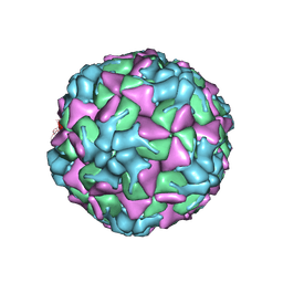 | | Echovirus 18 A-particle | | 分子名称: | Echovirus 18 capsid protein 1, Echovirus 18 capsid protein 2, Echovirus 18 capsid protein 3 | | 著者 | Buchta, D, Fuzik, T, Hrebik, D, Levdansky, Y, Moravcova, J, Plevka, P. | | 登録日 | 2018-08-10 | | 公開日 | 2019-03-20 | | 最終更新日 | 2019-12-11 | | 実験手法 | ELECTRON MICROSCOPY (3.36 Å) | | 主引用文献 | Enterovirus particles expel capsid pentamers to enable genome release.
Nat Commun, 10, 2019
|
|
4QZU
 | |
6HEB
 | | Influenza A Virus N9 Neuraminidase complex with Oseltamivir (Tern). | | 分子名称: | (3R,4R,5S)-4-(acetylamino)-5-amino-3-(pentan-3-yloxy)cyclohex-1-ene-1-carboxylic acid, 2-acetamido-2-deoxy-beta-D-glucopyranose-(1-4)-2-acetamido-2-deoxy-beta-D-glucopyranose, CALCIUM ION, ... | | 著者 | Salinger, M.T, Hobbs, J.R, Murray, J.W, Laver, W.G, Kuhn, P, Garman, E.F. | | 登録日 | 2018-08-20 | | 公開日 | 2018-08-29 | | 最終更新日 | 2024-01-17 | | 実験手法 | X-RAY DIFFRACTION (1.75 Å) | | 主引用文献 | High Resolution Structures of Viral Neuraminidase with Drugs Bound in the Active Site.
To Be Published
|
|
6HBW
 | |
6U6A
 | | Crystal structure of Yck2 from Candida albicans in complex with kinase inhibitor GW461484A | | 分子名称: | 2-(4-fluorophenyl)-6-methyl-3-(pyridin-4-yl)pyrazolo[1,5-a]pyridine, SULFATE ION, Serine/threonine protein kinase | | 著者 | Stogios, P.J, Evdokimova, E, Di Leo, R, Chang, C, Savchenko, A, Joachimiak, A, Satchell, K.J.F, Center for Structural Genomics of Infectious Diseases (CSGID) | | 登録日 | 2019-08-29 | | 公開日 | 2019-10-30 | | 最終更新日 | 2023-10-11 | | 実験手法 | X-RAY DIFFRACTION (2.45 Å) | | 主引用文献 | Overcoming Fungal Echinocandin Resistance through Inhibition of the Non-essential Stress Kinase Yck2.
Cell Chem Biol, 27, 2020
|
|
8PMF
 | |
8PMN
 | |
7ZXT
 | |
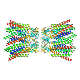
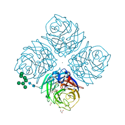
6U96
 | | Actin phalloidin at BeFx state | | 分子名称: | ADENOSINE-5'-DIPHOSPHATE, Actin, alpha skeletal muscle, ... | | 著者 | Das, S, Ge, P, Durer, Z.A.O, Grintsevich, E.E, Zhou, Z.H, Reisler, E. | | 登録日 | 2019-09-06 | | 公開日 | 2020-05-13 | | 最終更新日 | 2020-05-20 | | 実験手法 | ELECTRON MICROSCOPY (3.8 Å) | | 主引用文献 | D-loop Dynamics and Near-Atomic-Resolution Cryo-EM Structure of Phalloidin-Bound F-Actin.
Structure, 28, 2020
|
|
6HFC
 | | Influenza A Virus N9 Neuraminidase Native (Tern). | | 分子名称: | 2-acetamido-2-deoxy-beta-D-glucopyranose-(1-4)-2-acetamido-2-deoxy-beta-D-glucopyranose, CALCIUM ION, CARBON DIOXIDE, ... | | 著者 | Salinger, M.T, Hobbs, J.R, Murray, J.W, Laver, W.G, Kuhn, P, Garman, E.F. | | 登録日 | 2018-08-21 | | 公開日 | 2018-08-29 | | 最終更新日 | 2024-02-07 | | 実験手法 | X-RAY DIFFRACTION (1.29 Å) | | 主引用文献 | High Resolution Structures of Viral Neuraminidase with Drugs Bound in the Active Site. (In preparation)
To Be Published
|
|
4R5K
 | | Crystal structure of the DnaK C-terminus (Dnak-SBD-B) | | 分子名称: | CALCIUM ION, Chaperone protein DnaK, SULFATE ION | | 著者 | Leu, J.I, Zhang, P, Murphy, M.E, Marmorstein, R, George, D.L. | | 登録日 | 2014-08-21 | | 公開日 | 2014-09-10 | | 最終更新日 | 2024-02-28 | | 実験手法 | X-RAY DIFFRACTION (1.7469 Å) | | 主引用文献 | Structural Basis for the Inhibition of HSP70 and DnaK Chaperones by Small-Molecule Targeting of a C-Terminal Allosteric Pocket.
Acs Chem.Biol., 9, 2014
|
|


