3KUR
 
 | |
5GNV
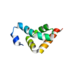
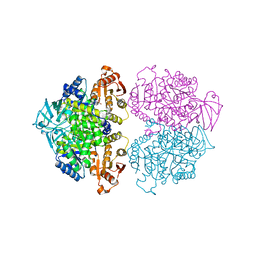
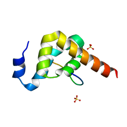
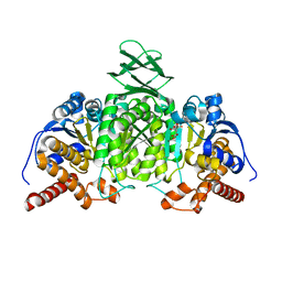
 | | Structure of PSD-95/MAP1A complex reveals unique target recognition mode of MAGUK GK domain | | 分子名称: | Disks large homolog 4, Microtubule-associated protein 1A, SULFATE ION | | 著者 | Shang, Y, Xia, Y, Zhu, R, Zhu, J. | | 登録日 | 2016-07-25 | | 公開日 | 2017-08-02 | | 最終更新日 | 2024-03-20 | | 実験手法 | X-RAY DIFFRACTION (2.596 Å) | | 主引用文献 | Structure of the PSD-95/MAP1A complex reveals a unique target recognition mode of the MAGUK GK domain
Biochem. J., 474, 2017
|
|
5GW0
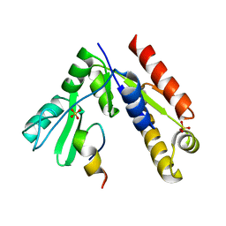
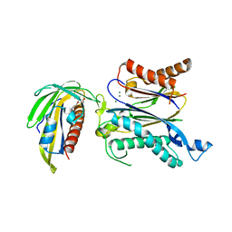
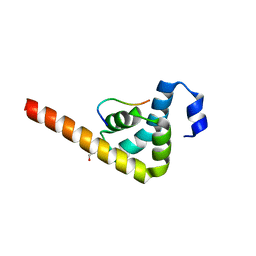
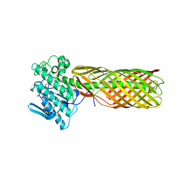
 | | Crystal structure of SNX16 PX-Coiled coil | | 分子名称: | Sorting nexin-16 | | 著者 | Xu, J, Liu, J. | | 登録日 | 2016-09-08 | | 公開日 | 2017-09-13 | | 実験手法 | X-RAY DIFFRACTION (3.3 Å) | | 主引用文献 | SNX16 Regulates the Recycling of E-Cadherin through a Unique Mechanism of Coordinated Membrane and Cargo Binding.
Structure, 25, 2017
|
|
5GNZ
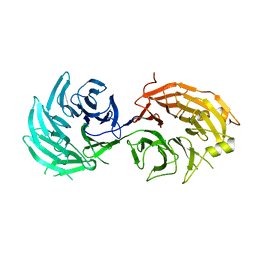
 | | The M3 mutant structure of Bgl6 | | 分子名称: | Beta-glucosidase, GLYCEROL, beta-D-glucopyranose | | 著者 | Xie, W, Pang, P, Cao, L.C, Liu, Y.H, Wang, Z. | | 登録日 | 2016-07-25 | | 公開日 | 2017-04-12 | | 最終更新日 | 2023-11-08 | | 実験手法 | X-RAY DIFFRACTION (2.2 Å) | | 主引用文献 | Structures of a glucose-tolerant beta-glucosidase provide insights into its mechanism.
J. Struct. Biol., 198, 2017
|
|
3KTX
 | | Crystal structure of Leishmania mexicana pyruvate kinase (LmPYK)in complex with 1,3,6,8-pyrenetetrasulfonic acid | | 分子名称: | GLYCEROL, Pyruvate kinase, pyrene-1,3,6,8-tetrasulfonic acid | | 著者 | Morgan, H.P, Walkinshaw, M.D. | | 登録日 | 2009-11-26 | | 公開日 | 2010-02-09 | | 最終更新日 | 2023-09-06 | | 実験手法 | X-RAY DIFFRACTION (2.1 Å) | | 主引用文献 | An improved strategy for the crystallization of Leishmania mexicana pyruvate kinase.
Acta Crystallogr.,Sect.F, 66, 2010
|
|
5GWP
 | | Crystal structure of RCAR3:PP2C wild-type with (+)-ABA | | 分子名称: | (2Z,4E)-5-[(1S)-1-hydroxy-2,6,6-trimethyl-4-oxocyclohex-2-en-1-yl]-3-methylpenta-2,4-dienoic acid, ABA receptor RCAR3, MAGNESIUM ION, ... | | 著者 | Han, S, Lee, S. | | 登録日 | 2016-09-12 | | 公開日 | 2017-09-13 | | 最終更新日 | 2023-11-08 | | 実験手法 | X-RAY DIFFRACTION (2.577 Å) | | 主引用文献 | Modulation of ABA Signaling by Altering VxG Phi L Motif of PP2Cs in Oryza sativa.
Mol Plant, 10, 2017
|
|
5GOW
 | |
5GXU
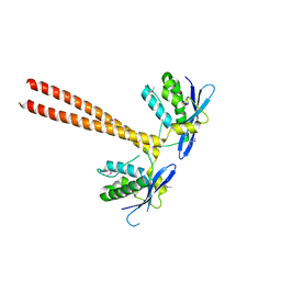
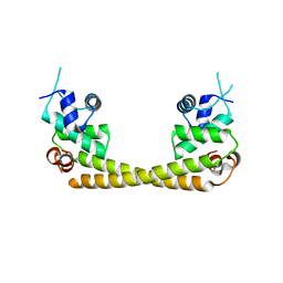
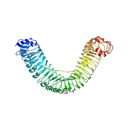
 | | Cystal structure of Arabidopsis ATR2 | | 分子名称: | FLAVIN MONONUCLEOTIDE, FLAVIN-ADENINE DINUCLEOTIDE, NADPH--cytochrome P450 reductase 2 | | 著者 | Niu, G, Liu, L. | | 登録日 | 2016-09-20 | | 公開日 | 2017-01-25 | | 最終更新日 | 2023-11-08 | | 実験手法 | X-RAY DIFFRACTION (2.3 Å) | | 主引用文献 | Structure of the Arabidopsis thaliana NADPH-cytochrome P450 reductase 2 (ATR2) provides insight into its function
FEBS J., 284, 2017
|
|
3KUJ
 | |
3KUS
 | |
5GPE
 | | Crystal structure of the transcription regulator PbrR691 from Ralstonia metallidurans CH34 in complex with Lead(II) | | 分子名称: | LEAD (II) ION, Transcriptional regulator, MerR-family | | 著者 | Huang, S.Q, Chen, W.Z, Wang, D, Hu, Q.Y, Liu, X.C, Gan, J.H, Chen, H. | | 登録日 | 2016-08-01 | | 公開日 | 2016-12-28 | | 最終更新日 | 2023-11-08 | | 実験手法 | X-RAY DIFFRACTION (2.01 Å) | | 主引用文献 | Structural Basis for the Selective Pb(II) Recognition of Metalloregulatory Protein PbrR691
Inorg Chem, 55, 2016
|
|
5H2K
 | | A three dimensional movie of structural changes in bacteriorhodopsin: structure obtained 2 us after photoexcitation | | 分子名称: | 2,3-DI-PHYTANYL-GLYCEROL, Bacteriorhodopsin, DECANE, ... | | 著者 | Royant, A, Nango, E, Nakane, T, Tanaka, T, Arima, T, Neutze, R, Iwata, S. | | 登録日 | 2016-10-15 | | 公開日 | 2016-12-21 | | 最終更新日 | 2023-11-08 | | 実験手法 | X-RAY DIFFRACTION (2.1 Å) | | 主引用文献 | A three-dimensional movie of structural changes in bacteriorhodopsin
Science, 354, 2016
|
|
5GQF
 | | Crystal structure of lacto-N-biosidase LnbX from Bifidobacterium longum subsp. longum, lacto-N-biose complex | | 分子名称: | CALCIUM ION, Lacto-N-biosidase, beta-D-galactopyranose-(1-3)-2-acetamido-2-deoxy-beta-D-glucopyranose | | 著者 | Yamada, C, Arakawa, T, Katayama, T, Fushinobu, S. | | 登録日 | 2016-08-07 | | 公開日 | 2017-04-19 | | 最終更新日 | 2024-03-20 | | 実験手法 | X-RAY DIFFRACTION (1.82 Å) | | 主引用文献 | Molecular Insight into Evolution of Symbiosis between Breast-Fed Infants and a Member of the Human Gut Microbiome Bifidobacterium longum
Cell Chem Biol, 24, 2017
|
|
5H30
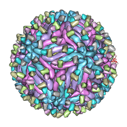 | | Cryo-EM structure of zika virus complexed with Fab C10 at pH 6.5 | | 分子名称: | IgG C10 heavy chain, IgG C10 light chain, structural protein E, ... | | 著者 | Zhang, S, Kostyuchenko, V, Ng, T.-S, Lok, S.-M. | | 登録日 | 2016-10-19 | | 公開日 | 2016-11-30 | | 最終更新日 | 2024-05-29 | | 実験手法 | ELECTRON MICROSCOPY (4.4 Å) | | 主引用文献 | Neutralization mechanism of a highly potent antibody against Zika virus
Nat Commun, 7, 2016
|
|
3KX1
 | |
6XQK
 | | Crystal structure of the D/D domain of PKA from S. cerevisiae | | 分子名称: | CHLORIDE ION, GLYCEROL, cAMP-dependent protein kinase regulatory subunit | | 著者 | Larrieux, N, Gonzalez Bardeci, N, Trajtenberg, F, Buschiazzo, A. | | 登録日 | 2020-07-09 | | 公開日 | 2021-04-14 | | 最終更新日 | 2024-03-06 | | 実験手法 | X-RAY DIFFRACTION (2.56 Å) | | 主引用文献 | The crystal structure of yeast regulatory subunit reveals key evolutionary insights into Protein Kinase A oligomerization.
J.Struct.Biol., 213, 2021
|
|
5GQW
 | |
3KV8
 | | Structural basis of the activity and substrate specificity of the fluoroacetyl-CoA thioesterase FlK - Wild type FlK in complex with fluoro-acetate | | 分子名称: | Fluoroacetyl-CoA thioesterase FlK, fluoroacetic acid | | 著者 | Dias, M.V.B, Huang, F, Chirgadze, D.Y, Tosin, M, Spiteller, D, Valentine, E.F, Leadlay, P.F, Spencer, J.B, Blundell, T.L. | | 登録日 | 2009-11-29 | | 公開日 | 2010-04-21 | | 最終更新日 | 2023-09-06 | | 実験手法 | X-RAY DIFFRACTION (1.85 Å) | | 主引用文献 | Structural basis for the activity and substrate specificity of fluoroacetyl-CoA thioesterase FlK.
J.Biol.Chem., 285, 2010
|
|
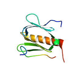
3KVH
 | | Crystal structure of human protein syndesmos (NUDT16-like protein) | | 分子名称: | CHLORIDE ION, GLYCEROL, Protein syndesmos | | 著者 | Tresaugues, L, Siponen, M.I, Arrowsmith, C.H, Berglund, H, Bountra, C, Collins, R, Edwards, A.M, Flodin, S, Flores, A, Graslund, S, Hammarstrom, M, Johansson, A, Johansson, I, Kallas, A, Karlberg, T, Kotenyova, T, Kotzsch, A, Kraulis, P, Moche, M, Nielsen, T.K, Nyman, T, Persson, C, Roos, A.K, Schuler, H, Schutz, P, Thorsell, A.G, Van Den Berg, S, Weigelt, J, Welin, M, Wisniewska, M, Nordlund, P, Structural Genomics Consortium (SGC) | | 登録日 | 2009-11-30 | | 公開日 | 2010-01-19 | | 最終更新日 | 2023-09-06 | | 実験手法 | X-RAY DIFFRACTION (1.7 Å) | | 主引用文献 | Crystal structure of human protein syndesmos (NUDT16L1)
To be Published
|
|
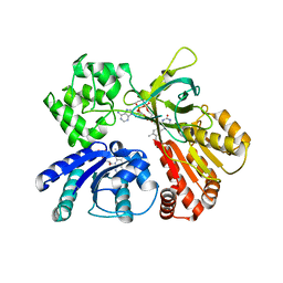
5H3F
 | | Crystal structure of mouse isocitrate dehydrogenases 2 complexed with isocitrate | | 分子名称: | ISOCITRIC ACID, Isocitrate dehydrogenase [NADP], mitochondrial, ... | | 著者 | Xu, Y, Liu, L, Miyakawa, T, Nakamura, A, Tanokura, M. | | 登録日 | 2016-10-23 | | 公開日 | 2017-08-30 | | 最終更新日 | 2023-11-08 | | 実験手法 | X-RAY DIFFRACTION (3.29 Å) | | 主引用文献 | Studies on the regulatory mechanism of isocitrate dehydrogenase 2 using acetylation mimics
Sci Rep, 7, 2017
|
|
3KVN
 | |
5H3S
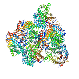
 | | apo form of GEMIN5-WD | | 分子名称: | GLYCEROL, Gem-associated protein 5 | | 著者 | Bharath, S.R, Tang, X, Song, H. | | 登録日 | 2016-10-27 | | 公開日 | 2016-12-28 | | 最終更新日 | 2023-11-08 | | 実験手法 | X-RAY DIFFRACTION (3 Å) | | 主引用文献 | Structural basis for specific recognition of pre-snRNA by Gemin5
Cell Res., 26, 2016
|
|
5GR8
 | | Crystal structure of PEPR1-AtPEP1 | | 分子名称: | 2-acetamido-2-deoxy-beta-D-glucopyranose, 2-acetamido-2-deoxy-beta-D-glucopyranose-(1-4)-2-acetamido-2-deoxy-beta-D-glucopyranose, Elicitor peptide 1, ... | | 著者 | Chai, J.J, Tang, J. | | 登録日 | 2016-08-08 | | 公開日 | 2016-12-14 | | 最終更新日 | 2023-11-08 | | 実験手法 | X-RAY DIFFRACTION (2.587 Å) | | 主引用文献 | Structural basis for recognition of an endogenous peptide by the plant receptor kinase PEPR1
Cell Res., 25, 2015
|
|
3KX7
 | | Structural basis of the activity and substrate specificity of the fluoroacetyl-CoA FlK - apo wild type FlK | | 分子名称: | Fluoroacetyl-CoA thioesterase FlK | | 著者 | Dias, M.V.B, Huang, F, Chirgadze, D.Y, Tosin, M, Spiteller, D, Valentine, E.F, Leadlay, P.F, Spencer, J.B, Blundell, T.L. | | 登録日 | 2009-12-02 | | 公開日 | 2010-04-21 | | 最終更新日 | 2023-09-06 | | 実験手法 | X-RAY DIFFRACTION (1.7 Å) | | 主引用文献 | Structural basis for the activity and substrate specificity of fluoroacetyl-CoA thioesterase FlK.
J.Biol.Chem., 285, 2010
|
|
5H4H
 | | Structure of PIN-domain protein (VapC4 toxin) from Pyrococcus horikoshii determined at 2.2 A resolution | | 分子名称: | CADMIUM ION, Ribonuclease VapC4 | | 著者 | Biswas, A, Hatti, K, Srinivasan, N, Murthy, M.R.N, Sekar, K. | | 登録日 | 2016-10-31 | | 公開日 | 2016-11-23 | | 最終更新日 | 2023-11-08 | | 実験手法 | X-RAY DIFFRACTION (2.23 Å) | | 主引用文献 | Structure determination of contaminant proteins using the MarathonMR procedure
J. Struct. Biol., 197, 2017
|
|


