4E0K
 
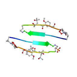
 | | Crystal Structure of the amyloid-fibril forming peptide KDWSFY derived from human Beta 2 Microglobulin (58-63) | | 分子名称: | 2-{2-[2-(2-{2-[2-(2-ETHOXY-ETHOXY)-ETHOXY]-ETHOXY}-ETHOXY)-ETHOXY]-ETHOXY}-ETHANOL, Amyloidogenic peptide segment KDWSFY, SODIUM ION | | 著者 | Zhao, M, Liu, C, Sawaya, M.R, Eisenberg, D. | | 登録日 | 2012-03-04 | | 公開日 | 2012-12-19 | | 最終更新日 | 2024-02-28 | | 実験手法 | X-RAY DIFFRACTION (0.97 Å) | | 主引用文献 | Out-of-register beta-sheets suggest a pathway to toxic amyloid aggregates.
Proc.Natl.Acad.Sci.USA, 109, 2012
|
|
4E0O
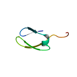
 | | SVQIVYK segment from human Tau (305-311) displayed on 54-membered macrocycle scaffold (form III) | | 分子名称: | (4S)-2-METHYL-2,4-PENTANEDIOL, Cyclic pseudo-peptide SVQIVYK(ORN)EF(HAO)(4BF)K(ORN), PHOSPHATE ION | | 著者 | Zhao, M, Liu, C, Sawaya, M.R, Eisenberg, D. | | 登録日 | 2012-03-04 | | 公開日 | 2012-12-19 | | 最終更新日 | 2023-11-15 | | 実験手法 | X-RAY DIFFRACTION (1.82 Å) | | 主引用文献 | Out-of-register beta-sheets suggest a pathway to toxic amyloid aggregates.
Proc.Natl.Acad.Sci.USA, 109, 2012
|
|
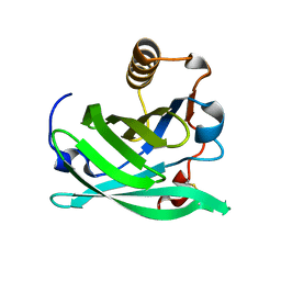
2NB9
 | | Solution structure of ZitP zinc finger | | 分子名称: | Uncharacterized protein, ZINC ION | | 著者 | Campagne, S, Berge, M, Viollier, P.H, Allain, F.H.-T. | | 登録日 | 2016-02-01 | | 公開日 | 2016-12-14 | | 最終更新日 | 2024-05-15 | | 実験手法 | SOLUTION NMR | | 主引用文献 | Modularity and determinants of a (bi-)polarization control system from free-living and obligate intracellular bacteria.
Elife, 5, 2016
|
|
1XFR
 | |
1KLQ
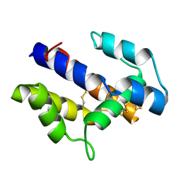
 | | The Mad2 Spindle Checkpoint Protein Undergoes Similar Major Conformational Changes upon Binding to Either Mad1 or Cdc20 | | 分子名称: | MITOTIC SPINDLE ASSEMBLY CHECKPOINT PROTEIN MAD2A, Mad2-binding peptide | | 著者 | Luo, X, Tang, Z, Rizo, J, Yu, H. | | 登録日 | 2001-12-12 | | 公開日 | 2002-01-25 | | 最終更新日 | 2024-05-22 | | 実験手法 | SOLUTION NMR | | 主引用文献 | The Mad2 spindle checkpoint protein undergoes similar major conformational changes upon binding to either Mad1 or Cdc20.
Mol.Cell, 9, 2002
|
|
2FFT
 | | NMR structure of Spinach Thylakoid Soluble Phosphoprotein of 9 kDa in SDS Micelles | | 分子名称: | thylakoid soluble phosphoprotein | | 著者 | Song, J, Carlberg, I, Lee, M.S, Markley, J.L, Center for Eukaryotic Structural Genomics (CESG) | | 登録日 | 2005-12-20 | | 公開日 | 2006-01-17 | | 最終更新日 | 2024-05-29 | | 実験手法 | SOLUTION NMR | | 主引用文献 | Micelle-induced folding of spinach thylakoid soluble phosphoprotein of 9 kDa and its functional implications.
Biochemistry, 45, 2006
|
|
1BUA
 | |
1BSU
 | |
2XND
 | | Crystal structure of bovine F1-c8 sub-complex of ATP Synthase | | 分子名称: | ATP SYNTHASE LIPID-BINDING PROTEIN, MITOCHONDRIAL, ATP SYNTHASE SUBUNIT ALPHA, ... | | 著者 | Watt, I.N, Montgomery, M.G, Runswick, M.J, Leslie, A.G.W, Walker, J.E. | | 登録日 | 2010-08-02 | | 公開日 | 2010-09-15 | | 最終更新日 | 2023-12-20 | | 実験手法 | X-RAY DIFFRACTION (3.5 Å) | | 主引用文献 | Bioenergetic Cost of Making an Adenosine Triphosphate Molecule in Animal Mitochondria.
Proc.Natl.Acad.Sci.USA, 107, 2010
|
|
2JR4
 | | NMR Solution Structure of the Anticodon of E.coli TRNA-VAL3 With no Modifications | | 分子名称: | 5'-R(*CP*CP*UP*CP*CP*CP*UP*UP*AP*CP*AP*AP*GP*GP*AP*GP*G)-3' | | 著者 | Vendeix, F.A.P, Dziergowska, A, Gustilo, E.M, Graham, W.D, Sproat, B, Malkiewicz, A, Agris, P.F. | | 登録日 | 2007-06-20 | | 公開日 | 2007-07-24 | | 最終更新日 | 2024-05-29 | | 実験手法 | SOLUTION NMR | | 主引用文献 | Anticodon domain modifications contribute order to tRNA for ribosome-mediated codon binding.
Biochemistry, 47, 2008
|
|
2JRG
 | | NMR solution structure of the anticodon of E. coli TRNA-VAL3 with 2 modifications (cmo5U34 M6A37) | | 分子名称: | 5'-R(*CP*CP*UP*CP*CP*CP*UP*(CM0)P*AP*CP*(6MZ)P*AP*GP*GP*AP*GP*G)-3' | | 著者 | Vendeix, F.A, Dziergowska, A, Gustilo, E.M, Graham, W.D, Sproat, B, Malkiewicz, A, Agris, P.F. | | 登録日 | 2007-06-26 | | 公開日 | 2007-07-24 | | 最終更新日 | 2023-12-20 | | 実験手法 | SOLUTION NMR | | 主引用文献 | Anticodon domain modifications contribute order to tRNA for ribosome-mediated codon binding.
Biochemistry, 47, 2008
|
|
6G04
 | | NMR Solution Structure of Yeast TSR2(1-152) in Complex with S26A(100-119) | | 分子名称: | 40S ribosomal protein S26-A, Pre-rRNA-processing protein TSR2 | | 著者 | Michel, E, Schuetz, S, Damberger, F.F, Allain, F.H.-T, Panse, V.G. | | 登録日 | 2018-03-16 | | 公開日 | 2018-09-19 | | 最終更新日 | 2024-05-15 | | 実験手法 | SOLUTION NMR | | 主引用文献 | Molecular basis for disassembly of an importin:ribosomal protein complex by the escortin Tsr2.
Nat Commun, 9, 2018
|
|
6G03
 | | NMR Solution Structure of yeast TSR2(1-152) | | 分子名称: | Pre-rRNA-processing protein TSR2 | | 著者 | Michel, E, Schuetz, S, Damberger, F.F, Allain, F.H.-T, Panse, V.G. | | 登録日 | 2018-03-16 | | 公開日 | 2018-09-19 | | 最終更新日 | 2024-06-19 | | 実験手法 | SOLUTION NMR | | 主引用文献 | Molecular basis for disassembly of an importin:ribosomal protein complex by the escortin Tsr2.
Nat Commun, 9, 2018
|
|
4B5Q
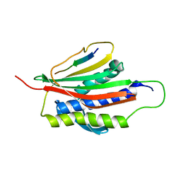
 | | The lytic polysaccharide monooxygenase GH61D structure from the basidiomycota fungus Phanerochaete chrysosporium | | 分子名称: | COPPER (II) ION, GLYCEROL, GLYCOSIDE HYDROLASE FAMILY 61 PROTEIN D, ... | | 著者 | Wu, M, Beckham, G.T, Larsson, A.M, Ishida, T, Kim, S, Crowley, M.F, Payne, C.M, Horn, S.J, Westereng, B, Stahlberg, J, Eijsink, V.G.H, Sandgren, M. | | 登録日 | 2012-08-07 | | 公開日 | 2013-04-03 | | 最終更新日 | 2023-12-20 | | 実験手法 | X-RAY DIFFRACTION (1.75 Å) | | 主引用文献 | Crystal Structure and Computational Characterization of the Lytic Polysaccharide Monooxygenase Gh61D from the Basidiomycota Fungus Phanerochaete Chrysosporium
J.Biol.Chem., 288, 2013
|
|
7M74
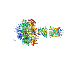
 | | ATP-bound AMP-activated protein kinase | | 分子名称: | 5'-AMP-activated protein kinase catalytic subunit alpha-1, 5'-AMP-activated protein kinase subunit beta-2, 5'-AMP-activated protein kinase subunit gamma-1, ... | | 著者 | Yan, Y, Mukherjee, S, Harikumar, K.G, Strutzenberg, T, Zhou, X.E, Powell, S.K, Xu, T, Sheldon, R, Lamp, J, Brunzelle, J.S, Radziwon, K, Ellis, A, Novick, S.J, Vega, I.E, Jones, R, Miller, L.J, Xu, H.E, Griffin, P.R, Kossiakoff, A.A, Melcher, K. | | 登録日 | 2021-03-26 | | 公開日 | 2021-12-15 | | 最終更新日 | 2021-12-22 | | 実験手法 | ELECTRON MICROSCOPY (3.93 Å) | | 主引用文献 | Structure of an AMPK complex in an inactive, ATP-bound state.
Science, 373, 2021
|
|
6VJV
 | | Crystal structure of the Prochlorococcus phage (myovirus P-SSM2) ferredoxin at 1.6 Angstroms | | 分子名称: | ACETATE ION, FE2/S2 (INORGANIC) CLUSTER, Ferredoxin, ... | | 著者 | Olmos Jr, J.L, Campbell, I.J, Miller, M.D, Xu, W, Kahanda, D, Atkinson, J.T, Sparks, N, Bennett, G.N, Silberg, J.J, Phillips Jr, G.N. | | 登録日 | 2020-01-17 | | 公開日 | 2020-02-19 | | 最終更新日 | 2023-10-11 | | 実験手法 | X-RAY DIFFRACTION (1.59 Å) | | 主引用文献 | Prochlorococcusphage ferredoxin: structural characterization and electron transfer to cyanobacterial sulfite reductases.
J.Biol.Chem., 295, 2020
|
|
2CH5
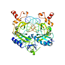
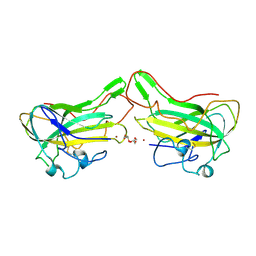
 | | Crystal structure of human N-acetylglucosamine kinase in complex with N-acetylglucosamine | | 分子名称: | 2-acetamido-2-deoxy-alpha-D-glucopyranose, 2-acetamido-2-deoxy-beta-D-glucopyranose, GLYCEROL, ... | | 著者 | Weihofen, W.A, Berger, M, Chen, H, Saenger, W, Hinderlich, S. | | 登録日 | 2006-03-13 | | 公開日 | 2006-09-18 | | 最終更新日 | 2024-05-01 | | 実験手法 | X-RAY DIFFRACTION (1.9 Å) | | 主引用文献 | Structures of Human N-Acetylglucosamine Kinase in Two Complexes with N-Acetylglucosamine and with Adp/Glucose: Insights Into Substrate Specificity and Regulation.
J.Mol.Biol., 364, 2006
|
|
1L9L
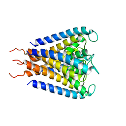
 | | GRANULYSIN FROM HUMAN CYTOLYTIC T LYMPHOCYTES | | 分子名称: | 3[N-MORPHOLINO]PROPANE SULFONIC ACID, ETHANOL, Granulysin, ... | | 著者 | Anderson, D.H, Sawaya, M.R, Cascio, D, Ernst, W, Krensky, A, Modlin, R, Eisenberg, D. | | 登録日 | 2002-03-25 | | 公開日 | 2002-11-06 | | 最終更新日 | 2017-09-13 | | 実験手法 | X-RAY DIFFRACTION (0.92 Å) | | 主引用文献 | Granulysin Crystal Structure and a Structure-Derived Lytic Mechanism
J.Mol.Biol., 325, 2002
|
|
1JVM
 | |
2CH6
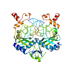
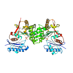 | | Crystal structure of human N-acetylglucosamine kinase in complex with ADP and glucose | | 分子名称: | ADENOSINE-5'-DIPHOSPHATE, N-ACETYL-D-GLUCOSAMINE KINASE, alpha-D-glucopyranose | | 著者 | Weihofen, W.A, Berger, M, Chen, H, Saenger, W, Hinderlich, S. | | 登録日 | 2006-03-13 | | 公開日 | 2006-09-18 | | 最終更新日 | 2024-05-08 | | 実験手法 | X-RAY DIFFRACTION (2.72 Å) | | 主引用文献 | Structures of Human N-Acetylglucosamine Kinase in Two Complexes with N-Acetylglucosamine and with Adp/Glucose: Insights Into Substrate Specificity and Regulation.
J.Mol.Biol., 364, 2006
|
|
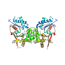
1MD4
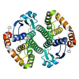 | | A folding mutant of human class pi glutathione transferase, created by mutating glycine 146 of the wild-type protein to valine | | 分子名称: | 2-(N-MORPHOLINO)-ETHANESULFONIC ACID, GLUTATHIONE, pi glutathione transferase | | 著者 | Kong, G.K.-W, Dragani, B, Aceto, A, Cocco, R, Mannervik, B, Stenberg, G, McKinstry, W.J, Polekhina, G, Parker, M.W. | | 登録日 | 2002-08-06 | | 公開日 | 2002-08-21 | | 最終更新日 | 2023-10-25 | | 実験手法 | X-RAY DIFFRACTION (2.1 Å) | | 主引用文献 | Contribution of Glycine 146 to a Conserved Folding Module Affecting Stability and Refolding of Human Glutathione Transferase P1-1
J.Biol.Chem., 278, 2003
|
|
1MD3
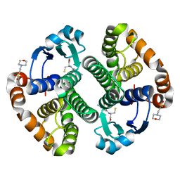 | | A folding mutant of human class pi glutathione transferase, created by mutating glycine 146 of the wild-type protein to alanine | | 分子名称: | 2-(N-MORPHOLINO)-ETHANESULFONIC ACID, GLUTATHIONE, pi glutathione transferase | | 著者 | Kong, G.K.-W, Dragani, B, Aceto, A, Cocco, R, Mannervik, B, Stenberg, G, McKinstry, W.J, Polekhina, G, Parker, M.W. | | 登録日 | 2002-08-06 | | 公開日 | 2002-08-21 | | 最終更新日 | 2023-10-25 | | 実験手法 | X-RAY DIFFRACTION (2.03 Å) | | 主引用文献 | Contribution of Glycine 146 to a Conserved Folding Module Affecting Stability and Refolding of Human Glutathione Transferase P1-1
J.Biol.Chem., 278, 2003
|
|
6NRE
 | |
1CLF
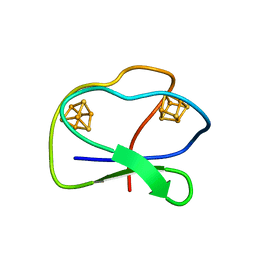 | | CLOSTRIDIUM PASTEURIANUM FERREDOXIN | | 分子名称: | FERREDOXIN, IRON/SULFUR CLUSTER | | 著者 | Bertini, I, Donaire, A, Feinberg, B.A, Luchinat, C, Piccioli, M, Yuan, H. | | 登録日 | 1995-06-21 | | 公開日 | 1996-01-29 | | 最終更新日 | 2024-05-22 | | 実験手法 | SOLUTION NMR | | 主引用文献 | Solution structure of the oxidized 2[4Fe-4S] ferredoxin from Clostridium pasteurianum.
Eur.J.Biochem., 232, 1995
|
|
2L8S
 | |


