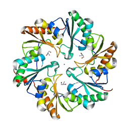
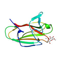
5JIM
 
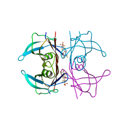 | | Crystal Structure of Human Transthyretin in Complex with Perfluoroktansulfonsyra (PFOS) | | 分子名称: | SODIUM ION, Transthyretin, heptadecafluoro-1-octanesulfonic acid | | 著者 | Begum, A, Zhang, J, Olofsson, A, Andersson, P, Sauer-Eriksson, A.E. | | 登録日 | 2016-04-22 | | 公開日 | 2016-10-05 | | 最終更新日 | 2024-01-10 | | 実験手法 | X-RAY DIFFRACTION (1.26 Å) | | 主引用文献 | Structure-Based Virtual Screening Protocol for in Silico Identification of Potential Thyroid Disrupting Chemicals Targeting Transthyretin.
Environ. Sci. Technol., 50, 2016
|
|
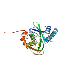
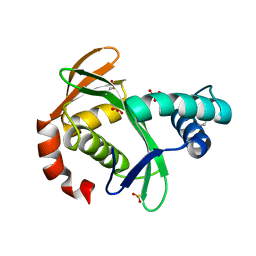
6MYD
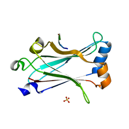 | | Structure of zebrafish TRAF6 in complex with STING CTT | | 分子名称: | STING CTT, Transmembrane protein 173, SULFATE ION, ... | | 著者 | de Oliveira Mann, C.C, Orzalli, M.H, King, D.S, Kagan, J.C, Lee, A.S.Y, Kranzusch, P.J. | | 登録日 | 2018-11-01 | | 公開日 | 2019-05-01 | | 最終更新日 | 2023-10-11 | | 実験手法 | X-RAY DIFFRACTION (1.399 Å) | | 主引用文献 | Modular Architecture of the STING C-Terminal Tail Allows Interferon and NF-kappa B Signaling Adaptation.
Cell Rep, 27, 2019
|
|
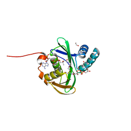
6T13
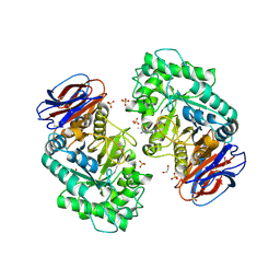 | | CRYSTAL STRUCTURE OF GLUCOCEREBROSIDASE IN COMPLEX WITH A PYRROLOPYRAZINE | | 分子名称: | 1,2-ETHANEDIOL, 1-[4-[2-(4-methoxyphenyl)-5-methyl-pyrrolo[2,3-b]pyrazin-6-yl]piperidin-1-yl]ethanone, 2-AMINO-2-HYDROXYMETHYL-PROPANE-1,3-DIOL, ... | | 著者 | Benz, J, Ehler, A, Hug, M, Huber, S, Rufer, A.C, Guba, W, Jagasia, R, Hofmann, E.C, Rodriguez Sarmiento, R.M. | | 登録日 | 2019-10-03 | | 公開日 | 2020-12-23 | | 最終更新日 | 2024-01-24 | | 実験手法 | X-RAY DIFFRACTION (1.85 Å) | | 主引用文献 | Novel beta-Glucocerebrosidase Activators That Bind to a New Pocket at a Dimer Interface and Induce Dimerization.
Angew.Chem.Int.Ed.Engl., 60, 2021
|
|
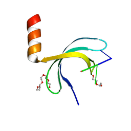
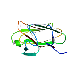
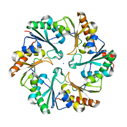
6JIQ
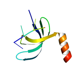 | | Crystal structure of Streptococcus pneumoniae SP_0782 (residues 7-79) in complex with single-stranded DNA dT6 | | 分子名称: | DNA (5'-D(*TP*TP*TP*TP*T)-3'), SP_0782 | | 著者 | Fang, X, Lu, G, Li, S, Zhu, J, Yang, Y, Gong, P. | | 登録日 | 2019-02-22 | | 公開日 | 2019-11-27 | | 最終更新日 | 2023-11-22 | | 実験手法 | X-RAY DIFFRACTION (1.67 Å) | | 主引用文献 | Structural insight into the length-dependent binding of ssDNA by SP_0782 from Streptococcus pneumoniae, reveals a divergence in the DNA-binding interface of PC4-like proteins.
Nucleic Acids Res., 48, 2020
|
|
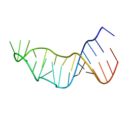
6JIP
 | | Crystal structure of Streptococcus pneumoniae SP_0782 (residues 7-79) in complex with single-stranded DNA dT6 | | 分子名称: | DNA (5'-D(*TP*TP*TP*TP*T)-3'), PENTAETHYLENE GLYCOL, SP_0782 | | 著者 | Fang, X, Lu, G, Li, S, Zhu, J, Yang, Y, Gong, P. | | 登録日 | 2019-02-22 | | 公開日 | 2019-11-27 | | 最終更新日 | 2024-03-27 | | 実験手法 | X-RAY DIFFRACTION (1.659 Å) | | 主引用文献 | Structural insight into the length-dependent binding of ssDNA by SP_0782 from Streptococcus pneumoniae, reveals a divergence in the DNA-binding interface of PC4-like proteins.
Nucleic Acids Res., 48, 2020
|
|
7ZQP
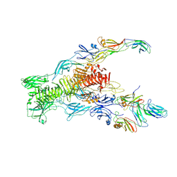
 | | Tail tip of siphophage T5 : open cone after interaction with bacterial receptor FhuA | | 分子名称: | Probable baseplate hub protein, Probable tape measure protein | | 著者 | Linares, R, Arnaud, C.A, Effantin, G, Darnault, C, Epalle, N, Boeri Erba, E, Schoehn, G, Breyton, C. | | 登録日 | 2022-05-02 | | 公開日 | 2023-02-08 | | 最終更新日 | 2023-07-05 | | 実験手法 | ELECTRON MICROSCOPY (3.6 Å) | | 主引用文献 | Structural basis of bacteriophage T5 infection trigger and E. coli cell wall perforation.
Sci Adv, 9, 2023
|
|
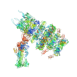
7ZHJ
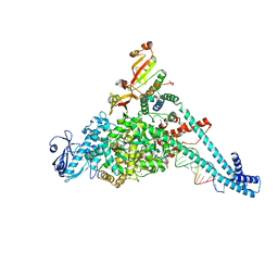
 | | Tail tip of siphophage T5 : tip proteins | | 分子名称: | Distal tail protein, L-shaped tail fiber protein p132, Minor tail protein, ... | | 著者 | Linares, R, Arnaud, C.A, Effantin, G, Darnault, C, Epalle, N, Boeri Erba, E, Schoehn, G, Breyton, C. | | 登録日 | 2022-04-06 | | 公開日 | 2023-02-08 | | 最終更新日 | 2023-07-05 | | 実験手法 | ELECTRON MICROSCOPY (3.53 Å) | | 主引用文献 | Structural basis of bacteriophage T5 infection trigger and E. coli cell wall perforation.
Sci Adv, 9, 2023
|
|
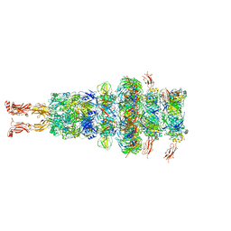
7ZN2
 | | Tail tip of siphophage T5 : full complex after interaction with its bacterial receptor FhuA | | 分子名称: | Distal tail protein, L-shaped tail fiber protein p132, Minor tail protein, ... | | 著者 | Linares, R, Arnaud, C.A, Effantin, G, Darnault, C, Epalle, N, Boeri Erba, E, Schoehn, G, Breyton, C. | | 登録日 | 2022-04-20 | | 公開日 | 2023-02-08 | | 最終更新日 | 2023-07-05 | | 実験手法 | ELECTRON MICROSCOPY (4.29 Å) | | 主引用文献 | Structural basis of bacteriophage T5 infection trigger and E. coli cell wall perforation.
Sci Adv, 9, 2023
|
|
7ZN4
 | | Tail tip of siphophage T5 : bent fibre after interaction with its bacterial receptor FhuA | | 分子名称: | Probable baseplate hub protein, Probable central straight fiber | | 著者 | Linares, R, Arnaud, C.A, Effantin, G, Darnault, C, Epalle, N, Boeri Erba, E, Schoehn, G, Breyton, C. | | 登録日 | 2022-04-20 | | 公開日 | 2023-02-08 | | 最終更新日 | 2024-07-24 | | 実験手法 | ELECTRON MICROSCOPY (4.32 Å) | | 主引用文献 | Structural basis of bacteriophage T5 infection trigger and E. coli cell wall perforation.
Sci Adv, 9, 2023
|
|
8URB
 | |
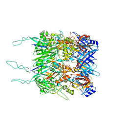
6GSD
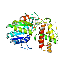 | | Plantago Major multifunctional oxidoreductase in complex with progesterone and NADP+ | | 分子名称: | NICOTINAMIDE-ADENINE-DINUCLEOTIDE, PROGESTERONE, Progesterone 5-beta-reductase | | 著者 | Fellows, R, Silva, C, Russo, C.M, Lee, S.G, Jez, J.M, Chisholm, J.D, Zubieta, C, Nanao, M. | | 登録日 | 2018-06-14 | | 公開日 | 2018-10-17 | | 最終更新日 | 2024-01-17 | | 実験手法 | X-RAY DIFFRACTION (2.7 Å) | | 主引用文献 | A multisubstrate reductase from Plantago major: structure-function in the short chain reductase superfamily.
Sci Rep, 8, 2018
|
|
7FEK
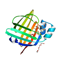 | | The 1.05 angstrom X-ray structure of the human heart fatty acid-binding protein complexed with perfluorooctanoic acid | | 分子名称: | DI(HYDROXYETHYL)ETHER, Fatty acid-binding protein, heart, ... | | 著者 | Sugiyama, S, Kakinouchi, K, Hara, T, Nakano, R, Matsuoka, S, Tsuchikawa, H, Sonoyama, M, Inoue, Y, Hayashi, F, Murata, M. | | 登録日 | 2021-07-21 | | 公開日 | 2022-07-27 | | 最終更新日 | 2023-11-29 | | 実験手法 | X-RAY DIFFRACTION (1.05 Å) | | 主引用文献 | The 1.05 angstrom X-ray structure of the human heart fatty acid-binding protein complexed with perfluorooctanoic acid
To Be Published
|
|
4TME
 | |
5KTC
 | | FdhC with bound products: Coenzyme A and 3-[(R)-3-hydroxybutanoylamino]-3,6-dideoxy-d-galactose | | 分子名称: | 1,2-ETHANEDIOL, COENZYME A, FdhC, ... | | 著者 | Salinger, A.J, Thoden, J.B, Holden, H.M. | | 登録日 | 2016-07-11 | | 公開日 | 2016-07-27 | | 最終更新日 | 2023-10-04 | | 実験手法 | X-RAY DIFFRACTION (1.8 Å) | | 主引用文献 | Structural and Functional Investigation of FdhC from Acinetobacter nosocomialis: A Sugar N-Acyltransferase Belonging to the GNAT Superfamily.
Biochemistry, 55, 2016
|
|
2VMH
 | | The structure of CBM51 from Clostridium perfringens GH95 | | 分子名称: | CALCIUM ION, FIBRONECTIN TYPE III DOMAIN PROTEIN | | 著者 | Gregg, K, Finn, R, Abbott, D.W, Boraston, A.B. | | 登録日 | 2008-01-25 | | 公開日 | 2008-02-19 | | 最終更新日 | 2024-05-08 | | 実験手法 | X-RAY DIFFRACTION (1.5 Å) | | 主引用文献 | Divergent Modes of Glycan Recognition by a New Family of Carbohydrate-Binding Modules
J.Biol.Chem., 283, 2008
|
|
6ZYB
 | |
2VMG
 | | The structure of CBM51 from Clostridium perfringens GH95 in complex with methyl-galactose | | 分子名称: | CALCIUM ION, FIBRONECTIN TYPE III DOMAIN PROTEIN, methyl beta-D-galactopyranoside | | 著者 | Gregg, K, Finn, R, Abbott, D.W, Boraston, A.B. | | 登録日 | 2008-01-25 | | 公開日 | 2008-02-19 | | 最終更新日 | 2024-05-08 | | 実験手法 | X-RAY DIFFRACTION (1.9 Å) | | 主引用文献 | Divergent Modes of Glycan Recognition by a New Family of Carbohydrate-Binding Modules
J.Biol.Chem., 283, 2008
|
|
5KTA
 | |
5KTD
 | | FdhC with bound products: Coenzyme A and dTDP-3-amino-3,6-dideoxy-d-glucose | | 分子名称: | 1,2-ETHANEDIOL, COENZYME A, FdhC, ... | | 著者 | Salinger, A.J, Thoden, J.B, Holden, H.M. | | 登録日 | 2016-07-11 | | 公開日 | 2016-07-27 | | 最終更新日 | 2023-10-04 | | 実験手法 | X-RAY DIFFRACTION (1.9 Å) | | 主引用文献 | Structural and Functional Investigation of FdhC from Acinetobacter nosocomialis: A Sugar N-Acyltransferase Belonging to the GNAT Superfamily.
Biochemistry, 55, 2016
|
|
4TM6
 | |
5E3E
 | | Crystal structure of CdiA-CT/CdiI complex from Y. kristensenii 33638 | | 分子名称: | CdiI immunity protein, Large exoprotein involved in heme utilization or adhesion, SODIUM ION | | 著者 | Michalska, K, Joachimiak, G, Jedrzejczak, R, Goulding, C.W, Joachimiak, A, Structure-Function Analysis of Polymorphic CDI Toxin-Immunity Protein Complexes (UC4CDI), Midwest Center for Structural Genomics (MCSG) | | 登録日 | 2015-10-02 | | 公開日 | 2015-11-25 | | 最終更新日 | 2019-12-25 | | 実験手法 | X-RAY DIFFRACTION (1.7 Å) | | 主引用文献 | The CDI toxin of Yersinia kristensenii is a novel bacterial member of the RNase A superfamily.
Nucleic Acids Res., 45, 2017
|
|
6ZXZ
 | |
7ZLV
 | | Tail tip of siphophage T5 : central fibre protein pb4 | | 分子名称: | Probable central straight fiber | | 著者 | Linares, R, Arnaud, C.A, Effantin, G, Epalle, N, Boeri Erba, E, Schoehn, G, Breyton, C. | | 登録日 | 2022-04-15 | | 公開日 | 2023-02-08 | | 最終更新日 | 2024-07-24 | | 実験手法 | ELECTRON MICROSCOPY (4.22 Å) | | 主引用文献 | Structural basis of bacteriophage T5 infection trigger and E. coli cell wall perforation.
Sci Adv, 9, 2023
|
|
6OID
 | | Redox Regulation of FN3K from Arabidopsis thaliana | | 分子名称: | 1,2-ETHANEDIOL, ADENOSINE-5'-DIPHOSPHATE, CHLORIDE ION, ... | | 著者 | Wood, Z.A, Kadirvelraj, R, Shrestha, S. | | 登録日 | 2019-04-09 | | 公開日 | 2020-05-20 | | 最終更新日 | 2023-10-11 | | 実験手法 | X-RAY DIFFRACTION (2.365 Å) | | 主引用文献 | A redox-active switch in fructosamine-3-kinases expands the regulatory repertoire of the protein kinase superfamily.
Sci.Signal., 13, 2020
|
|
2VMI
 | | The structure of seleno-methionine labelled CBM51 from Clostridium perfringens GH95 | | 分子名称: | CALCIUM ION, FIBRONECTIN TYPE III DOMAIN PROTEIN | | 著者 | Gregg, K, Finn, R, Abbott, D.W, Boraston, A.B. | | 登録日 | 2008-01-25 | | 公開日 | 2008-02-19 | | 最終更新日 | 2011-07-13 | | 実験手法 | X-RAY DIFFRACTION (1.7 Å) | | 主引用文献 | Divergent Modes of Glycan Recognition by a New Family of Carbohydrate-Binding Modules
J.Biol.Chem., 283, 2008
|
|


