2E92
 
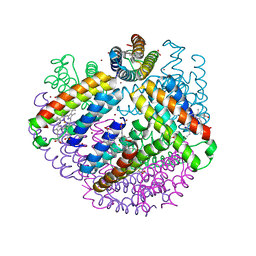
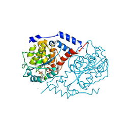
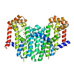 | | S. cerevisiae geranylgeranyl pyrophosphate synthase in complex with magnesium and BPH-261 | | 分子名称: | (1-HYDROXY-2-IMIDAZO[1,2-A]PYRIDIN-3-YLETHANE-1,1-DIYL)BIS(PHOSPHONIC ACID), Geranylgeranyl pyrophosphate synthetase, MAGNESIUM ION | | 著者 | Guo, R.T, Ko, T.P, Cao, R, Jeng, W.Y, Chen, C.K.-M, Chang, T.H, Liang, P.H, Oldfield, E, Wang, A.H.-J. | | 登録日 | 2007-01-24 | | 公開日 | 2007-06-12 | | 最終更新日 | 2023-10-25 | | 実験手法 | X-RAY DIFFRACTION (2.31 Å) | | 主引用文献 | Bisphosphonates target multiple sites in both cis- and trans-prenyltransferases
Proc.Natl.Acad.Sci.Usa, 104, 2007
|
|
3OS1
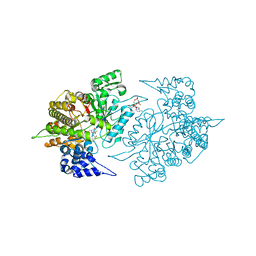
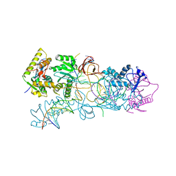 | | PFV target capture complex (TCC) at 2.97 A resolution | | 分子名称: | DNA (5'-D(*AP*TP*TP*GP*TP*CP*AP*TP*GP*GP*AP*AP*TP*TP*TP*CP*GP*CP*A)-3'), DNA (5'-D(*CP*CP*CP*GP*AP*GP*GP*CP*AP*CP*GP*TP*GP*CP*TP*AP*GP*CP*AP*CP*GP*TP*GP*CP*CP*TP*CP*GP*GP*G)-3'), DNA (5'-D(*TP*GP*CP*GP*AP*AP*AP*TP*TP*CP*CP*AP*TP*GP*AP*CP*(2DA))-3'), ... | | 著者 | Maertens, G.N, Hare, S, Cherepanov, P. | | 登録日 | 2010-09-08 | | 公開日 | 2010-11-17 | | 最終更新日 | 2023-11-01 | | 実験手法 | X-RAY DIFFRACTION (2.97 Å) | | 主引用文献 | The mechanism of retroviral integration from X-ray structures of its key intermediates
Nature, 468, 2010
|
|
2EI4
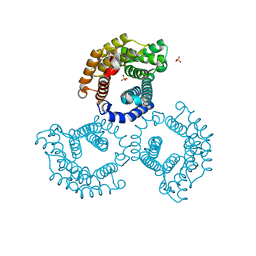
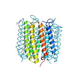 | | Trimeric complex of archaerhodopsin-2 | | 分子名称: | 2,3-DI-PHYTANYL-GLYCEROL, Archaerhodopsin-2, BACTERIORUBERIN, ... | | 著者 | Kouyama, T. | | 登録日 | 2007-03-11 | | 公開日 | 2008-01-01 | | 最終更新日 | 2023-10-25 | | 実験手法 | X-RAY DIFFRACTION (2.1 Å) | | 主引用文献 | Structural role of bacterioruberin in the trimeric structure of archaerhodopsin-2
J.Mol.Biol., 375, 2008
|
|
3BVN
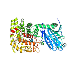
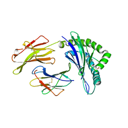 | | High resolution crystal structure of HLA-B*1402 in complex with the latent membrane protein 2 peptide (LMP2) of Epstein-Barr virus | | 分子名称: | Beta-2-microglobulin, HLA class I histocompatibility antigen, B*1402 alpha chain, ... | | 著者 | Kumar, P, Vahedi-Faridi, A, Saenger, W, Uchanska-Ziegler, B, Ziegler, A. | | 登録日 | 2008-01-07 | | 公開日 | 2009-02-03 | | 最終更新日 | 2023-08-30 | | 実験手法 | X-RAY DIFFRACTION (2.55 Å) | | 主引用文献 | Structural basis for T cell alloreactivity among three HLA-B14 and HLA-B27 antigens
J.Biol.Chem., 284, 2009
|
|
3M4B
 | | A Zn-mediated tetrahedral protein lattice cage | | 分子名称: | ACETATE ION, PROTOPORPHYRIN IX CONTAINING FE, Soluble cytochrome b562, ... | | 著者 | Tezcan, F.A, Ni, T.W. | | 登録日 | 2010-03-10 | | 公開日 | 2011-01-12 | | 最終更新日 | 2021-10-06 | | 実験手法 | X-RAY DIFFRACTION (2.5 Å) | | 主引用文献 | Structural characterization of a microperoxidase inside a metal-directed protein cage.
Angew.Chem.Int.Ed.Engl., 49, 2010
|
|
3ABE
 | | Structure of human REV7 in complex with a human REV3 fragment in a tetragonal crystal | | 分子名称: | DNA polymerase zeta catalytic subunit, Mitotic spindle assembly checkpoint protein MAD2B | | 著者 | Hara, K, Hashimoto, H, Murakumo, Y, Kobayashi, S, Kogame, T, Unzai, S, Akashi, S, Takeda, S, Shimizu, T, Sato, M. | | 登録日 | 2009-12-07 | | 公開日 | 2010-02-16 | | 最終更新日 | 2023-11-01 | | 実験手法 | X-RAY DIFFRACTION (2.6 Å) | | 主引用文献 | Crystal structure of human REV7 in complex with a human REV3 fragment and structural implication of the interaction between DNA polymerase {zeta} and REV1
J.Biol.Chem., 285, 2010
|
|
3BQT
 | |
1Y6I
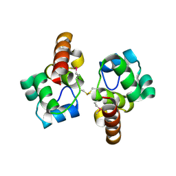
 | | Synechocystis GUN4 | | 分子名称: | Mg-chelatase cofactor GUN4 | | 著者 | Verdecia, M.A, Larkin, R.M, Ferrer, J.L, Riek, R, Chory, J, Noel, J.P. | | 登録日 | 2004-12-06 | | 公開日 | 2005-05-31 | | 最終更新日 | 2024-02-14 | | 実験手法 | X-RAY DIFFRACTION (1.78 Å) | | 主引用文献 | Structure of the Mg-chelatase cofactor GUN4 reveals a novel hand-shaped fold for porphyrin binding
Plos Biol., 3, 2005
|
|
3BXN
 | | The high resolution crystal structure of HLA-B*1402 complexed with a Cathepsin A signal sequence peptide, pCatA | | 分子名称: | Cathepsin A signal sequence octapeptide, GLYCEROL, HLA-B*1402 extracellular domain, ... | | 著者 | Kumar, P, Vahedi-Faridi, A, Saenger, W, Uchanska-Ziegler, B, Ziegler, A. | | 登録日 | 2008-01-14 | | 公開日 | 2009-02-03 | | 最終更新日 | 2023-08-30 | | 実験手法 | X-RAY DIFFRACTION (1.864 Å) | | 主引用文献 | Structural basis for T cell alloreactivity among three HLA-B14 and HLA-B27 antigens
J.Biol.Chem., 284, 2009
|
|
1JHR
 | | Three-dimensional Structure of CobT in Complex with Reaction Products of 2-hydroxypurine and NaMN | | 分子名称: | N7-(5'-PHOSPHO-ALPHA-RIBOSYL)-2-HYDROXYPURINE, NICOTINIC ACID, Nicotinate Mononucleotide:5,6-Dimethylbenzimidazole Phosphoribosyltransferase | | 著者 | Cheong, C.G, Escalante-Semerena, J, Rayment, I. | | 登録日 | 2001-06-28 | | 公開日 | 2001-10-18 | | 最終更新日 | 2023-08-16 | | 実験手法 | X-RAY DIFFRACTION (2 Å) | | 主引用文献 | Structural investigation of the biosynthesis of alternative lower ligands for cobamides by nicotinate mononucleotide: 5,6-dimethylbenzimidazole phosphoribosyltransferase from Salmonella enterica.
J.Biol.Chem., 276, 2001
|
|
2EUD
 | | Structures of Yeast Ribonucleotide Reductase I complexed with Ligands and Subunit Peptides | | 分子名称: | GEMCITABINE DIPHOSPHATE, MAGNESIUM ION, PHOSPHOAMINOPHOSPHONIC ACID-ADENYLATE ESTER, ... | | 著者 | Dealwis, C, Xu, H, Faber, C, Uchiki, T, Fairman, J.W, Racca, J. | | 登録日 | 2005-10-28 | | 公開日 | 2006-03-07 | | 最終更新日 | 2024-02-14 | | 実験手法 | X-RAY DIFFRACTION (2.3 Å) | | 主引用文献 | Structures of eukaryotic ribonucleotide reductase I define gemcitabine diphosphate binding and subunit assembly.
Proc.Natl.Acad.Sci.Usa, 103, 2006
|
|
3U64
 | | The Crystal Structure of Tat-T (Tp0956) | | 分子名称: | Protein TP_0956, SULFATE ION | | 著者 | Tomchick, D.R, Brautigam, C.A, Deka, R.K, Norgard, M.V. | | 登録日 | 2011-10-12 | | 公開日 | 2012-02-22 | | 最終更新日 | 2024-02-28 | | 実験手法 | X-RAY DIFFRACTION (2.3 Å) | | 主引用文献 | Structural, Bioinformatic, and In Vivo Analyses of Two Treponema pallidum Lipoproteins Reveal a Unique TRAP Transporter.
J.Mol.Biol., 416, 2012
|
|
1LF9
 | | CRYSTAL STRUCTURE OF BACTERIAL GLUCOAMYLASE COMPLEXED WITH ACARBOSE | | 分子名称: | 4,6-dideoxy-4-{[(1S,4R,5S,6S)-4,5,6-trihydroxy-3-(hydroxymethyl)cyclohex-2-en-1-yl]amino}-alpha-D-glucopyranose-(1-4)-alpha-D-glucopyranose-(1-4)-alpha-D-glucopyranose, GLUCOAMYLASE, SULFATE ION | | 著者 | Aleshin, A.E, Feng, P.-H, Honzatko, R.B, Reilly, P.J. | | 登録日 | 2002-04-10 | | 公開日 | 2003-02-25 | | 最終更新日 | 2023-08-16 | | 実験手法 | X-RAY DIFFRACTION (2.2 Å) | | 主引用文献 | Crystal structure and evolution of prokaryotic glucoamylase
J.Mol.Biol., 327, 2003
|
|
3Q36
 | |
2ET2
 | | Crystal structure of an Asn to Ala mutant of Winged Bean Chymotrypsin Inhibitor protein | | 分子名称: | Chymotrypsin inhibitor 3, NICKEL (II) ION, SULFATE ION | | 著者 | Dattagupta, J.K, Sen, U, Dasgupta, J, Khamrui, S. | | 登録日 | 2005-10-27 | | 公開日 | 2006-06-13 | | 最終更新日 | 2021-10-20 | | 実験手法 | X-RAY DIFFRACTION (2.1 Å) | | 主引用文献 | Spacer Asn Determines the Fate of Kunitz (STI) Inhibitors, as Revealed by Structural and Biochemical Studies on WCI Mutants.
Biochemistry, 45, 2006
|
|
3BP4
 | | The high resolution crystal structure of HLA-B*2705 in complex with a Cathepsin A signal sequence peptide pCatA | | 分子名称: | Beta-2-microglobulin, GLYCEROL, HLA class I histocompatibility antigen, ... | | 著者 | Kumar, P, Vahedi-Faridi, A, Saenger, W, Uchanska-Ziegler, B, Ziegler, A. | | 登録日 | 2007-12-18 | | 公開日 | 2008-12-23 | | 最終更新日 | 2023-11-01 | | 実験手法 | X-RAY DIFFRACTION (1.85 Å) | | 主引用文献 | Structural basis for T cell alloreactivity among three HLA-B14 and HLA-B27 antigens
J.Biol.Chem., 284, 2009
|
|
2Z9B
 | |
1KLL
 | | Molecular basis of mitomycin C resictance in streptomyces: Crystal structures of the MRD protein with and without a drug derivative | | 分子名称: | 1,2-CIS-1-HYDROXY-2,7-DIAMINO-MITOSENE, mitomycin-binding protein | | 著者 | Martin, T.W, Dauter, Z, Devedjiev, Y, Sheffield, P, Jelen, F, He, M, Sherman, D, Otlewski, J, Derewenda, Z.S, Derewenda, U. | | 登録日 | 2001-12-12 | | 公開日 | 2002-07-19 | | 最終更新日 | 2021-10-27 | | 実験手法 | X-RAY DIFFRACTION (1.5 Å) | | 主引用文献 | Molecular basis of mitomycin C resistance in streptomyces: structure and function of the MRD protein.
Structure, 10, 2002
|
|
3RE1
 | |
3PMB
 | |
2ZL3
 | | Crystal structure of H.pylori ClpP S99A | | 分子名称: | ATP-dependent Clp protease proteolytic subunit | | 著者 | Kim, D.Y, Kim, K.K. | | 登録日 | 2008-04-02 | | 公開日 | 2008-04-22 | | 最終更新日 | 2023-11-01 | | 実験手法 | X-RAY DIFFRACTION (2.81 Å) | | 主引用文献 | The structural basis for the activation and peptide recognition of bacterial ClpP
J.Mol.Biol., 379, 2008
|
|
2CIM
 | | Crystal structure of Methanosarcina barkeri seryl-tRNA synthetase | | 分子名称: | CHLORIDE ION, PLATINUM (II) ION, SERYL-TRNA SYNTHETASE, ... | | 著者 | Bilokapic, S, Maier, T, Ahel, D, Gruic-Sovulj, I, Soll, D, Weygand-Durasevic, I, Ban, N. | | 登録日 | 2006-03-24 | | 公開日 | 2006-06-26 | | 最終更新日 | 2024-05-08 | | 実験手法 | X-RAY DIFFRACTION (2.51 Å) | | 主引用文献 | Structure of the Unusual Seryl-tRNA Synthetase Reveals a Distinct Zinc-Dependent Mode of Substrate Recognition
Embo J., 25, 2006
|
|
3NFD
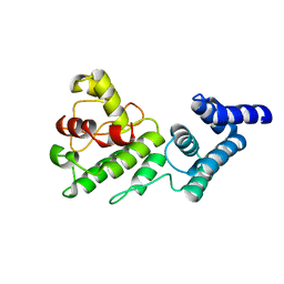
 | | Chronobacterium ammoniagenes ACPS-CoA complex | | 分子名称: | COENZYME A, Phosphopantetheine protein transferase, Ppt1p | | 著者 | Gokulan, K. | | 登録日 | 2010-06-10 | | 公開日 | 2011-01-05 | | 最終更新日 | 2019-07-17 | | 実験手法 | X-RAY DIFFRACTION (1.89 Å) | | 主引用文献 | Mycobacterium tuberculosis acyl carrier protein synthase adopts two different pH-dependent structural conformations.
Acta Crystallogr.,Sect.D, 67, 2011
|
|
1ULV
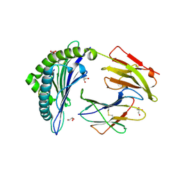
 | | Crystal Structure of Glucodextranase Complexed with Acarbose | | 分子名称: | 4,6-dideoxy-4-{[(1S,4R,5S,6S)-4,5,6-trihydroxy-3-(hydroxymethyl)cyclohex-2-en-1-yl]amino}-alpha-D-glucopyranose-(1-4)-alpha-D-glucopyranose, CALCIUM ION, glucodextranase | | 著者 | Mizuno, M, Tonozuka, T, Suzuki, S, Uotsu-Tomita, R, Kamitori, S, Nishikawa, A, Sakano, Y. | | 登録日 | 2003-09-16 | | 公開日 | 2003-12-09 | | 最終更新日 | 2023-12-27 | | 実験手法 | X-RAY DIFFRACTION (2.42 Å) | | 主引用文献 | Structural insights into substrate specificity and function of glucodextranase
J.Biol.Chem., 279, 2004
|
|
1JRN
 | |


