1CSK
 
 | |
1M3A
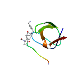
 | | Solution structure of a circular form of the truncated N-terminal SH3 domain from oncogene protein c-Crk. | | 分子名称: | Proto-oncogene C-crk | | 著者 | Schumann, F.H, Varadan, R, Tayakuniyil, P.P, Hall, J.B, Camarero, J.A, Fushman, D. | | 登録日 | 2002-06-27 | | 公開日 | 2003-08-05 | | 最終更新日 | 2021-10-27 | | 実験手法 | SOLUTION NMR | | 主引用文献 | Changing protein backbone topology: Structural and dynamic consequences of the backbone cyclization in SH3 domain
To be Published
|
|
1M8M
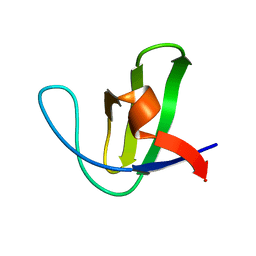
 | | SOLID-STATE MAS NMR STRUCTURE OF THE A-SPECTRIN SH3 DOMAIN | | 分子名称: | SPECTRIN ALPHA CHAIN, BRAIN | | 著者 | Castellani, F, Van Rossum, B, Diehl, A, Schubert, M, Rehbein, K, Oschkinat, H. | | 登録日 | 2002-07-25 | | 公開日 | 2002-11-20 | | 最終更新日 | 2024-05-22 | | 実験手法 | SOLID-STATE NMR | | 主引用文献 | Structure of a protein determined by solid-state magic-angle-spinning NMR spectroscopy
Nature, 420, 2002
|
|
1NEG
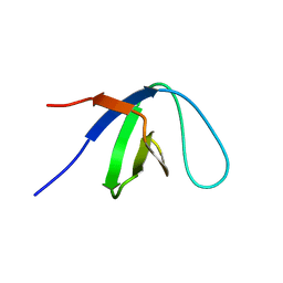
 | | Crystal Structure Analysis of N-and C-terminal labeled SH3-domain of alpha-Chicken Spectrin | | 分子名称: | AZIDE ION, Spectrin alpha chain, brain | | 著者 | Mueller, U, Buessow, K, Diehl, A, Niesen, F.H, Nyarsik, L, Heinemann, U. | | 登録日 | 2002-12-11 | | 公開日 | 2003-01-14 | | 最終更新日 | 2023-09-20 | | 実験手法 | X-RAY DIFFRACTION (2.3 Å) | | 主引用文献 | Rapid purification and crystal structure analysis of a small protein carrying two terminal affinity tags
J.STRUCT.FUNCT.GENOM., 4, 2003
|
|
1KIK
 | |
1M30
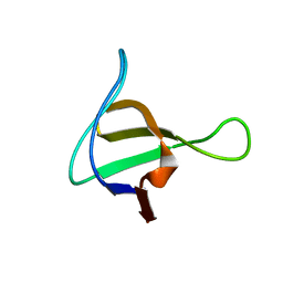
 | | Solution structure of N-terminal SH3 domain from oncogene protein c-Crk | | 分子名称: | Proto-oncogene C-crk | | 著者 | Schumann, F.H, Varadan, R, Tayakuniyil, P.P, Hall, J.B, Camarero, J.A, Fushman, D. | | 登録日 | 2002-06-26 | | 公開日 | 2003-08-05 | | 最終更新日 | 2024-05-22 | | 実験手法 | SOLUTION NMR | | 主引用文献 | Changing protein backbone topology: Structural and dynamic consequences of the backbone cyclization in SH3 domain
To be Published
|
|
1M3C
 | | Solution structure of a circular form of the N-terminal SH3 domain (E132C, E133G, R191G mutant) from oncogene protein c-Crk | | 分子名称: | Proto-oncogene C-crk | | 著者 | Schumann, F.H, Varadan, R, Tayakuniyil, P.P, Hall, J.B, Camarero, J.A, Fushman, D. | | 登録日 | 2002-06-27 | | 公開日 | 2003-08-05 | | 最終更新日 | 2021-10-27 | | 実験手法 | SOLUTION NMR | | 主引用文献 | Changing protein backbone topology: Structural and dynamic consequences of the backbone cyclization in SH3 domain
To be Published
|
|
1NYF
 | |
1M3B
 | | Solution structure of a circular form of the N-terminal SH3 domain (A134C, E135G, R191G mutant) from oncogene protein c-Crk. | | 分子名称: | Proto-oncogene C-crk | | 著者 | Schumann, F.H, Varadan, R, Tayakuniyil, P.P, Hall, J.B, Camarero, J.A, Fushman, D. | | 登録日 | 2002-06-27 | | 公開日 | 2003-08-05 | | 最終更新日 | 2021-10-27 | | 実験手法 | SOLUTION NMR | | 主引用文献 | Changing protein backbone topology: Structural and dynamic consequences of the backbone cyclization in SH3 domain
To be Published
|
|
1OEB
 | | Mona/Gads SH3C domain | | 分子名称: | CADMIUM ION, GRB2-RELATED ADAPTOR PROTEIN 2, LYMPHOCYTE CYTOSOLIC PROTEIN 2 | | 著者 | Harkiolaki, M, Lewitzky, M, Gilbert, R.J.C, Jones, E.Y, Bourette, R.P, Mouchiroud, G, Sondermann, H, Moarefi, I, Feller, S.M. | | 登録日 | 2003-03-24 | | 公開日 | 2003-04-02 | | 最終更新日 | 2024-05-08 | | 実験手法 | X-RAY DIFFRACTION (1.76 Å) | | 主引用文献 | Structural Basis for SH3 Domain-Mediated High-Affinity Binding between Mona/Gads and Slp-76
Embo J., 22, 2003
|
|
1N5Z
 | | Complex structure of Pex13p SH3 domain with a peptide of Pex14p | | 分子名称: | 14-mer peptide from Peroxisomal membrane protein PEX14, Peroxisomal membrane protein PAS20 | | 著者 | Douangamath, A, Filipp, F.V, Klein, A.T.J, Barnett, P, Zou, P, Voorn-Brouwer, T, Vega, M.C, Mayans, O.M, Sattler, M, Distel, B, Wilmanns, M. | | 登録日 | 2002-11-08 | | 公開日 | 2002-12-11 | | 最終更新日 | 2024-03-13 | | 実験手法 | X-RAY DIFFRACTION (2.7 Å) | | 主引用文献 | Topography for Independent Binding of alpha-Helical and PPII-Helical Ligands to a Peroxisomal SH3 Domain
MOL.CELL, 10, 2002
|
|
1NLP
 | | STRUCTURE OF SIGNAL TRANSDUCTION PROTEIN, NMR, MINIMIZED AVERAGE STRUCTURE | | 分子名称: | C-SRC, NL2 (MN8-MN1-PLPPLP) | | 著者 | Feng, S, Kapoor, T.M, Shirai, F, Combs, A.P, Schreiber, S.L. | | 登録日 | 1996-08-04 | | 公開日 | 1997-01-27 | | 最終更新日 | 2023-11-15 | | 実験手法 | SOLUTION NMR | | 主引用文献 | Molecular basis for the binding of SH3 ligands with non-peptide elements identified by combinatorial synthesis.
Chem.Biol., 3, 1996
|
|
1NLO
 | | STRUCTURE OF SIGNAL TRANSDUCTION PROTEIN, NMR, MINIMIZED AVERAGE STRUCTURE | | 分子名称: | C-SRC, NL1 (MN7-MN2-MN1-PLPPLP) | | 著者 | Feng, S, Kapoor, T.M, Shirai, F, Combs, A.P, Schreiber, S.L. | | 登録日 | 1996-08-04 | | 公開日 | 1997-01-27 | | 最終更新日 | 2023-11-15 | | 実験手法 | SOLUTION NMR | | 主引用文献 | Molecular basis for the binding of SH3 ligands with non-peptide elements identified by combinatorial synthesis.
Chem.Biol., 3, 1996
|
|
1NYG
 | |
1KFZ
 | | Solution Structure of C-terminal Sem-5 SH3 Domain (Ensemble of 16 Structures) | | 分子名称: | SEX MUSCLE ABNORMAL PROTEIN 5 | | 著者 | Ferreon, J.C, Volk, D.E, Luxon, B.A, Gorenstein, D, Hilser, V.J. | | 登録日 | 2001-11-24 | | 公開日 | 2003-05-20 | | 最終更新日 | 2024-05-22 | | 実験手法 | SOLUTION NMR | | 主引用文献 | Solution Structure, Dynamics and Thermodynamics of the Native State Ensemble
of Sem-5 C-Terminal SH3 Domain
Biochemistry, 42, 2003
|
|
1NM7
 | | Solution structure of the ScPex13p SH3 domain | | 分子名称: | Peroxisomal Membrane Protein PAS20 | | 著者 | Pires, J.R, Hong, X, Brockmann, C, Volkmer-Engert, R, Schneider-Mergener, J, Oschkinat, H, Erdmann, R. | | 登録日 | 2003-01-09 | | 公開日 | 2003-03-04 | | 最終更新日 | 2024-05-29 | | 実験手法 | SOLUTION NMR | | 主引用文献 | The ScPex13p SH3 Domain Exposes Two Distinct Binding Sites for Pex5p and Pex14p
J.Mol.Biol., 326, 2003
|
|
1OOT
 | |
4HCK
 | | HUMAN HCK SH3 DOMAIN, NMR, 25 STRUCTURES | | 分子名称: | HEMATOPOIETIC CELL KINASE | | 著者 | Horita, D.A, Baldisseri, D.M, Zhang, W, Altieri, A.S, Smithgall, T.E, Gmeiner, W.H, Byrd, R.A. | | 登録日 | 1998-03-09 | | 公開日 | 1998-06-17 | | 最終更新日 | 2024-05-01 | | 実験手法 | SOLUTION NMR | | 主引用文献 | Solution structure of the human Hck SH3 domain and identification of its ligand binding site.
J.Mol.Biol., 278, 1998
|
|
1QKX
 | |
4HXJ
 | | Crystal structure of SH3:RGT complex | | 分子名称: | C-terminal 3-mer peptide from Integrin beta-3, Proto-oncogene tyrosine-protein kinase Src | | 著者 | Xiao, R, Meng, G. | | 登録日 | 2012-11-11 | | 公開日 | 2012-11-28 | | 最終更新日 | 2023-11-08 | | 実験手法 | X-RAY DIFFRACTION (2 Å) | | 主引用文献 | Structural framework of c-Src activation by integrin beta 3
Blood, 121, 2013
|
|
4JJC
 | |
1QLY
 | | NMR Study of the SH3 Domain From Bruton's Tyrosine Kinase, 20 Structures | | 分子名称: | TYROSINE-PROTEIN KINASE BTK | | 著者 | Tzeng, S.R, Lou, Y.C, Pai, M.T, Chen, C, Chen, S.H, Cheng, J.Y. | | 登録日 | 1999-09-20 | | 公開日 | 1999-12-14 | | 最終更新日 | 2024-05-15 | | 実験手法 | SOLUTION NMR | | 主引用文献 | Solution Structure of the Human Btk SH3 Domain Complexed with a Proline-Rich Peptide from P120Cbl
J.Biomol.NMR, 16, 2000
|
|
1QKW
 | | Alpha-spectrin Src Homology 3 domain, N47G mutant in the distal loop. | | 分子名称: | ALPHA II SPECTRIN, GLYCEROL, SULFATE ION | | 著者 | Vega, M.C, Martinez, J, Serrano, L. | | 登録日 | 1999-08-16 | | 公開日 | 2000-08-18 | | 最終更新日 | 2023-12-13 | | 実験手法 | X-RAY DIFFRACTION (2 Å) | | 主引用文献 | Thermodynamic and structural characterization of Asn and Ala residues in the disallowed II' region of the Ramachandran plot.
Protein Sci., 9, 2000
|
|
4J9I
 | |
1QWF
 | | C-SRC SH3 DOMAIN COMPLEXED WITH LIGAND VSL12 | | 分子名称: | TYROSINE-PROTEIN KINASE TRANSFORMING PROTEIN SRC, VAL-SER-LEU-ALA-ARG-ARG-PRO-LEU-PRO-PRO-LEU-PRO | | 著者 | Feng, S, Chiyoshi, K, Rickles, R.J, Schreiber, S.L. | | 登録日 | 1995-11-09 | | 公開日 | 1996-03-08 | | 最終更新日 | 2024-05-22 | | 実験手法 | SOLUTION NMR | | 主引用文献 | Specific interactions outside the proline-rich core of two classes of Src homology 3 ligands.
Proc.Natl.Acad.Sci.USA, 92, 1995
|
|


