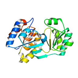
1YNM
 
 | | Crystal structure of restriction endonuclease HinP1I | | 分子名称: | R.HinP1I restriction endonuclease | | 著者 | Yang, Z, Horton, J.R, Maunus, R, Wilson, G.G, Roberts, R.J, Cheng, X. | | 登録日 | 2005-01-24 | | 公開日 | 2005-05-03 | | 最終更新日 | 2024-02-14 | | 実験手法 | X-RAY DIFFRACTION (2.65 Å) | | 主引用文献 | Structure of HinP1I endonuclease reveals a striking similarity to the monomeric restriction enzyme MspI
Nucleic Acids Res., 33, 2005
|
|
1YMK
 | | Crystal Structure of the CDC25B phosphatase catalytic domain in the apo form | | 分子名称: | CHLORIDE ION, M-phase inducer phosphatase 2 | | 著者 | Buhrman, G.K, Parker, B, Sohn, J, Rudolph, J, Mattos, C. | | 登録日 | 2005-01-21 | | 公開日 | 2005-04-12 | | 最終更新日 | 2023-08-23 | | 実験手法 | X-RAY DIFFRACTION (1.7 Å) | | 主引用文献 | Structural Mechanism of Oxidative Regulation of the Phosphatase Cdc25B via an Intramolecular Disulfide Bond
Biochemistry, 44, 2005
|
|
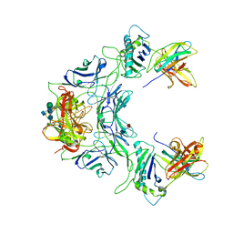
1YN5
 | | Crystal Structures of EAP Domains from Staphylococcus aureus Reveal an Unexpected Homology to Bacterial Superantigens | | 分子名称: | EapH2 | | 著者 | Geisbrecht, B.V, Hamaoka, B.Y, Perman, B, Zemla, A, Leahy, D.J. | | 登録日 | 2005-01-23 | | 公開日 | 2005-03-01 | | 最終更新日 | 2023-08-23 | | 実験手法 | X-RAY DIFFRACTION (2.2 Å) | | 主引用文献 | The Crystal Structures of EAP Domains from Staphylococcus aureus Reveal an Unexpected Homology to Bacterial Superantigens.
J.Biol.Chem., 280, 2005
|
|
1YS0
 | | Crystal Structure of the CDC25B phosphatase catalytic domain with the active site cysteine in the disulfide form | | 分子名称: | CHLORIDE ION, M-phase inducer phosphatase 2 | | 著者 | Buhrman, G.K, Parker, B, Sohn, J, Rudolph, J, Mattos, C. | | 登録日 | 2005-02-05 | | 公開日 | 2005-04-12 | | 最終更新日 | 2023-08-23 | | 実験手法 | X-RAY DIFFRACTION (2 Å) | | 主引用文献 | Structural Mechanism of Oxidative Regulation of the Phosphatase Cdc25B via an Intramolecular Disulfide Bond
Biochemistry, 44, 2005
|
|
1YTB
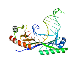 | | CRYSTAL STRUCTURE OF A YEAST TBP/TATA-BOX COMPLEX | | 分子名称: | DNA (29MER), PROTEIN (TATA BINDING PROTEIN (TBP)) | | 著者 | Kim, Y, Geiger, J.H, Hahn, S, Sigler, P.B. | | 登録日 | 1994-09-28 | | 公開日 | 1995-01-26 | | 最終更新日 | 2024-02-14 | | 実験手法 | X-RAY DIFFRACTION (1.8 Å) | | 主引用文献 | Crystal structure of a yeast TBP/TATA-box complex.
Nature, 365, 1993
|
|
1YLX
 | |
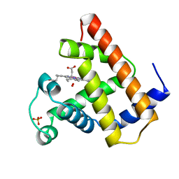
1YOH
 | | COBALT MYOGLOBIN (MET) | | 分子名称: | MYOGLOBIN, PROTOPORPHYRIN IX CONTAINING CO, SULFATE ION | | 著者 | Brucker, E.A, Phillips Jr, G.N. | | 登録日 | 1996-06-14 | | 公開日 | 1996-12-07 | | 最終更新日 | 2024-02-14 | | 実験手法 | X-RAY DIFFRACTION (1.65 Å) | | 主引用文献 | High resolution crystal structures of the deoxy, oxy, and aquomet forms of cobalt myoglobin.
J.Biol.Chem., 271, 1996
|
|
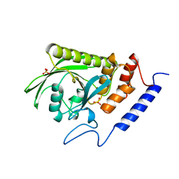
1YP2
 | | Crystal structure of potato tuber ADP-glucose pyrophosphorylase | | 分子名称: | Glucose-1-phosphate adenylyltransferase small subunit, PARA-MERCURY-BENZENESULFONIC ACID, SULFATE ION | | 著者 | Jin, X, Ballicora, M.A, Preiss, J, Geiger, J.H. | | 登録日 | 2005-01-29 | | 公開日 | 2005-03-15 | | 最終更新日 | 2011-07-13 | | 実験手法 | X-RAY DIFFRACTION (2.11 Å) | | 主引用文献 | Crystal structure of potato tuber ADP-glucose pyrophosphorylase.
Embo J., 24, 2005
|
|
1YP3
 | | Crystal structure of potato tuber ADP-glucose pyrophosphorylase in complex with ATP | | 分子名称: | ADENOSINE-5'-TRIPHOSPHATE, Glucose-1-phosphate adenylyltransferase small subunit, SULFATE ION | | 著者 | Jin, X, Ballicora, M.A, Preiss, J, Geiger, J.H. | | 登録日 | 2005-01-29 | | 公開日 | 2005-03-15 | | 最終更新日 | 2011-07-13 | | 実験手法 | X-RAY DIFFRACTION (2.6 Å) | | 主引用文献 | Crystal structure of potato tuber ADP-glucose pyrophosphorylase.
Embo J., 24, 2005
|
|
1YRV
 | | Novel Ubiquitin-Conjugating Enzyme | | 分子名称: | ubiquitin-conjugating ligase MGC351130 | | 著者 | Walker, J.R, Choe, J, Avvakumov, G.V, Newman, E.M, MacKenzie, F, Sundstrom, M, Arrowsmith, C, Edwards, A, Bochkarev, A, Dhe-Paganon, S, Structural Genomics Consortium (SGC) | | 登録日 | 2005-02-04 | | 公開日 | 2005-03-22 | | 最終更新日 | 2023-08-23 | | 実験手法 | X-RAY DIFFRACTION (2.18 Å) | | 主引用文献 | A human ubiquitin conjugating enzyme (E2)-HECT E3 ligase structure-function screen.
Mol Cell Proteomics, 11, 2012
|
|
1YT9
 | | HIV Protease with oximinoarylsulfonamide bound | | 分子名称: | (S)-N-((2S,3R)-3-HYDROXY-4-(4-((E)-(HYDROXYIMINO)METHYL)-N-ISOBUTYLPHENYLSULFONAMIDO)-1-PHENYLBUTAN-2-YL)-3-METHYL-2-(3 -((2-METHYLTHIAZOL-4-YL)METHYL)-2-OXOIMIDAZOLIDIN-1-YL)BUTANAMIDE, Pol polyprotein | | 著者 | Yeung, C.M, Klein, L.L, Flentge, C.A, Randolph, J.T, Zhao, C, Sun, M, Dekhtyar, T, Stoll, V.S, Kempf, D.J. | | 登録日 | 2005-02-10 | | 公開日 | 2005-04-12 | | 最終更新日 | 2024-02-14 | | 実験手法 | X-RAY DIFFRACTION (3 Å) | | 主引用文献 | Oximinoarylsulfonamides as potent HIV protease inhibitors.
Bioorg.Med.Chem.Lett., 15, 2005
|
|
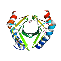
1YUH
 | | FAB FRAGMENT | | 分子名称: | 4-HYDROXY-3-NITROPHENYLACETYL-EPSILON-AMINOCAPROIC ACID, 88C6/12 FAB (HEAVY CHAIN), 88C6/12 FAB (LIGHT CHAIN) | | 著者 | Yuhasz, S.C, Amzel, L.M, Parry, C, Strand, M. | | 登録日 | 1996-01-30 | | 公開日 | 1996-07-11 | | 最終更新日 | 2018-03-21 | | 実験手法 | X-RAY DIFFRACTION (3 Å) | | 主引用文献 | Structural analysis of affinity maturation: the three-dimensional structures of complexes of an anti-nitrophenol antibody.
Mol.Immunol., 32, 1995
|
|
1YTI
 | | SIV PROTEASE CRYSTALLIZED WITH PEPTIDE PRODUCT | | 分子名称: | PEPTIDE PRODUCT, SIV PROTEASE | | 著者 | Rose, R.B, Craik, C.S, Douglas, N.L, Stroud, R.M. | | 登録日 | 1996-08-01 | | 公開日 | 1997-03-12 | | 最終更新日 | 2024-02-14 | | 実験手法 | X-RAY DIFFRACTION (2.2 Å) | | 主引用文献 | Three-dimensional structures of HIV-1 and SIV protease product complexes.
Biochemistry, 35, 1996
|
|
1YLW
 | | X-ray structure of CTX-M-16 beta-lactamase | | 分子名称: | CTX-M-16 beta-lactamase, SULFATE ION | | 著者 | Chen, Y, Delmas, J, Sirot, J, Shoichet, B, Bonnet, R. | | 登録日 | 2005-01-19 | | 公開日 | 2005-04-19 | | 最終更新日 | 2023-08-23 | | 実験手法 | X-RAY DIFFRACTION (1.74 Å) | | 主引用文献 | Atomic Resolution Structures of CTX-M beta-Lactamases: Extended Spectrum Activities from Increased Mobility and Decreased Stability.
J.Mol.Biol., 348, 2005
|
|
1YMQ
 | |
1YPZ
 | | Immune receptor | | 分子名称: | 2-acetamido-2-deoxy-beta-D-glucopyranose-(1-4)-2-acetamido-2-deoxy-beta-D-glucopyranose, 2-acetamido-2-deoxy-beta-D-glucopyranose-(1-4)-[alpha-L-fucopyranose-(1-6)]2-acetamido-2-deoxy-beta-D-glucopyranose, Beta-2-microglobulin, ... | | 著者 | Adams, E.J, Garcia, K.C. | | 登録日 | 2005-01-31 | | 公開日 | 2005-04-12 | | 最終更新日 | 2020-07-29 | | 実験手法 | X-RAY DIFFRACTION (3.4 Å) | | 主引用文献 | Structure of a gammadelta T cell receptor in complex with the nonclassical MHC T22.
Science, 308, 2005
|
|
1YOI
 | | COBALT MYOGLOBIN (OXY) | | 分子名称: | MYOGLOBIN, OXYGEN MOLECULE, PROTOPORPHYRIN IX CONTAINING CO, ... | | 著者 | Brucker, E.A, Phillips Jr, G.N. | | 登録日 | 1996-06-14 | | 公開日 | 1996-12-07 | | 最終更新日 | 2024-02-14 | | 実験手法 | X-RAY DIFFRACTION (1.65 Å) | | 主引用文献 | High resolution crystal structures of the deoxy, oxy, and aquomet forms of cobalt myoglobin.
J.Biol.Chem., 271, 1996
|
|
1YTW
 | |
2A2K
 | | Crystal Structure of an active site mutant, C473S, of Cdc25B Phosphatase Catalytic Domain | | 分子名称: | CHLORIDE ION, M-phase inducer phosphatase 2, SULFATE ION | | 著者 | Sohn, J, Parks, J, Buhrman, G, Brown, P, Kristjansdottir, K, Safi, A, Yang, W, Edelsbrunner, H, Rudolph, J. | | 登録日 | 2005-06-22 | | 公開日 | 2006-01-03 | | 最終更新日 | 2023-08-23 | | 実験手法 | X-RAY DIFFRACTION (1.52 Å) | | 主引用文献 | Experimental Validation of the Docking Orientation of Cdc25 with Its Cdk2-CycA Protein Substrate.
Biochemistry, 44, 2005
|
|
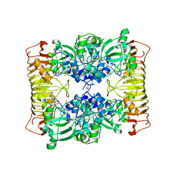
2BAT
 | | THE STRUCTURE OF THE COMPLEX BETWEEN INFLUENZA VIRUS NEURAMINIDASE AND SIALIC ACID, THE VIRAL RECEPTOR | | 分子名称: | 2-acetamido-2-deoxy-4-O-sulfo-alpha-D-galactopyranose-(1-4)-2-acetamido-2-deoxy-beta-D-glucopyranose-(1-4)-alpha-D-mannopyranose-(1-3)-beta-D-mannopyranose-(1-4)-2-acetamido-2-deoxy-beta-D-glucopyranose-(1-4)-[beta-L-fucopyranose-(1-6)]2-acetamido-2-deoxy-beta-D-glucopyranose, 2-acetamido-2-deoxy-beta-D-glucopyranose, CALCIUM ION, ... | | 著者 | Varghese, J.N, Colman, P.M. | | 登録日 | 1992-08-10 | | 公開日 | 1994-01-31 | | 最終更新日 | 2020-07-29 | | 実験手法 | X-RAY DIFFRACTION (2 Å) | | 主引用文献 | The structure of the complex between influenza virus neuraminidase and sialic acid, the viral receptor.
Proteins, 14, 1992
|
|
1ZZS
 | | Bovine eNOS N368D single mutant with L-N(omega)-Nitroarginine-(4R)-Amino-L-Proline Amide Bound | | 分子名称: | 5,6,7,8-TETRAHYDROBIOPTERIN, ACETATE ION, GLYCEROL, ... | | 著者 | Li, H, Flinspach, M.L, Igarashi, J, Jamal, J, Yang, W, Gomez-Vidal, J.A, Litzinger, E.A, Silverman, R.B, Poulos, T.L. | | 登録日 | 2005-06-14 | | 公開日 | 2005-12-06 | | 最終更新日 | 2021-10-20 | | 実験手法 | X-RAY DIFFRACTION (1.85 Å) | | 主引用文献 | Exploring the Binding Conformations of Bulkier Dipeptide Amide Inhibitors in Constitutive Nitric Oxide Synthases.
Biochemistry, 44, 2005
|
|
2ACH
 | | CRYSTAL STRUCTURE OF CLEAVED HUMAN ALPHA1-ANTICHYMOTRYPSIN AT 2.7 ANGSTROMS RESOLUTION AND ITS COMPARISON WITH OTHER SERPINS | | 分子名称: | 2-acetamido-2-deoxy-beta-D-glucopyranose-(1-4)-2-acetamido-2-deoxy-beta-D-glucopyranose, ALPHA 1-ANTICHYMOTRYPSIN, PHOSPHATE ION, ... | | 著者 | Baumann, U, Huber, R, Bode, W, Grosse, D, Lesjak, M, Laurell, C.B. | | 登録日 | 1993-04-26 | | 公開日 | 1993-07-15 | | 最終更新日 | 2020-07-29 | | 実験手法 | X-RAY DIFFRACTION (2.7 Å) | | 主引用文献 | Crystal structure of cleaved human alpha 1-antichymotrypsin at 2.7 A resolution and its comparison with other serpins.
J.Mol.Biol., 218, 1991
|
|
2CRK
 | |
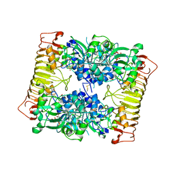
2AEY
 | | Crystal structure of fructan 1-exohydrolase IIa from Cichorium intybus in complex with 2,5 dideoxy-2,5-immino-D-mannitol | | 分子名称: | 2,5-DIDEOXY-2,5-IMINO-D-MANNITOL, 2-acetamido-2-deoxy-beta-D-glucopyranose-(1-4)-2-acetamido-2-deoxy-beta-D-glucopyranose, alpha-D-mannopyranose-(1-4)-2-acetamido-2-deoxy-beta-D-glucopyranose-(1-4)-2-acetamido-2-deoxy-beta-D-glucopyranose, ... | | 著者 | Verhaest, M, Le Roy, K, De Ranter, C.J, Van Laere, A, Van den Ende, W, Rabijns, A. | | 登録日 | 2005-07-25 | | 公開日 | 2006-08-29 | | 最終更新日 | 2023-08-23 | | 実験手法 | X-RAY DIFFRACTION (3.27 Å) | | 主引用文献 | Insights into the fine architecture of the active site of chicory fructan 1-exohydrolase: 1-kestose as substrate vs sucrose as inhibitor.
New Phytol, 174, 2007
|
|
2A3A
 | | Crystal structure of Aspergillus fumigatus chitinase B1 in complex with theophylline | | 分子名称: | SULFATE ION, THEOPHYLLINE, chitinase | | 著者 | Rao, F.V, Andersen, O.A, Vora, K.A, DeMartino, J.A, van Aalten, D.M.F. | | 登録日 | 2005-06-24 | | 公開日 | 2005-09-27 | | 最終更新日 | 2023-10-25 | | 実験手法 | X-RAY DIFFRACTION (2.1 Å) | | 主引用文献 | Methylxanthine drugs are chitinase inhibitors: investigation of inhibition and binding modes.
Chem.Biol., 12, 2005
|
|


