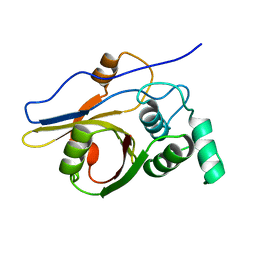
2YGX
 
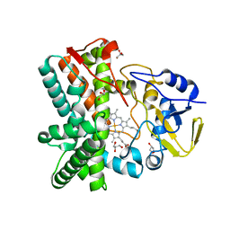 | | Structure of the mixed-function P450 MycG in P21 space group | | 分子名称: | GLYCEROL, P-450-LIKE PROTEIN, PROTOPORPHYRIN IX CONTAINING FE, ... | | 著者 | Li, S, Kells, P.M, Rutaganira, F.U, Sherman, D.H, Podust, L.M. | | 登録日 | 2011-04-22 | | 公開日 | 2012-02-29 | | 最終更新日 | 2023-12-20 | | 実験手法 | X-RAY DIFFRACTION (2.39 Å) | | 主引用文献 | Substrate Recognition by the Multifunctional Cytochrome P450 Mycg in Mycinamicin Hydroxylation and Epoxidation Reactions.
J.Biol.Chem., 287, 2012
|
|
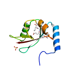
2YGG
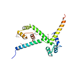 | | Complex of CaMBR and CaM | | 分子名称: | (4R)-2-METHYLPENTANE-2,4-DIOL, (4S)-2-METHYL-2,4-PENTANEDIOL, CALCIUM ION, ... | | 著者 | Koester, S, Yildiz, O. | | 登録日 | 2011-04-15 | | 公開日 | 2011-09-28 | | 最終更新日 | 2024-05-08 | | 実験手法 | X-RAY DIFFRACTION (2.227 Å) | | 主引用文献 | Structure of Human Na+/H+ Exchanger Nhe1 Regulatory Region in Complex with Cam and Ca2+
J.Biol.Chem., 286, 2011
|
|
2YQ1
 | |
2YBF
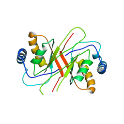
 | | Complex of Rad18 (Rad6 binding domain) with Rad6b | | 分子名称: | BETA-MERCAPTOETHANOL, E3 UBIQUITIN-PROTEIN LIGASE RAD18, SODIUM ION, ... | | 著者 | Hibbert, R.G, Sixma, T.K. | | 登録日 | 2011-03-08 | | 公開日 | 2011-04-20 | | 最終更新日 | 2023-12-20 | | 実験手法 | X-RAY DIFFRACTION (2 Å) | | 主引用文献 | E3 Ligase Rad18 Promotes Monoubiquitination Rather Than Ubiquitin Chain Formation by E2 Enzyme Rad6.
Proc.Natl.Acad.Sci.USA, 108, 2011
|
|
2YHN
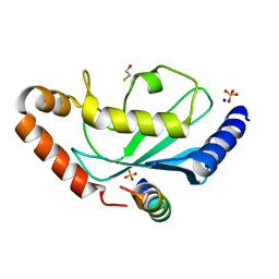
 | | The IDOL-UBE2D complex mediates sterol-dependent degradation of the LDL receptor | | 分子名称: | E3 UBIQUITIN-PROTEIN LIGASE MYLIP, ZINC ION | | 著者 | Fairall, L, Goult, B.T, Millard, C.J, Tontonoz, P, Schwabe, J.W.R. | | 登録日 | 2011-05-04 | | 公開日 | 2011-06-29 | | 最終更新日 | 2023-12-20 | | 実験手法 | X-RAY DIFFRACTION (3 Å) | | 主引用文献 | The Idol-Ube2D Complex Mediates Sterol-Dependent Degradation of the Ldl Receptor.
Genes Dev., 25, 2011
|
|
2YMK
 | |
2YEN
 | | Solution structure of the skeletal muscle and neuronal voltage gated sodium channel antagonist mu-conotoxin CnIIIC | | 分子名称: | Mu-conotoxin CnIIIC | | 著者 | Favreau, P, Benoit, E, Hocking, H.G, Carlier, L, D'hoedt, D, Leipold, E, Markgraf, R, Schlumberger, S, Cordova, M.A, Gaertner, H, Paolini-Bertrand, M, Hartley, O, Tytgat, J, Heinemann, S.H, Bertrand, D, Boelens, R, Stocklin, R, Molgo, J. | | 登録日 | 2011-03-28 | | 公開日 | 2012-02-08 | | 最終更新日 | 2023-06-14 | | 実験手法 | SOLUTION NMR | | 主引用文献 | A Novel Mu-Conopeptide, Cniiic, Exerts Potent and Preferential Inhibition of Na(V) 1.2/1.4 Channels and Blocks Neuronal Nicotinic Acetylcholine Receptors.
Br.J.Pharmacol., 166, 2012
|
|
2Y9T
 | |
2YDG
 | |
2YBN
 | |
2YCA
 | | Mixed-function P450 MycG in complex with mycinamicin III in P21212 space group | | 分子名称: | GLYCEROL, MYCINAMICIN III, P-450-LIKE PROTEIN, ... | | 著者 | Li, S, Kells, P.M, Sherman, D.H, Podust, L.M. | | 登録日 | 2011-03-12 | | 公開日 | 2012-03-28 | | 最終更新日 | 2023-12-20 | | 実験手法 | X-RAY DIFFRACTION (1.8 Å) | | 主引用文献 | Substrate Recognition by the Multifunctional Cytochrome P450 Mycg in Mycinamicin Hydroxylation and Epoxidation Reactions.
J.Biol.Chem., 287, 2012
|
|
2YCW
 | | TURKEY BETA1 ADRENERGIC RECEPTOR WITH STABILISING MUTATIONS AND BOUND ANTAGONIST CARAZOLOL | | 分子名称: | (2S)-1-(9H-Carbazol-4-yloxy)-3-(isopropylamino)propan-2-ol, BETA-1 ADRENERGIC RECEPTOR, HEGA-10, ... | | 著者 | Moukhametzianov, R, Warne, T, Edwards, P.C, Serrano-Vega, M.J, Leslie, A.G.W, Tate, C.G, Schertler, G.F.X. | | 登録日 | 2011-03-17 | | 公開日 | 2011-06-01 | | 最終更新日 | 2023-12-20 | | 実験手法 | X-RAY DIFFRACTION (3 Å) | | 主引用文献 | Two Distinct Conformations of Helix 6 Observed in Antagonist-Bound Structures of a Beta-1- Adrenergic Receptor.
Proc.Natl.Acad.Sci.USA, 108, 2011
|
|
2Z84
 | | Insights from crystal and solution structures of mouse UfSP1 | | 分子名称: | Ufm1-specific protease 1 | | 著者 | Ha, B.H, Ahn, H.C, Kang, S.H, Tanaka, K, Chung, C.H, Kim, E.E. | | 登録日 | 2007-08-30 | | 公開日 | 2008-03-04 | | 最終更新日 | 2024-03-13 | | 実験手法 | X-RAY DIFFRACTION (1.7 Å) | | 主引用文献 | Structural basis for Ufm1 processing by UfSP1
J. Biol. Chem., 283, 2008
|
|
2ZPN
 | |
376D
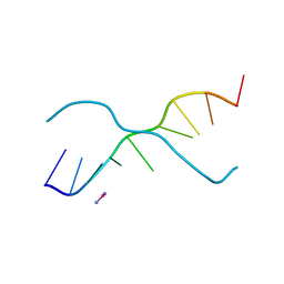 | | A ZIPPER-LIKE DNA DUPLEX D(GCGAAAGCT) | | 分子名称: | COBALT HEXAMMINE(III), DNA (5'-D(*GP*(CBR)P*GP*AP*AP*AP*GP*CP*T)-3') | | 著者 | Cruse, W.B.T, Shepard, W, Prange, T, delalFortelle, E, Fourme, R. | | 登録日 | 1998-01-22 | | 公開日 | 1999-10-26 | | 最終更新日 | 2024-04-03 | | 実験手法 | X-RAY DIFFRACTION (2.1 Å) | | 主引用文献 | A zipper-like duplex in DNA: the crystal structure of d(GCGAAAGCT) at 2.1 A resolution.
Structure, 6, 1998
|
|
3A0R
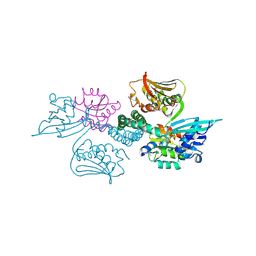 | | Crystal structure of histidine kinase ThkA (TM1359) in complex with response regulator protein TrrA (TM1360) | | 分子名称: | MERCURY (II) ION, Response regulator, Sensor protein | | 著者 | Yamada, S, Sugimoto, H, Kobayashi, M, Ohno, A, Nakamura, H, Shiro, Y. | | 登録日 | 2009-03-24 | | 公開日 | 2009-10-20 | | 最終更新日 | 2021-11-10 | | 実験手法 | X-RAY DIFFRACTION (3.8 Å) | | 主引用文献 | Structure of PAS-linked histidine kinase and the response regulator complex
Structure, 17, 2009
|
|
3A0V
 | | PAS domain of histidine kinase ThkA (TM1359) (SeMet, F486M/F489M) | | 分子名称: | ETHANOL, Sensor protein | | 著者 | Yamada, S, Sugimoto, H, Kobayashi, M, Ohno, A, Nakamura, H, Shiro, Y. | | 登録日 | 2009-03-24 | | 公開日 | 2009-10-20 | | 最終更新日 | 2021-11-10 | | 実験手法 | X-RAY DIFFRACTION (1.7 Å) | | 主引用文献 | Structure of PAS-linked histidine kinase and the response regulator complex
Structure, 17, 2009
|
|
3A4U
 | | Crystal structure of MCFD2 in complex with carbohydrate recognition domain of ERGIC-53 | | 分子名称: | CALCIUM ION, GLYCEROL, Multiple coagulation factor deficiency protein 2, ... | | 著者 | Nishio, M, Kamiya, Y, Mizushima, T, Wakatsuki, S, Sasakawa, H, Yamamoto, K, Uchiyama, S, Noda, M, McKay, A.R, Fukui, K, Hauri, H.P, Kato, K. | | 登録日 | 2009-07-17 | | 公開日 | 2010-01-05 | | 最終更新日 | 2023-11-01 | | 実験手法 | X-RAY DIFFRACTION (1.84 Å) | | 主引用文献 | Structural basis for the cooperative interplay between the two causative gene products of combined factor V and factor VIII deficiency.
Proc.Natl.Acad.Sci.USA, 107, 2010
|
|
3A0S
 | | PAS domain of histidine kinase ThkA (TM1359) | | 分子名称: | DI(HYDROXYETHYL)ETHER, Sensor protein, TETRAETHYLENE GLYCOL, ... | | 著者 | Yamada, S, Sugimoto, H, Kobayashi, M, Ohno, A, Nakamura, H, Shiro, Y. | | 登録日 | 2009-03-24 | | 公開日 | 2009-10-20 | | 最終更新日 | 2023-11-01 | | 実験手法 | X-RAY DIFFRACTION (1.47 Å) | | 主引用文献 | Structure of PAS-linked histidine kinase and the response regulator complex
Structure, 17, 2009
|
|
3A0Y
 | | Catalytic domain of histidine kinase ThkA (TM1359) (nucleotide free form 3: 1,2-propanediol, orthorombic) | | 分子名称: | Sensor protein | | 著者 | Yamada, S, Sugimoto, H, Kobayashi, M, Ohno, A, Nakamura, H, Shiro, Y. | | 登録日 | 2009-03-25 | | 公開日 | 2009-10-20 | | 最終更新日 | 2023-11-01 | | 実験手法 | X-RAY DIFFRACTION (1.57 Å) | | 主引用文献 | Structure of PAS-linked histidine kinase and the response regulator complex
Structure, 17, 2009
|
|
3A1E
 | | Crystal structure of the P- and N-domains of His462Gln mutant CopA, a copper-transporting P-type ATPase, bound with AMPPCP-Mg | | 分子名称: | MAGNESIUM ION, PHOSPHOMETHYLPHOSPHONIC ACID ADENYLATE ESTER, Probable copper-exporting P-type ATPase A | | 著者 | Tsuda, T, Toyoshima, C. | | 登録日 | 2009-03-31 | | 公開日 | 2009-07-21 | | 最終更新日 | 2023-11-01 | | 実験手法 | X-RAY DIFFRACTION (1.95 Å) | | 主引用文献 | Nucleotide recognition by CopA, a Cu+-transporting P-type ATPase.
Embo J., 28, 2009
|
|
3A0W
 | | Catalytic domain of histidine kinase ThkA (TM1359) for MAD phasing (nucleotide free form 2, orthorombic) | | 分子名称: | (4S)-2-METHYL-2,4-PENTANEDIOL, 2-(ETHYLMERCURI-THIO)-BENZOIC ACID, ETHYL MERCURY ION, ... | | 著者 | Yamada, S, Sugimoto, H, Kobayashi, M, Ohno, A, Nakamura, H, Shiro, Y. | | 登録日 | 2009-03-24 | | 公開日 | 2009-10-20 | | 最終更新日 | 2024-03-13 | | 実験手法 | X-RAY DIFFRACTION (1.69 Å) | | 主引用文献 | Structure of PAS-linked histidine kinase and the response regulator complex
Structure, 17, 2009
|
|
3A4C
 | | Crystal structure of cdt1 C terminal domain | | 分子名称: | DNA replication factor Cdt1 | | 著者 | Cho, Y, Lee, J.H. | | 登録日 | 2009-07-06 | | 公開日 | 2009-10-13 | | 最終更新日 | 2024-03-13 | | 実験手法 | X-RAY DIFFRACTION (1.889 Å) | | 主引用文献 | Structure of the Cdt1 C-terminal domain: Conservation of the winged helix fold in replication licensing factors
Protein Sci., 18, 2009
|
|
3A1C
 | | crystal structure of the P- and N-domains of CopA, a copper-transporting P-type ATPase, bound with AMPPCP-Mg | | 分子名称: | MAGNESIUM ION, PHOSPHOMETHYLPHOSPHONIC ACID ADENYLATE ESTER, Probable copper-exporting P-type ATPase A | | 著者 | Tsuda, T, Toyoshima, C. | | 登録日 | 2009-03-31 | | 公開日 | 2009-07-21 | | 最終更新日 | 2023-11-01 | | 実験手法 | X-RAY DIFFRACTION (1.85 Å) | | 主引用文献 | Nucleotide recognition by CopA, a Cu+-transporting P-type ATPase.
Embo J., 28, 2009
|
|
3A0X
 | | Catalytic domain of histidine kinase ThkA (TM1359) (nucleotide free form 1: ammomium phosphate, monoclinic) | | 分子名称: | Sensor protein | | 著者 | Yamada, S, Sugimoto, H, Kobayashi, M, Ohno, A, Nakamura, H, Shiro, Y. | | 登録日 | 2009-03-25 | | 公開日 | 2009-10-20 | | 最終更新日 | 2023-11-01 | | 実験手法 | X-RAY DIFFRACTION (1.89 Å) | | 主引用文献 | Structure of PAS-linked histidine kinase and the response regulator complex
Structure, 17, 2009
|
|


