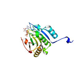
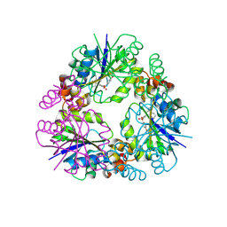
5NRD
 
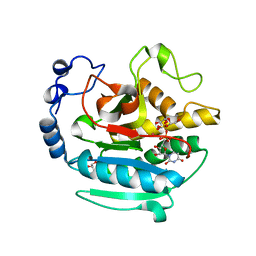 | | A Native Ternary Complex of Alpha-1,3-Galactosyltransferase (a-3GalT) Supports a Conserved Reaction Mechanism for Retaining Glycosyltransferases - alpha-3GalT in complex with Co2+, UDP-Gal and lactose - a3GalT-Co2+-UDP-Gal-LAT-2 | | 分子名称: | COBALT (II) ION, GALACTOSE-URIDINE-5'-DIPHOSPHATE, GLYCEROL, ... | | 著者 | Albesa-Jove, D, Marina, A, Sainz-Polo, M.A, Guerin, M.E. | | 登録日 | 2017-04-22 | | 公開日 | 2017-10-11 | | 最終更新日 | 2024-01-17 | | 実験手法 | X-RAY DIFFRACTION (2.12 Å) | | 主引用文献 | Structural Snapshots of alpha-1,3-Galactosyltransferase with Native Substrates: Insight into the Catalytic Mechanism of Retaining Glycosyltransferases.
Angew. Chem. Int. Ed. Engl., 56, 2017
|
|
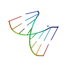
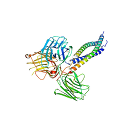
1HAA
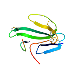 | | A beta-Hairpin Structure in a 13-mer Peptide that Binds a-Bungarotoxin with High Affinity and Neutralizes its Toxicity | | 分子名称: | ALPHA-BUNGAROTOXIN, PEPTIDE | | 著者 | Scherf, T, Kasher, R, Balass, M, Fridkin, M, Fuchs, S, Katchalski-Katzir, E. | | 登録日 | 2001-04-05 | | 公開日 | 2001-05-25 | | 最終更新日 | 2017-02-08 | | 実験手法 | SOLUTION NMR | | 主引用文献 | A Beta-Hairpin Structure in a 13-mer Peptide that Binds Alpha-Bungarotoxin with High Affinity and Neutralizes its Toxicity
Proc.Natl.Acad.Sci.USA, 98, 2001
|
|
1H56
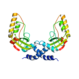 | | Structural and biochemical characterization of a new magnesium ion binding site near Tyr94 in the restriction endonuclease PvuII | | 分子名称: | MAGNESIUM ION, TYPE II RESTRICTION ENZYME PVUII | | 著者 | Spyrida, A, Matzen, C, Lanio, T, Jeltsch, A, Simoncsits, A, Athanasiadis, A, Scheuring-Vanamee, E, Kokkinidis, M, Pingoud, A. | | 登録日 | 2001-05-20 | | 公開日 | 2003-08-07 | | 最終更新日 | 2023-12-13 | | 実験手法 | X-RAY DIFFRACTION (3 Å) | | 主引用文献 | Structural and Biochemical Characterization of a New Mg(2+) Binding Site Near Tyr94 in the Restriction Endonuclease PvuII.
J.Mol.Biol., 331, 2003
|
|
5D3D
 | |
1GQV
 | | Atomic Resolution (0.98A) Structure of Eosinophil-Derived Neurotoxin | | 分子名称: | ACETATE ION, EOSINOPHIL-DERIVED NEUROTOXIN | | 著者 | Swaminathan, G.J, Holloway, D.E, Veluraja, K, Acharya, K.R. | | 登録日 | 2001-12-05 | | 公開日 | 2002-03-08 | | 最終更新日 | 2023-12-13 | | 実験手法 | X-RAY DIFFRACTION (0.98 Å) | | 主引用文献 | Atomic Resolution (0.98 A) Structure of Eosinophil-Derived Neurotoxin
Biochemistry, 41, 2002
|
|
2PLO
 | |
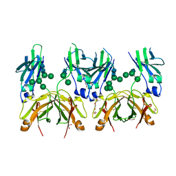
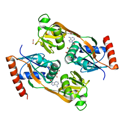
5MO2
 | | Neutron structure of cationic trypsin in complex with N-amidinopiperidine | | 分子名称: | CALCIUM ION, Cationic trypsin, SULFATE ION, ... | | 著者 | Schiebel, J, Schrader, T.E, Ostermann, A, Heine, A, Klebe, G. | | 登録日 | 2016-12-13 | | 公開日 | 2018-02-28 | | 最終更新日 | 2024-01-17 | | 実験手法 | NEUTRON DIFFRACTION (1.5 Å) | | 主引用文献 | Intriguing role of water in protein-ligand binding studied by neutron crystallography on trypsin complexes.
Nat Commun, 9, 2018
|
|
3OTD
 | | Crystal structure of human tRNAHis guanylyltransferase (Thg1)- NaI derivative | | 分子名称: | IODIDE ION, tRNA(His) guanylyltransferase | | 著者 | Hyde, S.J, Eckenroth, B.E, Doublie, S. | | 登録日 | 2010-09-11 | | 公開日 | 2010-11-17 | | 最終更新日 | 2024-02-21 | | 実験手法 | X-RAY DIFFRACTION (2.28 Å) | | 主引用文献 | tRNAHis guanylyltransferase (THG1), a unique 3'-5' nucleotidyl transferase, shares unexpected structural homology with canonical 5'-3' DNA polymerases.
Proc.Natl.Acad.Sci.USA, 107, 2010
|
|
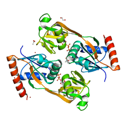
5D3C
 | | Crystal structure of a double mutant catalytic domain of Human MMP12 in complex with an hydroxamate analogue of RXP470 | | 分子名称: | CALCIUM ION, Macrophage metalloelastase, N-[(2R)-2-{[3-(3'-chlorobiphenyl-4-yl)-1,2-oxazol-5-yl]methyl}-4-(hydroxyamino)-4-oxobutanoyl]-L-alpha-glutamyl-L-alpha-glutamine, ... | | 著者 | Rouanet-Mehouas, C, Devel, L, Dive, V, Stura, E.A. | | 登録日 | 2015-08-06 | | 公開日 | 2016-08-17 | | 最終更新日 | 2024-01-10 | | 実験手法 | X-RAY DIFFRACTION (1.314 Å) | | 主引用文献 | Zinc-Metalloproteinase Inhibitors: Evaluation of the Complex Role Played by the Zinc-Binding Group on Potency and Selectivity.
J. Med. Chem., 60, 2017
|
|
5MOK
 | | Crystal structure of human IgE-Fc epsilon 3-4 | | 分子名称: | 1,2-ETHANEDIOL, DI(HYDROXYETHYL)ETHER, Ig epsilon chain C region, ... | | 著者 | Dore, K.A, Davies, A.M, Drinkwater, N, Beavil, A.J, McDonnell, J.M, Sutton, B.J. | | 登録日 | 2016-12-14 | | 公開日 | 2018-01-10 | | 最終更新日 | 2024-01-17 | | 実験手法 | X-RAY DIFFRACTION (2 Å) | | 主引用文献 | Thermal sensitivity and flexibility of the C epsilon 3 domains in immunoglobulin E.
Biochim. Biophys. Acta, 1865, 2017
|
|
5MPD
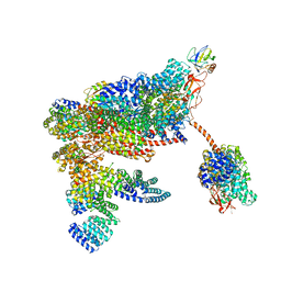 | | 26S proteasome in presence of ATP (s1) | | 分子名称: | 26S proteasome complex subunit SEM1, 26S proteasome regulatory subunit RPN1, 26S proteasome regulatory subunit RPN10, ... | | 著者 | Wehmer, M, Rudack, T, Beck, F, Aufderheide, A, Pfeifer, G, Plitzko, J.M, Foerster, F, Schulten, K, Baumeister, W, Sakata, E. | | 登録日 | 2016-12-16 | | 公開日 | 2017-03-08 | | 最終更新日 | 2024-05-08 | | 実験手法 | ELECTRON MICROSCOPY (4.1 Å) | | 主引用文献 | Structural insights into the functional cycle of the ATPase module of the 26S proteasome.
Proc. Natl. Acad. Sci. U.S.A., 114, 2017
|
|
5MQ7
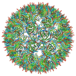 | | Structure of AaLS-13 | | 分子名称: | 6,7-dimethyl-8-ribityllumazine synthase | | 著者 | Sasaki, E, Boehringer, D, Leibundgut, M, Ban, N, Hilvert, D. | | 登録日 | 2016-12-20 | | 公開日 | 2017-03-22 | | 最終更新日 | 2024-05-15 | | 実験手法 | ELECTRON MICROSCOPY (5.2 Å) | | 主引用文献 | Structure and assembly of scalable porous protein cages.
Nat Commun, 8, 2017
|
|
5M81
 | | Translation initiation factor 4E in complex with (SP)-iPr-m7GppSpG mRNA 5' cap analog | | 分子名称: | Eukaryotic translation initiation factor 4E, GLYCEROL, [[[(3~{a}~{R},4~{R},6~{R},6~{a}~{R})-4-(2-azanyl-7-methyl-6-oxidanylidene-1~{H}-purin-7-ium-9-yl)-2,2-dimethyl-3~{a},4,6,6~{a}-tetrahydrofuro[3,4-d][1,3]dioxol-6-yl]methoxy-oxidanyl-phosphoryl]oxy-sulfanyl-phosphoryl] [(2~{R},3~{S},4~{R},5~{R})-5-(2-azanyl-6-oxidanylidene-3~{H}-purin-9-yl)-3,4-bis(oxidanyl)oxolan-2-yl]methyl hydrogen phosphate | | 著者 | Warminski, M, Nowak, E, Kowalska, J, Jemielity, J, Nowotny, M. | | 登録日 | 2016-10-28 | | 公開日 | 2017-12-20 | | 最終更新日 | 2024-01-17 | | 実験手法 | X-RAY DIFFRACTION (1.9 Å) | | 主引用文献 | Translation initiation factor 4E in complex with (SP)-iPr-m7GppSpG mRNA 5' cap analog
To Be Published
|
|
3P87
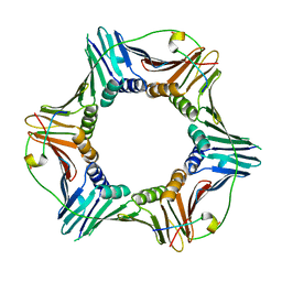 | | Structure of human PCNA bound to RNASEH2B PIP box peptide | | 分子名称: | Proliferating cell nuclear antigen, Ribonuclease H2 subunit B | | 著者 | Bubeck, D, Reijns, M.A, Graham, S.C, Astell, K.R, Jones, E.Y, Jackson, A.P. | | 登録日 | 2010-10-13 | | 公開日 | 2011-02-02 | | 最終更新日 | 2023-09-06 | | 実験手法 | X-RAY DIFFRACTION (2.99 Å) | | 主引用文献 | PCNA directs type 2 RNase H activity on DNA replication and repair substrates.
Nucleic Acids Res., 39, 2011
|
|
8QIX
 | |
5MC9
 | |
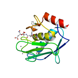
5UBH
 | | Catalytic core domain of Adenosine triphosphate phosphoribosyltransferase from Campylobacter jejuni with bound ATP | | 分子名称: | ACETATE ION, ADENOSINE-5'-TRIPHOSPHATE, ATP phosphoribosyltransferase, ... | | 著者 | Mittelstaedt, G, Jiao, W, Livingstone, E.K, Parker, E.J. | | 登録日 | 2016-12-20 | | 公開日 | 2017-12-20 | | 最終更新日 | 2023-10-04 | | 実験手法 | X-RAY DIFFRACTION (2 Å) | | 主引用文献 | A dimeric catalytic core relates the short and long forms of ATP-phosphoribosyltransferase.
Biochem. J., 475, 2018
|
|
5UBI
 | | Catalytic core domain of Adenosine triphosphate phosphoribosyltransferase from Campylobacter jejuni with bound PRPP | | 分子名称: | 1,2-ETHANEDIOL, 1-O-pyrophosphono-5-O-phosphono-alpha-D-ribofuranose, ACETATE ION, ... | | 著者 | Mittelstaedt, G, Jiao, W, Livingstone, E.K, Parker, E.J. | | 登録日 | 2016-12-20 | | 公開日 | 2017-12-20 | | 最終更新日 | 2023-10-04 | | 実験手法 | X-RAY DIFFRACTION (2.14 Å) | | 主引用文献 | A dimeric catalytic core relates the short and long forms of ATP-phosphoribosyltransferase.
Biochem. J., 475, 2018
|
|
6E28
 | | The CARD9 CARD domain-swapped dimer | | 分子名称: | Caspase recruitment domain-containing protein 9 | | 著者 | Holliday, M.J, Ferrao, R, Boenig, G, Deuber, E.C, Fairbrother, W.J. | | 登録日 | 2018-07-10 | | 公開日 | 2018-09-26 | | 最終更新日 | 2023-10-11 | | 実験手法 | X-RAY DIFFRACTION (1.36 Å) | | 主引用文献 | Picomolar zinc binding modulates formation of Bcl10-nucleating assemblies of the caspase recruitment domain (CARD) of CARD9.
J. Biol. Chem., 293, 2018
|
|
8RWL
 | | Crystal structure of Methanopyrus kandleri malate dehydrogenase mutant 1 | | 分子名称: | CHLORIDE ION, GLYCEROL, Malate dehydrogenase, ... | | 著者 | Coquille, S, Roche, J, Engilberge, S, Girard, E, Madern, D. | | 登録日 | 2024-02-05 | | 公開日 | 2024-07-10 | | 実験手法 | X-RAY DIFFRACTION (2.3 Å) | | 主引用文献 | Navigating the conformational landscape of an enzyme. Stabilization of a low populated conformer by evolutionary mutations triggers Allostery into a non-allosteric enzyme.
To Be Published
|
|
8R0S
 | | Structure of reverse transcriptase from Cauliflower Mosaic Virus in complex with RNA/DNA hybrid | | 分子名称: | DNA (5'-D(*GP*CP*TP*AP*CP*GP*CP*AP*CP*TP*GP*CP*TP*GP*GP*A)-3'), Enzymatic polyprotein, RNA (5'-R(*GP*UP*CP*CP*AP*GP*CP*AP*GP*UP*GP*CP*GP*UP*AP*GP*C)-3') | | 著者 | Prabaharan, C, Figiel, M, Chamera, S, Szczepanowski, R, Nowak, E, Nowotny, M. | | 登録日 | 2023-10-31 | | 公開日 | 2024-07-24 | | 実験手法 | X-RAY DIFFRACTION (2.35 Å) | | 主引用文献 | Structural and biochemical characterization of cauliflower mosaic virus reverse transcriptase.
J.Biol.Chem., 2024
|
|
1GNZ
 | | LECTIN I-B4 FROM GRIFFONIA SIMPLICIFOLIA (GS I-B4)METAL FREE FORM | | 分子名称: | 2-acetamido-2-deoxy-beta-D-glucopyranose, GSI-B4 ISOLECTIN, PHOSPHATE ION | | 著者 | Lescar, J, Loris, R, Mitchell, E, Gautier, C, Imberty, A. | | 登録日 | 2001-10-11 | | 公開日 | 2001-11-29 | | 最終更新日 | 2023-12-13 | | 実験手法 | X-RAY DIFFRACTION (2.5 Å) | | 主引用文献 | Isolectins I-A and I-B of Griffonia (Bandeiraea) Simplicifolia. Crystal Structure of Metal-Free Gs I-B(4) and Molecular Basis for Metal Binding and Monosaccharide Specificity.
J.Biol.Chem., 277, 2002
|
|
6H5X
 | |
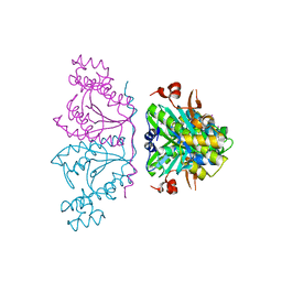
6C24
 | | Cryo-EM structure of PRC2 bound to cofactors AEBP2 and JARID2 in the Extended Active State | | 分子名称: | Histone-binding protein RBBP4, Histone-lysine N-methyltransferase EZH2, JARID2-substrate, ... | | 著者 | Kasinath, V, Faini, M, Poepsel, S, Reif, D, Feng, A, Stjepanovic, G, Aebersold, R, Nogales, E. | | 登録日 | 2018-01-06 | | 公開日 | 2018-01-24 | | 最終更新日 | 2019-12-18 | | 実験手法 | ELECTRON MICROSCOPY (3.5 Å) | | 主引用文献 | Structures of human PRC2 with its cofactors AEBP2 and JARID2.
Science, 359, 2018
|
|
8QID
 | |


