6K39
 
 | |
6NU6
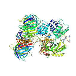
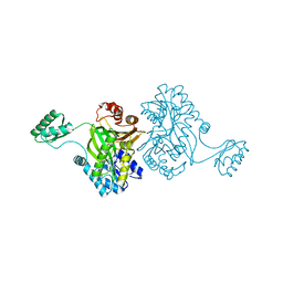
 | | Crystal structure of Mycobacterium tuberculosis dethiobiotin synthetase in complex with fragment analogue 5 | | 分子名称: | 4-[(1R,2S)-2-(carboxymethyl)cyclopentane-1-carbonyl]benzoic acid, 4-[(1S,2R)-2-(carboxymethyl)cyclopentane-1-carbonyl]benzoic acid, ATP-dependent dethiobiotin synthetase BioD, ... | | 著者 | Thompson, A.P, Polyak, S.W, Wegener, K.L, Bruning, J.B. | | 登録日 | 2019-01-31 | | 公開日 | 2020-02-05 | | 最終更新日 | 2023-10-11 | | 実験手法 | X-RAY DIFFRACTION (2.437 Å) | | 主引用文献 | Crystal structure of Mycobacterium tuberculosis dethiobiotin synthetase in complex with fragment analogue 5
To Be Published
|
|
6NWN
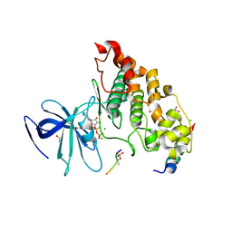
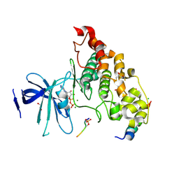
 | | Crystal structure of Mycobacterium tuberculosis dethiobiotin synthetase in complex with glutamic acid linked compound 10 | | 分子名称: | 5'-{[N-({(1S,2R)-2-[4-(carboxymethyl)benzene-1-carbonyl]cyclopentyl}acetyl)-L-gamma-glutamyl]amino}-2',5'-dideoxycytidine, ATP-dependent dethiobiotin synthetase BioD, CITRIC ACID | | 著者 | Thompson, A.P, Polyak, S.W, Wegener, K.L, Bruning, J.B. | | 登録日 | 2019-02-06 | | 公開日 | 2020-02-12 | | 最終更新日 | 2023-10-11 | | 実験手法 | X-RAY DIFFRACTION (2.005 Å) | | 主引用文献 | Crystal structure of Mycobacterium tuberculosis dethiobiotin synthetase in complex with glutamic acid linked compound 10
To Be Published
|
|
6NVE
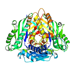
 | | Crystal structure of Mycobacterium tuberculosis dethiobiotin synthetase in complex with fragment analogue 7 | | 分子名称: | 4-{[(1S,2S)-2-(carboxymethyl)cyclopentyl]methyl}benzoic acid, ATP-dependent dethiobiotin synthetase BioD, SULFATE ION | | 著者 | Thompson, A.P, Polyak, S.W, Wegener, K.L, Bruning, J.B. | | 登録日 | 2019-02-05 | | 公開日 | 2020-02-12 | | 最終更新日 | 2023-10-11 | | 実験手法 | X-RAY DIFFRACTION (2 Å) | | 主引用文献 | Crystal structure of Mycobacterium tuberculosis dethiobiotin synthetase in complex with fragment analogue 7
To Be Published
|
|
7QNZ
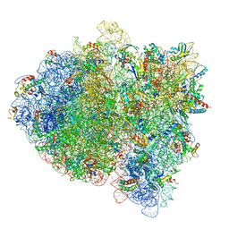
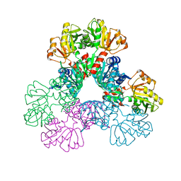
 | | human Lig1-DNA-PCNA complex reconstituted in absence of ATP | | 分子名称: | ADENOSINE MONOPHOSPHATE, DNA ligase 1, Oligo13P, ... | | 著者 | Blair, K, Tehseen, M, Raducanu, V.S, Shahid, T, Lancey, C, Cruehet, R, Hamdan, S, De Biasio, A. | | 登録日 | 2021-12-23 | | 公開日 | 2023-01-11 | | 最終更新日 | 2024-07-17 | | 実験手法 | ELECTRON MICROSCOPY (4.58 Å) | | 主引用文献 | Mechanism of human Lig1 regulation by PCNA in Okazaki fragment sealing.
Nat Commun, 13, 2022
|
|
4NM5
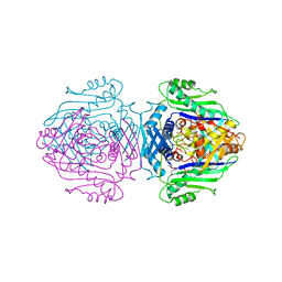
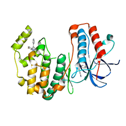
 | | Crystal structure of GSK-3/Axin complex bound to phosphorylated Wnt receptor LRP6 c-motif | | 分子名称: | ADENOSINE-5'-DIPHOSPHATE, Axin-1, CHLORIDE ION, ... | | 著者 | Stamos, J.L, Chu, M.L.-H, Enos, M.D, Shah, N, Weis, W.I. | | 登録日 | 2013-11-14 | | 公開日 | 2014-03-26 | | 最終更新日 | 2023-09-20 | | 実験手法 | X-RAY DIFFRACTION (2.3 Å) | | 主引用文献 | Structural basis of GSK-3 inhibition by N-terminal phosphorylation and by the Wnt receptor LRP6.
Elife, 3, 2014
|
|
5L3P
 | |
1QM4
 | | Methionine Adenosyltransferase Complexed with a L-Methionine Analogue | | 分子名称: | L-2-AMINO-4-METHOXY-CIS-BUT-3-ENOIC ACID, MAGNESIUM ION, METHIONINE ADENOSYLTRANSFERASE, ... | | 著者 | Gonzalez, B, Pajares, M.A, Hermoso, J.A, Sanz-Aparicio, J. | | 登録日 | 1999-09-20 | | 公開日 | 2000-09-21 | | 最終更新日 | 2023-12-13 | | 実験手法 | X-RAY DIFFRACTION (2.66 Å) | | 主引用文献 | The Crystal Structure of Tetrameric Methionine Adenosyltransferase from Rat Liver Reveals the Methionine-Binding Site
J.Mol.Biol., 300, 2000
|
|
1ULZ
 | | Crystal structure of the biotin carboxylase subunit of pyruvate carboxylase | | 分子名称: | pyruvate carboxylase n-terminal domain | | 著者 | Kondo, S, Nakajima, Y, Sugio, S, Yong-Biao, J, Sueda, S, Kondo, H. | | 登録日 | 2003-09-18 | | 公開日 | 2004-03-09 | | 最終更新日 | 2023-12-27 | | 実験手法 | X-RAY DIFFRACTION (2.2 Å) | | 主引用文献 | Structure of the biotin carboxylase subunit of pyruvate carboxylase from Aquifex aeolicus at 2.2 A resolution.
Acta Crystallogr.,Sect.D, 60, 2004
|
|
4NM7
 | | Crystal structure of GSK-3/Axin complex bound to phosphorylated Wnt receptor LRP6 e-motif | | 分子名称: | 2,3-DIHYDROXY-1,4-DITHIOBUTANE, ADENOSINE-5'-DIPHOSPHATE, Axin-1, ... | | 著者 | Stamos, J.L, Chu, M.L.-H, Enos, M.D, Shah, N, Weis, W.I. | | 登録日 | 2013-11-14 | | 公開日 | 2014-03-26 | | 最終更新日 | 2023-09-20 | | 実験手法 | X-RAY DIFFRACTION (2.3 Å) | | 主引用文献 | Structural basis of GSK-3 inhibition by N-terminal phosphorylation and by the Wnt receptor LRP6.
Elife, 3, 2014
|
|
6LTW
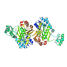
 | | Crystal structure of Apo form of I122A/I330A variant of S-adenosylmethionine synthetase from Cryptosporidium hominis | | 分子名称: | MAGNESIUM ION, PHOSPHATE ION, S-adenosylmethionine synthase | | 著者 | Singh, R.K, Michailidou, F, Rentmeister, A, Kuemmel, D. | | 登録日 | 2020-01-23 | | 公開日 | 2020-10-21 | | 最終更新日 | 2023-11-29 | | 実験手法 | X-RAY DIFFRACTION (1.65 Å) | | 主引用文献 | Engineered SAM Synthetases for Enzymatic Generation of AdoMet Analogs with Photocaging Groups and Reversible DNA Modification in Cascade Reactions.
Angew.Chem.Int.Ed.Engl., 60, 2021
|
|
6LTV
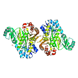
 | | Crystal Structure of I122A/I330A variant of S-adenosylmethionine synthetase from Cryptosporidium hominis in complex with ONB-SAM (2-nitro benzyme S-adenosyl-methionine) | | 分子名称: | MAGNESIUM ION, S-adenosylmethionine synthase, TRIPHOSPHATE, ... | | 著者 | Singh, R.K, Michailidou, F, Rentmeister, A, Kuemmel, D. | | 登録日 | 2020-01-23 | | 公開日 | 2020-10-21 | | 最終更新日 | 2023-11-29 | | 実験手法 | X-RAY DIFFRACTION (1.87 Å) | | 主引用文献 | Engineered SAM Synthetases for Enzymatic Generation of AdoMet Analogs with Photocaging Groups and Reversible DNA Modification in Cascade Reactions.
Angew.Chem.Int.Ed.Engl., 60, 2021
|
|
6ASV
 | | E. coli PRPP Synthetase | | 分子名称: | CHLORIDE ION, PHOSPHATE ION, Ribose-phosphate pyrophosphokinase | | 著者 | French, J.B, Zhou, W. | | 登録日 | 2017-08-25 | | 公開日 | 2019-01-23 | | 最終更新日 | 2023-10-04 | | 実験手法 | X-RAY DIFFRACTION (2.21 Å) | | 主引用文献 | Crystal structure of E. coli PRPP synthetase.
BMC Struct. Biol., 19, 2019
|
|
3HVC
 | | Crystal structure of human p38alpha MAP kinase | | 分子名称: | 4-[3-(4-FLUOROPHENYL)-1H-PYRAZOL-4-YL]PYRIDINE, Mitogen-activated protein kinase 14 | | 著者 | Perry, J.J, Tainer, J.A. | | 登録日 | 2009-06-15 | | 公開日 | 2009-06-30 | | 最終更新日 | 2023-09-06 | | 実験手法 | X-RAY DIFFRACTION (2.1 Å) | | 主引用文献 | p38alpha MAP kinase C-terminal domain binding pocket characterized by crystallographic and computational analyses.
J.Mol.Biol., 391, 2009
|
|
7KC7
 | | Biotin Carboxylase domain of Thermophilic 2-Oxoglutarate Carboxylase bound to ADP without Magnesium with disordered phosphate tail | | 分子名称: | 2-oxoglutarate carboxylase small subunit, ADENOSINE-5'-DIPHOSPHATE, PHOSPHATE ION | | 著者 | Buhrman, G.K, Rose, R.B, Enriquez, P, Truong, V. | | 登録日 | 2020-10-05 | | 公開日 | 2021-01-27 | | 最終更新日 | 2023-10-18 | | 実験手法 | X-RAY DIFFRACTION (2.2 Å) | | 主引用文献 | Structure, Function, and Thermal Adaptation of the Biotin Carboxylase Domain Dimer from Hydrogenobacter thermophilus 2-Oxoglutarate Carboxylase.
Biochemistry, 60, 2021
|
|
7KCT
 | | Crystal Structure of the Hydrogenobacter thermophilus 2-Oxoglutarate Carboxylase (OGC) Biotin Carboxylase (BC) Domain Dimer in Complex with Adenosine 5'-Diphosphate Magnesium Salt (MgADP), Adenosine 5'-Diphosphate (ADP, and Bicarbonate Anion (Hydrogen Carbonate/HCO3-) | | 分子名称: | 2-oxoglutarate carboxylase small subunit, ADENOSINE-5'-DIPHOSPHATE, BICARBONATE ION, ... | | 著者 | Buhrman, G.K, Rose, R.B, Enriquez, P, Truong, V. | | 登録日 | 2020-10-07 | | 公開日 | 2021-01-27 | | 最終更新日 | 2023-10-18 | | 実験手法 | X-RAY DIFFRACTION (2.02 Å) | | 主引用文献 | Structure, Function, and Thermal Adaptation of the Biotin Carboxylase Domain Dimer from Hydrogenobacter thermophilus 2-Oxoglutarate Carboxylase.
Biochemistry, 60, 2021
|
|
7KBL
 | | Biotin Carboxylase domain of Thermophilic 2-Oxoglutarate Carboxylase bound to Bicarbonate | | 分子名称: | 2-oxoglutarate carboxylase small subunit, BICARBONATE ION | | 著者 | Buhrman, G.K, Rose, R.B, Enriquez, P, Truong, V. | | 登録日 | 2020-10-02 | | 公開日 | 2021-01-27 | | 最終更新日 | 2023-10-18 | | 実験手法 | X-RAY DIFFRACTION (2.3 Å) | | 主引用文献 | Structure, Function, and Thermal Adaptation of the Biotin Carboxylase Domain Dimer from Hydrogenobacter thermophilus 2-Oxoglutarate Carboxylase.
Biochemistry, 60, 2021
|
|
4J1R
 | | Crystal Structure of GSK3b in complex with inhibitor 15R | | 分子名称: | (2R)-2-(1H-indol-3-ylmethyl)-1,4-dihydropyrido[2,3-b]pyrazin-3(2H)-one, Glycogen synthase kinase-3 beta, PHOSPHATE ION, ... | | 著者 | Zhan, C, Wang, Y, Wach, J, Sheehan, P, Zhong, C, Harris, R, Patskovsky, Y, Bishop, J, Haggarty, S, Ramek, A, Berry, K, O'Herin, C, Koehler, A.N, Hung, A.W, Young, D.W, Almo, S.C, New York Structural Genomics Research Consortium (NYSGRC) | | 登録日 | 2013-02-01 | | 公開日 | 2013-03-20 | | 最終更新日 | 2023-12-06 | | 実験手法 | X-RAY DIFFRACTION (2.702 Å) | | 主引用文献 | Fragment-based approach using diversity-oriented synthesis yields a GSK3b inhibitor
To be Published
|
|
4J71
 | | Crystal Structure of GSK3b in complex with inhibitor 1R | | 分子名称: | (2R)-2-methyl-1,4-dihydropyrido[2,3-b]pyrazin-3(2H)-one, CHLORIDE ION, Glycogen synthase kinase-3 beta, ... | | 著者 | Zhan, C, Wang, Y, Wach, J, Sheehan, P, Zhong, C, Harris, R, Patskovsky, Y, Bishop, J, Haggarty, S, Ramek, A, Berry, K, O'Herin, C, Koehler, A.N, Hung, A.W, Young, D.W, Almo, S.C, New York Structural Genomics Research Consortium (NYSGRC) | | 登録日 | 2013-02-12 | | 公開日 | 2013-03-20 | | 最終更新日 | 2023-12-06 | | 実験手法 | X-RAY DIFFRACTION (2.31 Å) | | 主引用文献 | Fragment-based approach using diversity-oriented synthesis yields a GSK3b inhibitor
To be Published
|
|
4KRD
 | | Crystal Structure of Pho85-Pcl10 Complex | | 分子名称: | Cyclin-dependent protein kinase PHO85, PHO85 cyclin-10 | | 著者 | Quiocho, F.A, Zheng, F. | | 登録日 | 2013-05-16 | | 公開日 | 2013-09-18 | | 最終更新日 | 2023-09-20 | | 実験手法 | X-RAY DIFFRACTION (1.952 Å) | | 主引用文献 | New Structural Insights into Phosphorylation-free Mechanism for Full Cyclin-dependent Kinase (CDK)-Cyclin Activity and Substrate Recognition.
J.Biol.Chem., 288, 2013
|
|
1KQP
 | | NH3-DEPENDENT NAD+ SYNTHETASE FROM BACILLUS SUBTILIS AT 1 A RESOLUTION | | 分子名称: | 1,2-ETHANEDIOL, MAGNESIUM ION, NH(3)-dependent NAD(+) synthetase, ... | | 著者 | Symersky, J, Devedjiev, Y, Moore, K, Brouillette, C, DeLucas, L. | | 登録日 | 2002-01-07 | | 公開日 | 2002-06-28 | | 最終更新日 | 2023-08-16 | | 実験手法 | X-RAY DIFFRACTION (1.03 Å) | | 主引用文献 | NH3-dependent NAD+ synthetase from Bacillus subtilis at 1 A resolution.
Acta Crystallogr.,Sect.D, 58, 2002
|
|
3CQU
 | | Crystal Structure of Akt-1 complexed with substrate peptide and inhibitor | | 分子名称: | Glycogen synthase kinase-3 beta, N-[2-(5-methyl-4H-1,2,4-triazol-3-yl)phenyl]-7H-pyrrolo[2,3-d]pyrimidin-4-amine, RAC-alpha serine/threonine-protein kinase | | 著者 | Pandit, J. | | 登録日 | 2008-04-03 | | 公開日 | 2008-05-27 | | 最終更新日 | 2021-10-20 | | 実験手法 | X-RAY DIFFRACTION (2.2 Å) | | 主引用文献 | Synthesis and structure based optimization of novel Akt inhibitors
Bioorg.Med.Chem.Lett., 18, 2008
|
|
3CQW
 | | Crystal Structure of Akt-1 complexed with substrate peptide and inhibitor | | 分子名称: | 5-(5-chloro-7H-pyrrolo[2,3-d]pyrimidin-4-yl)-4,5,6,7-tetrahydro-1H-imidazo[4,5-c]pyridine, Glycogen synthase kinase-3 beta, MANGANESE (II) ION, ... | | 著者 | Pandit, J. | | 登録日 | 2008-04-03 | | 公開日 | 2008-05-27 | | 最終更新日 | 2021-10-20 | | 実験手法 | X-RAY DIFFRACTION (2 Å) | | 主引用文献 | Synthesis and structure based optimization of novel Akt inhibitors
Bioorg.Med.Chem.Lett., 18, 2008
|
|
3C8Z
 | | The 1.6 A Crystal Structure of MshC: The Rate Limiting Enzyme in the Mycothiol Biosynthetic Pathway | | 分子名称: | 4-(2-HYDROXYETHYL)-1-PIPERAZINE ETHANESULFONIC ACID, 5'-O-(N-(L-CYSTEINYL)-SULFAMOYL)ADENOSINE, Cysteinyl-tRNA synthetase, ... | | 著者 | Tremblay, L.W, Fan, F, Vetting, M.W, Blanchard, J.S. | | 登録日 | 2008-02-14 | | 公開日 | 2008-12-30 | | 最終更新日 | 2024-04-03 | | 実験手法 | X-RAY DIFFRACTION (1.6 Å) | | 主引用文献 | The 1.6 A crystal structure of Mycobacterium smegmatis MshC: the penultimate enzyme in the mycothiol biosynthetic pathway.
Biochemistry, 47, 2008
|
|
3G8C
 | | Crystal Structure of Biotin Carboxylase in Complex with Biotin, Bicarbonate, ADP and Mg Ion | | 分子名称: | ADENOSINE-5'-DIPHOSPHATE, BICARBONATE ION, BIOTIN, ... | | 著者 | Chou, C.Y, Yu, L.P, Tong, L. | | 登録日 | 2009-02-12 | | 公開日 | 2009-03-03 | | 最終更新日 | 2024-02-21 | | 実験手法 | X-RAY DIFFRACTION (2 Å) | | 主引用文献 | Crystal structure of biotin carboxylase in complex with substrates and implications for its catalytic mechanism.
J.Biol.Chem., 284, 2009
|
|


