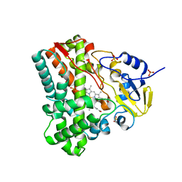
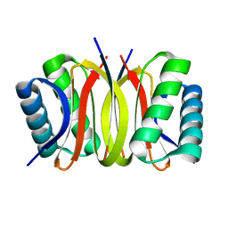
3FSM
 
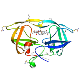 | |
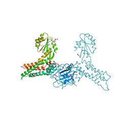
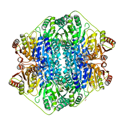
3JQ3
 | | Crystal Structure of Lombricine Kinase, complexed with substrate ADP | | 分子名称: | ADENOSINE-5'-DIPHOSPHATE, Lombricine kinase | | 著者 | Bush, D.J, Kirillova, O, Clark, S.A, Fabiola, F, Somasundaram, T, Chapman, M.S. | | 登録日 | 2009-09-05 | | 公開日 | 2010-09-15 | | 最終更新日 | 2023-09-06 | | 実験手法 | X-RAY DIFFRACTION (2.503 Å) | | 主引用文献 | The structure of lombricine kinase: implications for phosphagen kinase conformational changes.
J.Biol.Chem., 286, 2011
|
|
3JQO
 | | Crystal structure of the outer membrane complex of a type IV secretion system | | 分子名称: | (4S)-2-METHYL-2,4-PENTANEDIOL, LAURYL DIMETHYLAMINE-N-OXIDE, TraF protein, ... | | 著者 | Chandran, V, Fronzes, R, Duquerroy, S, Cronin, N, Navaza, J, Waksman, G. | | 登録日 | 2009-09-07 | | 公開日 | 2009-12-01 | | 最終更新日 | 2024-04-03 | | 実験手法 | X-RAY DIFFRACTION (2.6 Å) | | 主引用文献 | Structure of the outer membrane complex of a type IV secretion system
Nature, 462, 2009
|
|
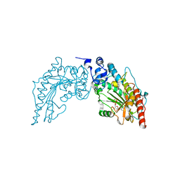
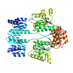
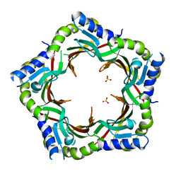
3KA2
 | |
3FYG
 | | CRYSTAL STRUCTURE OF TETRADECA-(3-FLUOROTYROSYL)-GLUTATHIONE S-TRANSFERASE | | 分子名称: | (9R,10R)-9-(S-GLUTATHIONYL)-10-HYDROXY-9,10-DIHYDROPHENANTHRENE, MU CLASS TETRADECA-(3-FLUOROTYROSYL)-GLUTATHIONE S-TRANSFERASE OF ISOENZYME | | 著者 | Xiao, G, Parsons, J.F, Armstrong, R.N, Gilliland, G.L. | | 登録日 | 1997-08-07 | | 公開日 | 1999-06-01 | | 最終更新日 | 2023-08-09 | | 実験手法 | X-RAY DIFFRACTION (2.2 Å) | | 主引用文献 | Conformational changes in the crystal structure of rat glutathione transferase M1-1 with global substitution of 3-fluorotyrosine for tyrosine.
J.Mol.Biol., 281, 1998
|
|
3JSC
 | | CcdBVfi-FormI-pH7.0 | | 分子名称: | CcdB, SULFATE ION | | 著者 | De Jonge, N, Buts, L, Loris, R. | | 登録日 | 2009-09-10 | | 公開日 | 2009-12-01 | | 最終更新日 | 2023-11-01 | | 実験手法 | X-RAY DIFFRACTION (1.5 Å) | | 主引用文献 | Structural and thermodynamic characterization of vibrio fischeri CCDB
J.Biol.Chem., 285, 2010
|
|
3FZ2
 | | Crystal structure of the tail terminator protein from phage lambda (gpU-D74A) | | 分子名称: | Minor tail protein U, SULFATE ION | | 著者 | Pell, L.G, Liu, A, Edmonds, E, Donaldson, L.W, Howell, P.L, Davidson, A.R. | | 登録日 | 2009-01-23 | | 公開日 | 2009-05-26 | | 最終更新日 | 2021-10-20 | | 実験手法 | X-RAY DIFFRACTION (2.7 Å) | | 主引用文献 | The X-ray crystal structure of the phage lambda tail terminator protein reveals the biologically relevant hexameric ring structure and demonstrates a conserved mechanism of tail termination among diverse long-tailed phages.
J.Mol.Biol., 389, 2009
|
|
3GEH
 | | Crystal structure of MnmE from Nostoc in complex with GDP, FOLINIC ACID and ZN | | 分子名称: | GUANOSINE-5'-DIPHOSPHATE, N-{[4-({[(6R)-2-amino-5-formyl-4-oxo-1,4,5,6,7,8-hexahydropteridin-6-yl]methyl}amino)phenyl]carbonyl}-L-glutamic acid, ZINC ION, ... | | 著者 | Meyer, S, Wittinghofer, A. | | 登録日 | 2009-02-25 | | 公開日 | 2009-10-27 | | 最終更新日 | 2023-11-01 | | 実験手法 | X-RAY DIFFRACTION (3.2 Å) | | 主引用文献 | Kissing G domains of MnmE monitored by X-ray crystallography and pulse electron paramagnetic resonance spectroscopy
Plos Biol., 7, 2009
|
|
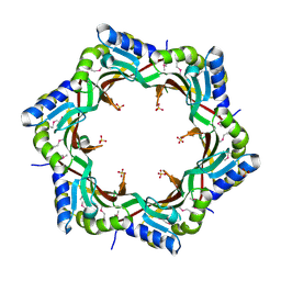
3K41
 | | Crystal structure of sCD-MPR mutant E19Q/K137M bound to Man-6-P | | 分子名称: | 2-acetamido-2-deoxy-beta-D-glucopyranose-(1-4)-2-acetamido-2-deoxy-beta-D-glucopyranose, 6-O-phosphono-beta-D-mannopyranose, Cation-dependent mannose-6-phosphate receptor, ... | | 著者 | Olson, L.J, Sun, G, Bohnsack, R.N, Peterson, F.C, Dahms, N.M, Kim, J.J.P. | | 登録日 | 2009-10-05 | | 公開日 | 2009-11-24 | | 最終更新日 | 2023-09-06 | | 実験手法 | X-RAY DIFFRACTION (1.9 Å) | | 主引用文献 | Intermonomer interactions are essential for lysosomal enzyme binding by the cation-dependent mannose 6-phosphate receptor.
Biochemistry, 49, 2010
|
|
3G5F
 | | Crystallographic analysis of cytochrome P450 cyp121 | | 分子名称: | Cytochrome P450 121, PROTOPORPHYRIN IX CONTAINING FE, SULFATE ION | | 著者 | Belin, P, Le Du, M.H, Gondry, M. | | 登録日 | 2009-02-05 | | 公開日 | 2009-04-21 | | 最終更新日 | 2023-11-01 | | 実験手法 | X-RAY DIFFRACTION (1.4 Å) | | 主引用文献 | Identification and structural basis of the reaction catalyzed by CYP121, an essential cytochrome P450 in Mycobacterium tuberculosis.
Proc.Natl.Acad.Sci.USA, 106, 2009
|
|
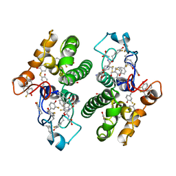
3GEE
 | | Crystal structure of MnmE from Chlorobium tepidum in complex with GDP and FOLINIC ACID | | 分子名称: | GUANOSINE-5'-DIPHOSPHATE, N-{[4-({[(6R)-2-amino-5-formyl-4-oxo-1,4,5,6,7,8-hexahydropteridin-6-yl]methyl}amino)phenyl]carbonyl}-L-glutamic acid, tRNA modification GTPase mnmE | | 著者 | Meyer, S, Wittinghofer, A. | | 登録日 | 2009-02-25 | | 公開日 | 2009-10-27 | | 最終更新日 | 2023-11-01 | | 実験手法 | X-RAY DIFFRACTION (2.95 Å) | | 主引用文献 | Kissing G domains of MnmE monitored by X-ray crystallography and pulse electron paramagnetic resonance spectroscopy
Plos Biol., 7, 2009
|
|
3DZD
 | | Crystal structure of sigma54 activator NTRC4 in the inactive state | | 分子名称: | ADENOSINE-5'-DIPHOSPHATE, SODIUM ION, Transcriptional regulator (NtrC family) | | 著者 | Batchelor, J.D, Doucleff, M, Lee, C.-J, Matsubara, K, De Carlo, S, Heideker, J, Lamers, M.M, Pelton, J.G, Wemmer, D.E. | | 登録日 | 2008-07-29 | | 公開日 | 2008-11-25 | | 最終更新日 | 2023-11-15 | | 実験手法 | X-RAY DIFFRACTION (2.4 Å) | | 主引用文献 | Structure and regulatory mechanism of Aquifex aeolicus NtrC4: variability and evolution in bacterial transcriptional regulation.
J.Mol.Biol., 384, 2008
|
|
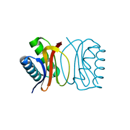
3KU7
 | | Crystal structure of Helicobacter pylori MinE, a cell division topological specificity factor | | 分子名称: | Cell division topological specificity factor | | 著者 | Kang, G.B, Song, H.E, Kim, M.K, Eom, S.H. | | 登録日 | 2009-11-26 | | 公開日 | 2010-05-05 | | 最終更新日 | 2024-03-20 | | 実験手法 | X-RAY DIFFRACTION (2.8 Å) | | 主引用文献 | Crystal structure of Helicobacter pylori MinE, a cell division topological specificity factor
Mol.Microbiol., 76, 2010
|
|
3DXS
 | | Crystal structure of a copper binding domain from HMA7, a P-type ATPase | | 分子名称: | Copper-transporting ATPase RAN1, LITHIUM ION, ZINC ION | | 著者 | Zimmermann, M, Xiao, Z, Clarke, O.B, Gulbis, J.M, Wedd, A.G. | | 登録日 | 2008-07-25 | | 公開日 | 2009-08-11 | | 最終更新日 | 2024-05-29 | | 実験手法 | X-RAY DIFFRACTION (1.7 Å) | | 主引用文献 | Metal binding affinities of Arabidopsis zinc and copper transporters: selectivities match the relative, but not the absolute, affinities of their amino-terminal domains.
Biochemistry, 48, 2009
|
|
3L59
 | | Structure of BACE Bound to SCH710413 | | 分子名称: | (2Z)-3-(3-chlorobenzyl)-2-imino-5,5-dimethylimidazolidin-4-one, Beta-secretase 1, D(-)-TARTARIC ACID | | 著者 | Strickland, C, Zhu, Z. | | 登録日 | 2009-12-21 | | 公開日 | 2010-02-16 | | 最終更新日 | 2018-01-24 | | 実験手法 | X-RAY DIFFRACTION (2 Å) | | 主引用文献 | Discovery of Cyclic Acylguanidines as Highly Potent and Selective beta-Site Amyloid Cleaving Enzyme (BACE) Inhibitors: Part I-Inhibitor Design and Validation
J.Med.Chem., 53, 2010
|
|
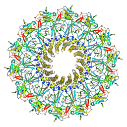
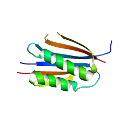
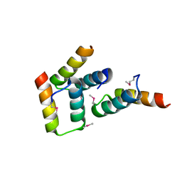
3DVT
 | |
3DVH
 | | LC8 Point mutant K36P | | 分子名称: | Dynein light chain 1, cytoplasmic | | 著者 | Lightcap, C.M, Williams, J.C. | | 登録日 | 2008-07-18 | | 公開日 | 2009-01-20 | | 最終更新日 | 2023-08-30 | | 実験手法 | X-RAY DIFFRACTION (2 Å) | | 主引用文献 | Biochemical and structural characterization of the Pak1-LC8 interaction.
J.Biol.Chem., 283, 2008
|
|
3DVP
 | | Pak1 peptide bound LC8 | | 分子名称: | Dynein light chain 1, cytoplasmic, P21 activated Kinase peptide | | 著者 | Lightcap, C.M, Williams, J.C. | | 登録日 | 2008-07-18 | | 公開日 | 2009-01-20 | | 最終更新日 | 2024-02-21 | | 実験手法 | X-RAY DIFFRACTION (2.5 Å) | | 主引用文献 | Biochemical and structural characterization of the Pak1-LC8 interaction.
J.Biol.Chem., 283, 2008
|
|
3FZB
 | | Crystal structure of the tail terminator protein from phage lambda (gpU-WT) | | 分子名称: | Minor tail protein U, SULFATE ION | | 著者 | Pell, L.G, Liu, A, Edmonds, E, Donaldson, L.W, Howell, P.L, Davidson, A.R. | | 登録日 | 2009-01-24 | | 公開日 | 2009-05-26 | | 最終更新日 | 2024-02-21 | | 実験手法 | X-RAY DIFFRACTION (2.8 Å) | | 主引用文献 | The X-ray crystal structure of the phage lambda tail terminator protein reveals the biologically relevant hexameric ring structure and demonstrates a conserved mechanism of tail termination among diverse long-tailed phages.
J.Mol.Biol., 389, 2009
|
|
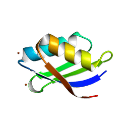
3FYR
 | | Crystal structure of the sporulation histidine kinase inhibitor Sda from Bacillus subtilis | | 分子名称: | Sporulation inhibitor sda | | 著者 | Jacques, D.A, Streamer, M, King, G.F, Guss, J.M, Trewhella, J, Langley, D.B. | | 登録日 | 2009-01-23 | | 公開日 | 2009-06-23 | | 最終更新日 | 2017-11-01 | | 実験手法 | X-RAY DIFFRACTION (1.97 Å) | | 主引用文献 | Structure of the sporulation histidine kinase inhibitor Sda from Bacillus subtilis and insights into its solution state
Acta Crystallogr.,Sect.D, 65, 2009
|
|
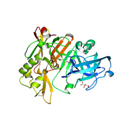
3FZN
 | | Intermediate analogue in benzoylformate decarboxylase | | 分子名称: | 3-[(4-amino-2-methylpyrimidin-5-yl)methyl]-2-{(S)-hydroxy[(R)-hydroxy(methoxy)phosphoryl]phenylmethyl}-5-(2-{[(R)-hydroxy(phosphonooxy)phosphoryl]oxy}ethyl)-4-methyl-1,3-thiazol-3-ium, Benzoylformate decarboxylase, CHLORIDE ION, ... | | 著者 | Bruning, M, Berheide, M, Meyer, D, Golbik, R, Bartunik, H, Liese, A, Tittmann, K. | | 登録日 | 2009-01-26 | | 公開日 | 2009-05-05 | | 最終更新日 | 2023-11-01 | | 実験手法 | X-RAY DIFFRACTION (1.62 Å) | | 主引用文献 | Structural and kinetic studies on native intermediates and an intermediate analogue in benzoylformate decarboxylase reveal a least motion mechanism with an unprecedented short-lived predecarboxylation intermediate.
Biochemistry, 48, 2009
|
|
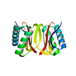
3K42
 | | Crystal structure of sCD-MPR mutant E19Q/K137M pH 7.0 | | 分子名称: | 2-acetamido-2-deoxy-beta-D-glucopyranose, Cation-dependent mannose-6-phosphate receptor, SN-GLYCEROL-1-PHOSPHATE, ... | | 著者 | Olson, L.J, Sun, G, Bohnsack, R.N, Peterson, F.C, Dahms, N.M, Kim, J.J.P. | | 登録日 | 2009-10-05 | | 公開日 | 2009-11-24 | | 最終更新日 | 2023-09-06 | | 実験手法 | X-RAY DIFFRACTION (2.3 Å) | | 主引用文献 | Intermonomer interactions are essential for lysosomal enzyme binding by the cation-dependent mannose 6-phosphate receptor.
Biochemistry, 49, 2010
|
|
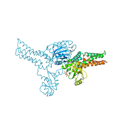
3J6D
 | | Model of the PrgH-PrgK periplasmic rings | | 分子名称: | Pathogenicity 1 island effector protein, Protein PrgH | | 著者 | Bergeron, J.R.C, Strynadka, N.C.J. | | 登録日 | 2014-02-14 | | 公開日 | 2015-01-14 | | 最終更新日 | 2024-02-21 | | 実験手法 | ELECTRON MICROSCOPY (11.7 Å) | | 主引用文献 | The Modular Structure of the Inner-Membrane Ring Component PrgK Facilitates Assembly of the Type III Secretion System Basal Body.
Structure, 23, 2015
|
|
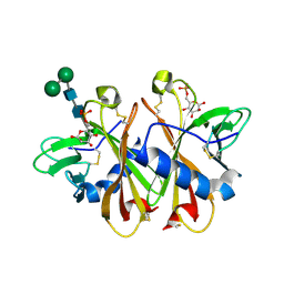
3K5V
 | | Structure of Abl kinase in complex with imatinib and GNF-2 | | 分子名称: | 3-(6-{[4-(trifluoromethoxy)phenyl]amino}pyrimidin-4-yl)benzamide, 4-(4-METHYL-PIPERAZIN-1-YLMETHYL)-N-[4-METHYL-3-(4-PYRIDIN-3-YL-PYRIMIDIN-2-YLAMINO)-PHENYL]-BENZAMIDE, CHLORIDE ION, ... | | 著者 | Cowan-Jacob, S.W, Fendrich, G, Rummel, G, Strauss, A. | | 登録日 | 2009-10-08 | | 公開日 | 2010-01-19 | | 最終更新日 | 2023-09-06 | | 実験手法 | X-RAY DIFFRACTION (1.74 Å) | | 主引用文献 | Targeting Bcr-Abl by combining allosteric with ATP-binding-site inhibitors.
Nature, 463, 2010
|
|
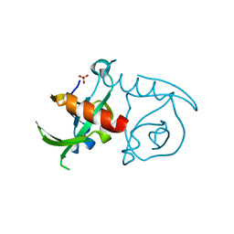
3K77
 | | X-ray crystal structure of XRCC1 | | 分子名称: | DNA repair protein XRCC1 | | 著者 | Cuneo, M.J, London, R.E. | | 登録日 | 2009-10-12 | | 公開日 | 2010-04-28 | | 最終更新日 | 2023-09-06 | | 実験手法 | X-RAY DIFFRACTION (2.597 Å) | | 主引用文献 | Oxidation state of the XRCC1 N-terminal domain regulates DNA polymerase beta binding affinity.
Proc.Natl.Acad.Sci.USA, 107, 2010
|
|


