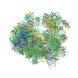
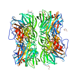
4U4N
 
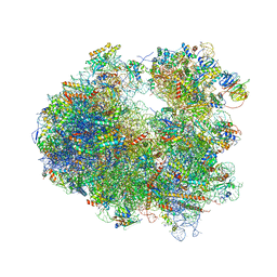
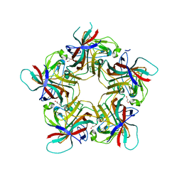
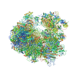 | | Crystal structure of Edeine bound to the yeast 80S ribosome | | 分子名称: | 18S ribosomal RNA, 25S ribosomal RNA, 40S ribosomal protein S0-A, ... | | 著者 | Garreau de Loubresse, N, Prokhorova, I, Yusupova, G, Yusupov, M. | | 登録日 | 2014-07-24 | | 公開日 | 2014-10-22 | | 最終更新日 | 2024-11-13 | | 実験手法 | X-RAY DIFFRACTION (3.1 Å) | | 主引用文献 | Structural basis for the inhibition of the eukaryotic ribosome.
Nature, 513, 2014
|
|
4U6A
 | |
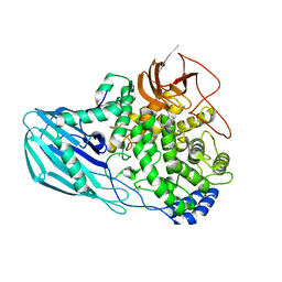
4U8O
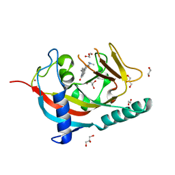
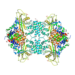
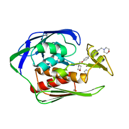
 | | Structure of Aspergillus fumigatus UDP-Galactopyranose mutase mutant N207A complexed with UDP | | 分子名称: | 1,2-ETHANEDIOL, DIHYDROFLAVINE-ADENINE DINUCLEOTIDE, SULFATE ION, ... | | 著者 | Qureshi, I.A, Chaudhary, R, Tanner, J.J. | | 登録日 | 2014-08-03 | | 公開日 | 2014-12-03 | | 最終更新日 | 2023-09-27 | | 実験手法 | X-RAY DIFFRACTION (2.3 Å) | | 主引用文献 | Contributions of Unique Active Site Residues of Eukaryotic UDP-Galactopyranose Mutases to Substrate Recognition and Active Site Dynamics.
Biochemistry, 53, 2014
|
|
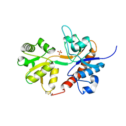
4U8N
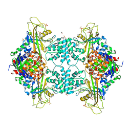
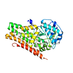
 | | Structure of Aspergillus fumigatus UDP-Galactopyranose mutase mutant F66A complexed with UDP | | 分子名称: | 1,2-ETHANEDIOL, DIHYDROFLAVINE-ADENINE DINUCLEOTIDE, SULFATE ION, ... | | 著者 | Qureshi, I.A, Chaudhary, R, Tanner, J.J. | | 登録日 | 2014-08-03 | | 公開日 | 2014-12-03 | | 最終更新日 | 2023-09-27 | | 実験手法 | X-RAY DIFFRACTION (2.3 Å) | | 主引用文献 | Contributions of Unique Active Site Residues of Eukaryotic UDP-Galactopyranose Mutases to Substrate Recognition and Active Site Dynamics.
Biochemistry, 53, 2014
|
|
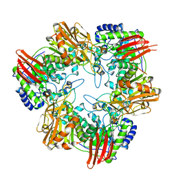
4UBD
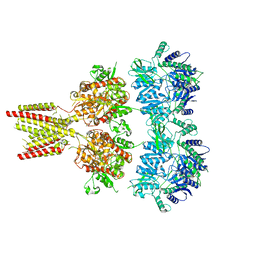
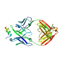
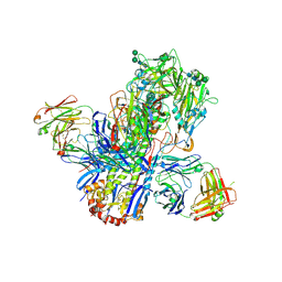 | | Crystal structure of a neutralizing human monoclonal antibody with 1968 H3 HA | | 分子名称: | 2-acetamido-2-deoxy-beta-D-glucopyranose, 2-acetamido-2-deoxy-beta-D-glucopyranose-(1-4)-2-acetamido-2-deoxy-beta-D-glucopyranose, Hemagglutinin HA1 chain, ... | | 著者 | Shore, D.A, Yang, H, Cho, M, Donis, R.O, Stevens, J. | | 登録日 | 2014-08-12 | | 公開日 | 2015-06-24 | | 最終更新日 | 2024-11-13 | | 実験手法 | X-RAY DIFFRACTION (3.5 Å) | | 主引用文献 | A potent broad-spectrum protective human monoclonal antibody crosslinking two haemagglutinin monomers of influenza A virus.
Nat Commun, 6, 2015
|
|
4U3M
 | | Crystal structure of Anisomycin bound to the yeast 80S ribosome | | 分子名称: | 18S rRNA, 25s rRNA, 40S ribosomal protein S0-A, ... | | 著者 | Garreau de Loubresse, N, Prokhorova, I, Yusupova, G, Yusupov, M. | | 登録日 | 2014-07-22 | | 公開日 | 2014-10-22 | | 最終更新日 | 2024-10-23 | | 実験手法 | X-RAY DIFFRACTION (3 Å) | | 主引用文献 | Structural basis for the inhibition of the eukaryotic ribosome.
Nature, 513, 2014
|
|
4U5D
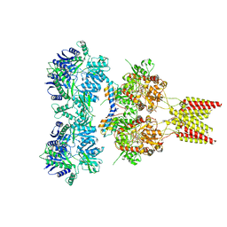 | | Crystal structure of GluA2, con-ikot-ikot snail toxin, partial agonist KA and postitive modulator (R,R)-2b complex | | 分子名称: | 2-acetamido-2-deoxy-beta-D-glucopyranose, 3-(CARBOXYMETHYL)-4-ISOPROPENYLPROLINE, Con-ikot-ikot, ... | | 著者 | Chen, L, Gouaux, E. | | 登録日 | 2014-07-25 | | 公開日 | 2014-08-13 | | 最終更新日 | 2024-10-23 | | 実験手法 | X-RAY DIFFRACTION (3.5757 Å) | | 主引用文献 | X-ray structures of AMPA receptor-cone snail toxin complexes illuminate activation mechanism.
Science, 345, 2014
|
|
4TUO
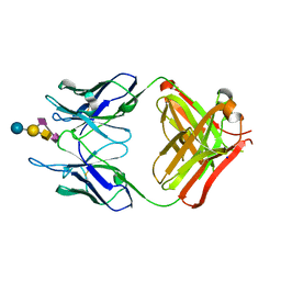 | | Crystal structure of monoclonal antibody against neuroblastoma associated antigen. | | 分子名称: | Heavy chain of monoclonal antibody against neuroblastoma associated antigen, Light chain of monoclonal antibody against neuroblastoma associated antigen, N-acetyl-alpha-neuraminic acid-(2-8)-N-acetyl-alpha-neuraminic acid-(2-3)-[2-acetamido-2-deoxy-beta-D-galactopyranose-(1-4)]beta-D-galactopyranose-(1-4)-alpha-D-glucopyranose, ... | | 著者 | Golik, P, Grudnik, P, Horwacik, I, Zdzalik, M, Rokita, H, Dubin, G. | | 登録日 | 2014-06-24 | | 公開日 | 2015-07-15 | | 最終更新日 | 2024-11-06 | | 実験手法 | X-RAY DIFFRACTION (1.55 Å) | | 主引用文献 | Structural Basis of GD2 Ganglioside and Mimetic Peptide Recognition by 14G2a Antibody.
Mol.Cell Proteomics, 14, 2015
|
|
4U52
 | | Crystal structure of Nagilactone C bound to the yeast 80S ribosome | | 分子名称: | 18S ribosomal RNA, 25S ribosomal RNA, 40S ribosomal protein S0-A, ... | | 著者 | Garreau de Loubresse, N, Prokhorova, I, Yusupova, G, Yusupov, M. | | 登録日 | 2014-07-24 | | 公開日 | 2014-10-22 | | 最終更新日 | 2024-10-16 | | 実験手法 | X-RAY DIFFRACTION (3 Å) | | 主引用文献 | Structural basis for the inhibition of the eukaryotic ribosome.
Nature, 513, 2014
|
|
4U8L
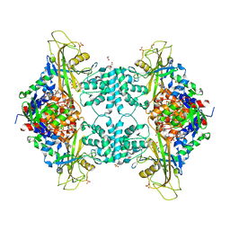
 | | Structure of Aspergillus fumigatus UDP-Galactopyranose mutase mutant N207A | | 分子名称: | 1,2-ETHANEDIOL, DIHYDROFLAVINE-ADENINE DINUCLEOTIDE, SULFATE ION, ... | | 著者 | Qureshi, I.A, Chaudhary, R, Tanner, J.J. | | 登録日 | 2014-08-03 | | 公開日 | 2014-12-03 | | 最終更新日 | 2023-09-27 | | 実験手法 | X-RAY DIFFRACTION (2.3 Å) | | 主引用文献 | Contributions of Unique Active Site Residues of Eukaryotic UDP-Galactopyranose Mutases to Substrate Recognition and Active Site Dynamics.
Biochemistry, 53, 2014
|
|
4U1Y
 | | Full length GluA2-FW-(R,R)-2b complex | | 分子名称: | 2-AMINO-3-(5-FLUORO-2,4-DIOXO-3,4-DIHYDRO-2H-PYRIMIDIN-1-YL)-PROPIONIC ACID, 2-acetamido-2-deoxy-beta-D-glucopyranose, Glutamate receptor 2, ... | | 著者 | Chen, L, Gouaux, E. | | 登録日 | 2014-07-16 | | 公開日 | 2014-08-20 | | 最終更新日 | 2024-11-06 | | 実験手法 | X-RAY DIFFRACTION (3.8999 Å) | | 主引用文献 | Structure and Dynamics of AMPA Receptor GluA2 in Resting, Pre-Open, and Desensitized States.
Cell, 158, 2014
|
|
4UFI
 | | Mouse Galactocerebrosidase complexed with aza-galacto-fagomine AGF | | 分子名称: | (3R,4S,5R)-3-(hydroxymethyl)-1,2-diazinane-4,5-diol, 2-acetamido-2-deoxy-beta-D-glucopyranose, 2-acetamido-2-deoxy-beta-D-glucopyranose-(1-4)-2-acetamido-2-deoxy-beta-D-glucopyranose, ... | | 著者 | Hill, C.H, Viuff, A.H, Spratley, S.J, Salamone, S, Christensen, S.H, Read, R.J, Moriarty, N.W, Jensen, H.H, Deane, J.E. | | 登録日 | 2015-03-17 | | 公開日 | 2015-03-25 | | 最終更新日 | 2024-11-13 | | 実験手法 | X-RAY DIFFRACTION (2.4 Å) | | 主引用文献 | Azasugar Inhibitors as Pharmacological Chaperones for Krabbe Disease.
Chem.Sci., 6, 2015
|
|
4U8U
 | | The Crystallographic structure of the giant hemoglobin from Glossoscolex paulistus at 3.2 A resolution. | | 分子名称: | 2-acetamido-2-deoxy-beta-D-glucopyranose, CALCIUM ION, CYANIDE ION, ... | | 著者 | Bachega, J.F.R, Maluf, F.V, Andi, B, D'Muniz Pereira, H, Carazzollea, M.F, Orville, A, Tabak, M, Garratt, R.C, Horjales, E. | | 登録日 | 2014-08-04 | | 公開日 | 2015-06-10 | | 最終更新日 | 2024-10-16 | | 実験手法 | X-RAY DIFFRACTION (3.2 Å) | | 主引用文献 | The structure of the giant haemoglobin from Glossoscolex paulistus.
Acta Crystallogr.,Sect.D, 71, 2015
|
|
4U3D
 | |
4U74
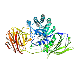
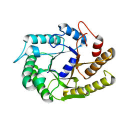
 | | Crystal structure of 4-phenylimidazole bound form of human indoleamine 2,3-dioxygenase (G262A mutant) | | 分子名称: | 2-[N-CYCLOHEXYLAMINO]ETHANE SULFONIC ACID, 4-PHENYL-1H-IMIDAZOLE, Indoleamine 2,3-dioxygenase 1, ... | | 著者 | Sugimoto, H, Horitani, M, Kometani, E, Shiro, Y. | | 登録日 | 2014-07-30 | | 公開日 | 2015-09-02 | | 最終更新日 | 2024-11-20 | | 実験手法 | X-RAY DIFFRACTION (2.31 Å) | | 主引用文献 | Conformation and Mobility of Active Site Loop is Critical for Substrate Binding and Inhibition in Human Indoleamine 2,3-Dioxygenase
to be published
|
|
4U5I
 | |
4U5K
 | |
4TXN
 | | Crystal structure of uridine phosphorylase from Schistosoma mansoni in complex with 5-fluorouracil | | 分子名称: | 5-FLUOROURACIL, SULFATE ION, Uridine phosphorylase | | 著者 | Marinho, A, Torini, J, Romanello, L, Cassago, A, DeMarco, R, Brandao-Neto, J, Pereira, H.M. | | 登録日 | 2014-07-03 | | 公開日 | 2015-10-14 | | 最終更新日 | 2023-09-27 | | 実験手法 | X-RAY DIFFRACTION (2 Å) | | 主引用文献 | Analysis of two Schistosoma mansoni uridine phosphorylases isoforms suggests the emergence of a protein with a non-canonical function.
Biochimie, 125, 2016
|
|
4UDJ
 | | Crystal structure of b-1,4-mannopyranosyl-chitobiose phosphorylase at 1.60 Angstrom in complex with beta-D-mannopyranose and inorganic phosphate | | 分子名称: | 1,2-ETHANEDIOL, PHOSPHATE ION, POTASSIUM ION, ... | | 著者 | Ladeveze, S, Cioci, G, Potocki-Veronese, G, Tranier, S, Mourey, L. | | 登録日 | 2014-12-10 | | 公開日 | 2015-05-27 | | 最終更新日 | 2023-12-20 | | 実験手法 | X-RAY DIFFRACTION (1.94 Å) | | 主引用文献 | Structural Bases for N-Glycan Processing by Mannoside Phosphorylase.
Acta Crystallogr.,Sect.D, 71, 2015
|
|
4UFC
 | | Crystal structure of the GH95 enzyme BACOVA_03438 | | 分子名称: | CACODYLATE ION, CALCIUM ION, GH95, ... | | 著者 | Rogowski, A, Briggs, J.A, Mortimer, J.C, Tryfona, T, Terrapon, N, Lowe, E.C, Basle, A, Morland, C, Day, A.M, Zheng, H, Rogers, T.E, Thompson, P, Hawkins, A.R, Yadav, M.P, Henrissat, B, Martens, E.C, Dupree, P, Gilbert, H.J, Bolam, D.N. | | 登録日 | 2015-03-16 | | 公開日 | 2015-07-08 | | 最終更新日 | 2023-12-20 | | 実験手法 | X-RAY DIFFRACTION (2.81 Å) | | 主引用文献 | Glycan Complexity Dictates Microbial Resource Allocation in the Large Intestine.
Nat.Commun., 6, 2015
|
|
4U2R
 | | Crystal structure of the GLUR2 ligand binding core (S1S2J, flip variant) in the apo state | | 分子名称: | Glutamate receptor 2, SULFATE ION | | 著者 | Duerr, K.L, Chen, L, Gouaux, E. | | 登録日 | 2014-07-17 | | 公開日 | 2014-08-20 | | 最終更新日 | 2024-11-06 | | 実験手法 | X-RAY DIFFRACTION (1.4114 Å) | | 主引用文献 | Structure and Dynamics of AMPA Receptor GluA2 in Resting, Pre-Open, and Desensitized States.
Cell, 158, 2014
|
|
4UC0
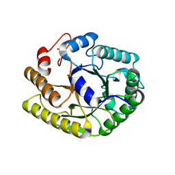
 | | Crystal Structure Of a purine nucleoside phosphorylase (PSI-NYSGRC-029736) from Agrobacterium vitis | | 分子名称: | HYPOXANTHINE, Purine nucleoside phosphorylase | | 著者 | Cameron, S.A, Sampathkumar, P, Ramagopal, U.A, Attonito, J, Ahmed, M, Bhosle, R, Bonanno, J, Chamala, S, Chowdhury, S, Glenn, A.S, Hammonds, J, Hillerich, B, Love, J.D, Seidel, R, Stead, M, Toro, R, Wasserman, S.R, Schramm, V.L, Almo, S.C, New York Structural Genomics Research Consortium (NYSGRC) | | 登録日 | 2014-08-13 | | 公開日 | 2014-10-08 | | 最終更新日 | 2024-10-16 | | 実験手法 | X-RAY DIFFRACTION (2.4 Å) | | 主引用文献 | Crystal Structure Of a purine nucleoside phosphorylase (PSI-NYSGRC-029736) from Agrobacterium vitis
To be published
|
|
4UCF
 | | Crystal structure of Bifidobacterium bifidum beta-galactosidase in complex with alpha-galactose | | 分子名称: | BETA-GALACTOSIDASE, DI(HYDROXYETHYL)ETHER, N-PROPANOL, ... | | 著者 | Godoy, A.S, Murakami, M.T, Camilo, C.M, Bernardes, A, Polikarpov, I. | | 登録日 | 2014-12-03 | | 公開日 | 2016-01-20 | | 最終更新日 | 2023-12-20 | | 実験手法 | X-RAY DIFFRACTION (1.94 Å) | | 主引用文献 | Crystal Structure of Beta1-6-Galactosidase from Bifidobacterium Bifidum S17: Trimeric Architecture, Molecular Determinants of the Enzymatic Activity and its Inhibition by Alpha-Galactose.
FEBS J., 283, 2016
|
|
4U62
 | |
4U6G
 | | Crystal structure of 10E8 Fab in complex with a hydrocarbon-stapled HIV-1 gp41 MPER peptide | | 分子名称: | 10E8 Fab Heavy Chain, 10E8 Fab Light Chain, SAH-MPER(662-683KKK)(B,q) | | 著者 | Ofek, G, Bird, G.H, Walensky, L.D, Kwong, P.D. | | 登録日 | 2014-07-28 | | 公開日 | 2014-12-03 | | 最終更新日 | 2023-12-27 | | 実験手法 | X-RAY DIFFRACTION (4.2012 Å) | | 主引用文献 | Structural Fidelity and Neutralizing Antibody Binding Capacity of
Hydrocarbon-Stapled HIV-1 Antigens
To be published
|
|


