5EH7
 
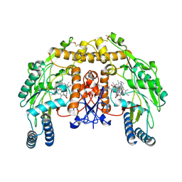
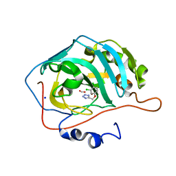 | | human carbonic anhydrase II in complex with ligand | | 分子名称: | 5-[[3,4-bis(chloranyl)phenoxy]methyl]-1~{H}-1,2,3,4-tetrazole, Carbonic anhydrase 2, FORMIC ACID, ... | | 著者 | Ren, B. | | 登録日 | 2015-10-28 | | 公開日 | 2016-03-09 | | 最終更新日 | 2024-03-06 | | 実験手法 | X-RAY DIFFRACTION (1.425 Å) | | 主引用文献 | Native State Mass Spectrometry, Surface Plasmon Resonance, and X-ray Crystallography Correlate Strongly as a Fragment Screening Combination.
J.Med.Chem., 59, 2016
|
|
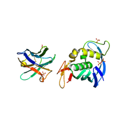
8BM2
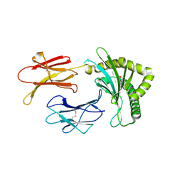
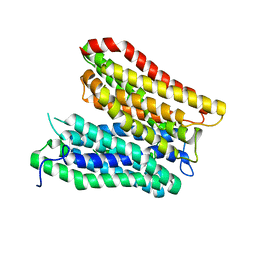
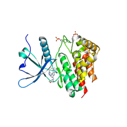 | | Crystal structure of JAK2 JH1 in complex with gandotinib | | 分子名称: | 3-[(4-chloranyl-2-fluoranyl-phenyl)methyl]-2-methyl-~{N}-(5-methyl-1~{H}-pyrazol-3-yl)-8-(morpholin-4-ylmethyl)imidazo[1,2-b]pyridazin-6-amine, Tyrosine-protein kinase JAK2 | | 著者 | Miao, Y, Haikarainen, T. | | 登録日 | 2022-11-10 | | 公開日 | 2023-11-22 | | 最終更新日 | 2024-07-10 | | 実験手法 | X-RAY DIFFRACTION (1.5 Å) | | 主引用文献 | Functional and Structural Characterization of Clinical-Stage Janus Kinase 2 Inhibitors Identifies Determinants for Drug Selectivity.
J.Med.Chem., 67, 2024
|
|
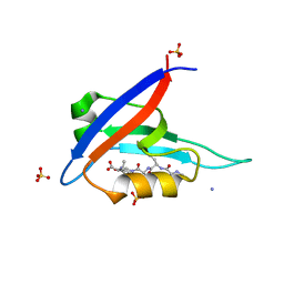
7PR4
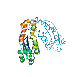
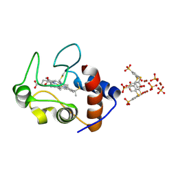 | | Cocrystal Form II of a cytochrome c, sulfonato-thiacalix[4]arene - zinc cluster | | 分子名称: | Cytochrome c iso-1, HEME C, PHOSPHATE ION, ... | | 著者 | Flood, R.J, Ramberg, K, Guagnini, F, Crowley, P.B. | | 登録日 | 2021-09-20 | | 公開日 | 2022-03-02 | | 最終更新日 | 2024-01-31 | | 実験手法 | X-RAY DIFFRACTION (1.32 Å) | | 主引用文献 | Protein Frameworks with Thiacalixarene and Zinc.
Cryst.Growth Des., 22, 2022
|
|
8BN2
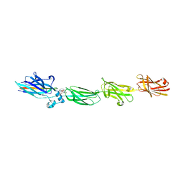
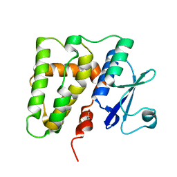
 | | Crystal structure of the ligand-binding domain (LBD) of human iGluR Delta-1 (GluD1) in complex with D-Serine | | 分子名称: | 1,2-ETHANEDIOL, 2-acetamido-2-deoxy-beta-D-glucopyranose, CALCIUM ION, ... | | 著者 | Heroven, C, Malinauskas, T, Aricescu, A.R. | | 登録日 | 2022-11-11 | | 公開日 | 2023-11-22 | | 最終更新日 | 2024-01-03 | | 実験手法 | X-RAY DIFFRACTION (1.63 Å) | | 主引用文献 | GluD1 binds GABA and controls inhibitory plasticity.
Science, 382, 2023
|
|
8K7O
 | |
7NFT
 | |
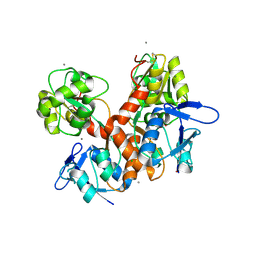
5EJ9
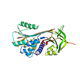
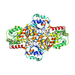 | | EcMenD-ThDP-Mn2+ complex soaked with 2-ketoglutarate for 2 min and isochorismate for 13 min | | 分子名称: | 2-succinyl-5-enolpyruvyl-6-hydroxy-3-cyclohexene-1-carboxylate synthase, FORMIC ACID, GLYCEROL, ... | | 著者 | Song, H.G, Dong, C, Chen, Y.Z, Sun, Y.R, Guo, Z.H. | | 登録日 | 2015-11-01 | | 公開日 | 2016-06-01 | | 最終更新日 | 2023-11-08 | | 実験手法 | X-RAY DIFFRACTION (1.72 Å) | | 主引用文献 | A Thiamine-Dependent Enzyme Utilizes an Active Tetrahedral Intermediate in Vitamin K Biosynthesis
J.Am.Chem.Soc., 138, 2016
|
|
8BP4
 | |
8BP6
 | | Structure of MHC-class I related molecule MR1 with bound M3Ade | | 分子名称: | (1R,5S)-8-(9H-purin-6-yl)-2-oxa-8-azabicyclo[3.3.1]nona-3,6-diene-4,6-dicarbaldehyde, Beta-2-microglobulin,Major histocompatibility complex class I-related gene protein | | 著者 | Berloffa, G, Jakob, R.P, Maier, T. | | 登録日 | 2022-11-16 | | 公開日 | 2023-11-29 | | 実験手法 | X-RAY DIFFRACTION (2.8 Å) | | 主引用文献 | The carbonyl nucleoside adduct M3Ade stabilizes MR1 and activates MR1-restricted self- and tumor-reactive T cells
To Be Published
|
|
7VCN
 | |
5EI0
 | | Structure of RCL-cleaved vaspin (serpinA12) | | 分子名称: | Serpin A12 | | 著者 | Pippel, J, Kuettner, B.E, Ulbricht, D, Daberger, J, Schultz, S, Heiker, J.T, Strater, N. | | 登録日 | 2015-10-29 | | 公開日 | 2015-11-11 | | 最終更新日 | 2024-01-10 | | 実験手法 | X-RAY DIFFRACTION (2.5 Å) | | 主引用文献 | Crystal structure of cleaved vaspin (serpinA12).
Biol.Chem., 397, 2016
|
|
8BTT
 | | Structure of human RTCB | | 分子名称: | MANGANESE (II) ION, RNA-splicing ligase RtcB homolog | | 著者 | Kopp, J, Gerber, J.L, Peschek, J. | | 登録日 | 2022-11-30 | | 公開日 | 2023-12-13 | | 最終更新日 | 2024-04-17 | | 実験手法 | X-RAY DIFFRACTION (2.6 Å) | | 主引用文献 | Structural and mechanistic insights into activation of the human RNA ligase RTCB by Archease.
Nat Commun, 15, 2024
|
|
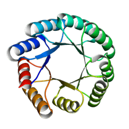
5VV7
 | | Structure of bovine endothelial nitric oxide synthase heme domain in complex with 7-(((3-(Pyridin-3-yl)propyl)amino)methyl)quinolin-2-amine | | 分子名称: | 5,6,7,8-TETRAHYDROBIOPTERIN, 7-(((3-(PYRIDIN-3-YL)PROPYL)AMINO)METHYL)QUINOLIN-2-, GLYCEROL, ... | | 著者 | Li, H, Poulos, T.L. | | 登録日 | 2017-05-19 | | 公開日 | 2017-08-16 | | 最終更新日 | 2023-10-04 | | 実験手法 | X-RAY DIFFRACTION (2.2 Å) | | 主引用文献 | Hydrophilic, Potent, and Selective 7-Substituted 2-Aminoquinolines as Improved Human Neuronal Nitric Oxide Synthase Inhibitors.
J. Med. Chem., 60, 2017
|
|
7Q0L
 | | Crystal structure of the peptide transporter YePEPT-K314A at 2.93 A | | 分子名称: | Peptide transporter YePEPT | | 著者 | Jeckelmann, J.M, Stauffer, M, Ilgue, H, Boggavarapu, R, Fotiadis, D. | | 登録日 | 2021-10-15 | | 公開日 | 2022-03-09 | | 最終更新日 | 2024-02-07 | | 実験手法 | X-RAY DIFFRACTION (2.71 Å) | | 主引用文献 | Peptide transporter structure reveals binding and action mechanism of a potent PEPT1 and PEPT2 inhibitor.
Commun Chem, 5, 2022
|
|
5KLE
 | | Structure of CBM_E1, a novel carbohydrate-binding module found by sugar cane soil metagenome, complexed with cellopentaose | | 分子名称: | Carbohydrate binding module E1, beta-D-glucopyranose-(1-4)-beta-D-glucopyranose-(1-4)-beta-D-glucopyranose-(1-4)-beta-D-glucopyranose-(1-4)-beta-D-glucopyranose | | 著者 | Liberato, M.V, Campos, B.M, Zeri, A.C.M, Squina, F.M. | | 登録日 | 2016-06-24 | | 公開日 | 2016-09-21 | | 最終更新日 | 2023-09-27 | | 実験手法 | X-RAY DIFFRACTION (1.5 Å) | | 主引用文献 | A Novel Carbohydrate-binding Module from Sugar Cane Soil Metagenome Featuring Unique Structural and Carbohydrate Affinity Properties.
J.Biol.Chem., 291, 2016
|
|
7VH4
 | |
5EL8
 | |
8BV7
 | |
6LLB
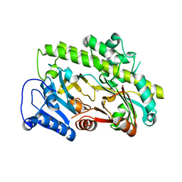
 | | Crystal structure of mpy-RNase J (mutant S247A), an archaeal RNase J from Methanolobus psychrophilus R15, in complex with 6 nt RNA | | 分子名称: | MPY-RNase J, RNA (5'-R(P*AP*AP*AP*AP*AP*A)-3'), SULFATE ION, ... | | 著者 | Li, D.F, Hou, Y.J, Guo, L. | | 登録日 | 2019-12-22 | | 公開日 | 2020-01-01 | | 最終更新日 | 2023-11-22 | | 実験手法 | X-RAY DIFFRACTION (2.6 Å) | | 主引用文献 | A newly identified duplex RNA unwinding activity of archaeal RNase J depends on processive exoribonucleolysis coupled steric occlusion by its structural archaeal loops.
Rna Biol., 17, 2020
|
|
7VEO
 | | Crystal structure of juvenile hormone acid methyltransferase silkworm JHAMT isoform3 complex with S-Adenosyl-L-homocysteine | | 分子名称: | Methyltranfer_dom domain-containing protein, S-ADENOSYL-L-HOMOCYSTEINE | | 著者 | Guo, P.C, Zhang, Y.S, Zhang, l, Xu, H.Y. | | 登録日 | 2021-09-09 | | 公開日 | 2022-09-28 | | 最終更新日 | 2023-11-29 | | 実験手法 | X-RAY DIFFRACTION (2.53 Å) | | 主引用文献 | Structural characterization and functional analysis of juvenile hormone acid methyltransferase JHAMT3 from the silkworm, Bombyx mori.
Insect Biochem.Mol.Biol., 151, 2022
|
|
7PR3
 | | Cocrystal Form I of a cytochrome c, sulfonato-thiacalix[4]arene - zinc cluster | | 分子名称: | Cytochrome c iso-1, HEME C, PHOSPHATE ION, ... | | 著者 | Flood, R.J, Ramberg, K, Guagnini, F, Crowley, P.B. | | 登録日 | 2021-09-20 | | 公開日 | 2022-03-02 | | 最終更新日 | 2024-01-31 | | 実験手法 | X-RAY DIFFRACTION (2.37 Å) | | 主引用文献 | Protein Frameworks with Thiacalixarene and Zinc.
Cryst.Growth Des., 22, 2022
|
|
8K56
 | |
8BRF
 | |
7Q0M
 | | Crystal structure of the peptide transporter YePEPT-K314A in complex with LZNV at 2.66 A | | 分子名称: | (2~{S})-2-[[(2~{S})-2-azanyl-6-[(4-nitrophenyl)methoxycarbonylamino]hexanoyl]amino]-3-methyl-butanoic acid, Peptide transporter YePEPT, UNDECYL-MALTOSIDE | | 著者 | Jeckelmann, J.M, Stauffer, M, Ilgue, H, Fotiadis, D. | | 登録日 | 2021-10-15 | | 公開日 | 2022-03-09 | | 最終更新日 | 2024-02-07 | | 実験手法 | X-RAY DIFFRACTION (2.54 Å) | | 主引用文献 | Peptide transporter structure reveals binding and action mechanism of a potent PEPT1 and PEPT2 inhibitor.
Commun Chem, 5, 2022
|
|
5VUR
 | | Structure of rat neuronal nitric oxide synthase heme domain in complex with 4-(2-(((2-Aminoquinolin-7-yl)methyl)amino)ethyl)-2-methylbenzonitrile | | 分子名称: | 4-(2-{[(2-aminoquinolin-7-yl)methyl]amino}ethyl)-2-methylbenzonitrile, 5,6,7,8-TETRAHYDROBIOPTERIN, ACETATE ION, ... | | 著者 | Li, H, Poulos, T.L. | | 登録日 | 2017-05-19 | | 公開日 | 2017-08-16 | | 最終更新日 | 2023-10-04 | | 実験手法 | X-RAY DIFFRACTION (1.97 Å) | | 主引用文献 | Hydrophilic, Potent, and Selective 7-Substituted 2-Aminoquinolines as Improved Human Neuronal Nitric Oxide Synthase Inhibitors.
J. Med. Chem., 60, 2017
|
|


