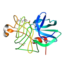
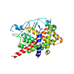
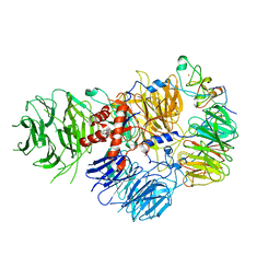
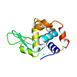
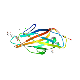
6WTS
 
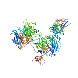 | | CryoEM structure of the C. sordellii lethal toxin TcsL in complex with SEMA6A | | 分子名称: | 2-acetamido-2-deoxy-beta-D-glucopyranose, 2-acetamido-2-deoxy-beta-D-glucopyranose-(1-4)-2-acetamido-2-deoxy-beta-D-glucopyranose, SEMA6A, ... | | 著者 | Kucharska, I, Rubinstein, J.L, Julien, J.P. | | 登録日 | 2020-05-03 | | 公開日 | 2020-07-08 | | 最終更新日 | 2020-08-05 | | 実験手法 | ELECTRON MICROSCOPY (3.3 Å) | | 主引用文献 | Recognition of Semaphorin Proteins by P. sordellii Lethal Toxin Reveals Principles of Receptor Specificity in Clostridial Toxins.
Cell, 182, 2020
|
|
6DBN
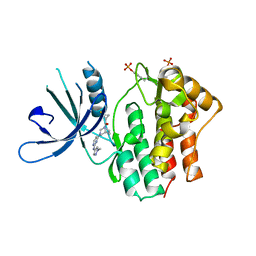 | | Jak1 with compound 23 | | 分子名称: | Tyrosine-protein kinase JAK1, [(1S)-2,2-difluorocyclopropyl][(1R,5S)-3-{2-[(1-methyl-1H-pyrazol-4-yl)amino]pyrimidin-4-yl}-3,8-diazabicyclo[3.2.1]octan-8-yl]methanone | | 著者 | Vajdos, F.F. | | 登録日 | 2018-05-03 | | 公開日 | 2018-08-29 | | 最終更新日 | 2018-11-07 | | 実験手法 | X-RAY DIFFRACTION (2.48 Å) | | 主引用文献 | Dual Inhibition of TYK2 and JAK1 for the Treatment of Autoimmune Diseases: Discovery of (( S)-2,2-Difluorocyclopropyl)((1 R,5 S)-3-(2-((1-methyl-1 H-pyrazol-4-yl)amino)pyrimidin-4-yl)-3,8-diazabicyclo[3.2.1]octan-8-yl)methanone (PF-06700841).
J. Med. Chem., 61, 2018
|
|
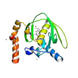
6DLS
 | |
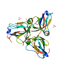
6QZ3
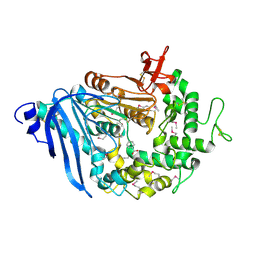 | | Structure of MHETase from Ideonella sakaiensis | | 分子名称: | BENZOIC ACID, CALCIUM ION, Mono(2-hydroxyethyl) terephthalate hydrolase | | 著者 | Allen, M.D, Johnson, C.W, Knott, B.C, Beckham, G.T, McGeehan, J.E. | | 登録日 | 2019-03-11 | | 公開日 | 2020-09-30 | | 最終更新日 | 2020-10-21 | | 実験手法 | X-RAY DIFFRACTION (1.6 Å) | | 主引用文献 | Characterization and engineering of a two-enzyme system for plastics depolymerization.
Proc.Natl.Acad.Sci.USA, 117, 2020
|
|
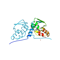
3LPR
 | |
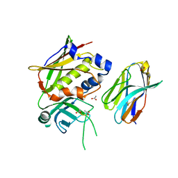
3M0X
 | | Crystal structure of Pseudomonas stutzeri L-rhamnose isomerase mutant S329L in complex with D-psicose | | 分子名称: | D-psicose, L-rhamnose isomerase, MANGANESE (II) ION | | 著者 | Yoshida, H, Takeda, K, Izumori, K, Kamitori, S. | | 登録日 | 2010-03-03 | | 公開日 | 2010-11-10 | | 最終更新日 | 2023-11-01 | | 実験手法 | X-RAY DIFFRACTION (1.79 Å) | | 主引用文献 | Elucidation of the role of Ser329 and the C-terminal region in the catalytic activity of Pseudomonas stutzeri L-rhamnose isomerase
Protein Eng.Des.Sel., 23, 2010
|
|
3M19
 | |
3M1J
 | |
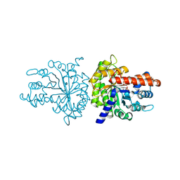
6DHM
 | | Bovine glutamate dehydrogenase complexed with zinc | | 分子名称: | GLUTAMIC ACID, GUANOSINE-5'-TRIPHOSPHATE, Glutamate dehydrogenase 1, ... | | 著者 | Smith, T.J. | | 登録日 | 2018-05-20 | | 公開日 | 2018-07-25 | | 最終更新日 | 2024-03-13 | | 実験手法 | X-RAY DIFFRACTION (3 Å) | | 主引用文献 | A novel mechanism of V-type zinc inhibition of glutamate dehydrogenase results from disruption of subunit interactions necessary for efficient catalysis.
FEBS J., 278, 2011
|
|
3M31
 | |
3M6Q
 | |
6DWU
 | |
3M4T
 | |
6QPK
 | |
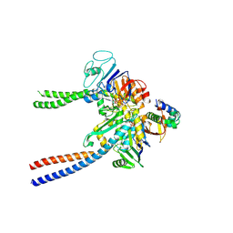
6DNH
 | | Cryo-EM structure of human CPSF-160-WDR33-CPSF-30-PAS RNA complex at 3.4 A resolution | | 分子名称: | Cleavage and polyadenylation specificity factor subunit 1, Cleavage and polyadenylation specificity factor subunit 4, RNA (5'-R(P*AP*AP*UP*AP*AP*AP*C)-3'), ... | | 著者 | Sun, Y, Zhang, Y, Hamilton, K, Walz, T, Tong, L. | | 登録日 | 2018-06-06 | | 公開日 | 2018-06-27 | | 最終更新日 | 2024-03-13 | | 実験手法 | ELECTRON MICROSCOPY (3.4 Å) | | 主引用文献 | Molecular basis for the recognition of the human AAUAAA polyadenylation signal.
Proc. Natl. Acad. Sci. U.S.A., 115, 2018
|
|
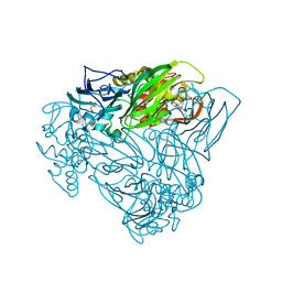
6QPW
 | | Structural basis of cohesin ring opening | | 分子名称: | MAGNESIUM ION, PHOSPHOTHIOPHOSPHORIC ACID-ADENYLATE ESTER, Sister chromatid cohesion protein 1, ... | | 著者 | Panne, D, Muir, K.W, Li, Y, Weis, F. | | 登録日 | 2019-02-15 | | 公開日 | 2020-02-05 | | 最終更新日 | 2020-03-18 | | 実験手法 | ELECTRON MICROSCOPY (3.3 Å) | | 主引用文献 | The structure of the cohesin ATPase elucidates the mechanism of SMC-kleisin ring opening.
Nat.Struct.Mol.Biol., 27, 2020
|
|
6QQ1
 | | Crystal structure of as isolated Y323F mutant of haem-Cu containing nitrite reductase from Ralstonia pickettii | | 分子名称: | COPPER (II) ION, Copper-containing nitrite reductase, GLYCEROL, ... | | 著者 | Antonyuk, S.V, Shenoy, R.T, Hedison, T.M, Eady, R.R, Hasnain, S.S, Scrutton, N.S. | | 登録日 | 2019-02-16 | | 公開日 | 2019-11-06 | | 最終更新日 | 2020-02-26 | | 実験手法 | X-RAY DIFFRACTION (1.75 Å) | | 主引用文献 | Unexpected Roles of a Tether Harboring a Tyrosine Gatekeeper Residue in Modular Nitrite Reductase Catalysis.
Acs Catalysis, 9, 2019
|
|
3MAL
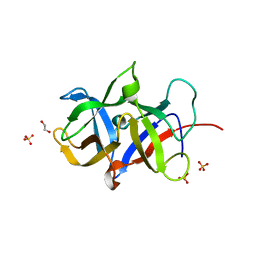 | | Crystal structure of the SDF2-like protein from Arabidopsis thaliana | | 分子名称: | 1,2-ETHANEDIOL, SULFATE ION, Stromal cell-derived factor 2-like protein | | 著者 | Ravaud, S, Radzimanowski, J, Sinning, I. | | 登録日 | 2010-03-24 | | 公開日 | 2010-04-07 | | 最終更新日 | 2023-09-06 | | 実験手法 | X-RAY DIFFRACTION (1.95 Å) | | 主引用文献 | Arabidopsis stromal-derived Factor2 (SDF2) is a crucial target of the unfolded protein response in the endoplasmic reticulum.
J.Biol.Chem., 285, 2010
|
|
6QQ9
 | |
6QQE
 | |
3MBQ
 | |
3MC0
 | | Crystal Structure of Staphylococcal Enterotoxin G (SEG) in Complex with a Mouse T-cell Receptor beta Chain | | 分子名称: | ACETATE ION, Enterotoxin SEG, variable beta 8.2 mouse T cell receptor | | 著者 | Fernandez, M.M, Cho, S, Robinson, H, Mariuzza, R.A, Malchiodi, E.L. | | 登録日 | 2010-03-26 | | 公開日 | 2010-10-13 | | 最終更新日 | 2023-09-06 | | 実験手法 | X-RAY DIFFRACTION (2 Å) | | 主引用文献 | Crystal structure of staphylococcal enterotoxin G (SEG) in complex with a mouse T-cell receptor {beta} chain.
J.Biol.Chem., 286, 2011
|
|
3MBF
 | |
3MCY
 | |
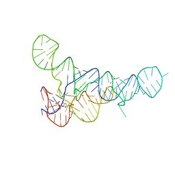
3MBU
 | | Structure of a bipyridine-modified PNA duplex | | 分子名称: | 1,2-ETHANEDIOL, Bipyridine-PNA, CARBONATE ION, ... | | 著者 | Yeh, J.I, Pohl, E, Truan, D, He, W, Sheldrick, G.M, Du, S, Achim, C. | | 登録日 | 2010-03-26 | | 公開日 | 2011-03-30 | | 最終更新日 | 2023-11-15 | | 実験手法 | X-RAY DIFFRACTION (1.05 Å) | | 主引用文献 | The crystal structure of non-modified and bipyridine-modified PNA duplexes.
Chemistry, 16, 2010
|
|


