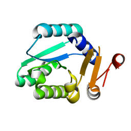
6KKN
 
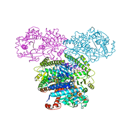
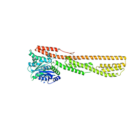
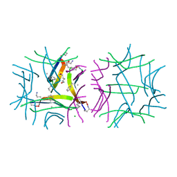
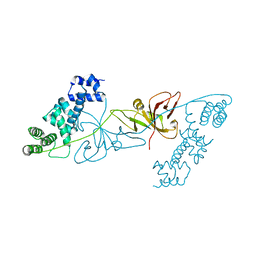 | | Crystal structure of RuBisCO accumulation factor Raf1 from Anabaena sp. PCC 7120 | | 分子名称: | All5250 protein | | 著者 | Xia, L.Y, Jiang, Y.L, Kong, W.W, Chen, Y, Zhou, C.Z. | | 登録日 | 2019-07-26 | | 公開日 | 2020-05-13 | | 最終更新日 | 2023-11-22 | | 実験手法 | X-RAY DIFFRACTION (2.85 Å) | | 主引用文献 | Molecular basis for the assembly of RuBisCO assisted by the chaperone Raf1.
Nat.Plants, 6, 2020
|
|
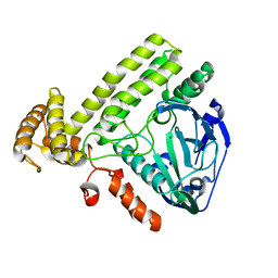
6KL1
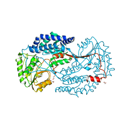
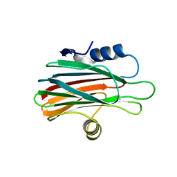
 | | Crystal structure of the S65T/F99S/M153T/V163A variant of non-deuterated GFP at pD 8.5 | | 分子名称: | Green fluorescent protein | | 著者 | Tai, Y, Takaba, K, Hanazono, Y, Dao, H.A, Miki, K, Takeda, K. | | 登録日 | 2019-07-28 | | 公開日 | 2019-12-11 | | 最終更新日 | 2023-11-22 | | 実験手法 | X-RAY DIFFRACTION (0.851 Å) | | 主引用文献 | X-ray crystallographic studies on the hydrogen isotope effects of green fluorescent protein at sub-angstrom resolutions
Acta Crystallogr.,Sect.D, 75, 2019
|
|
6K0Z
 | |
5SUH
 | |
6K19
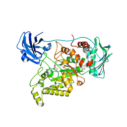
 | | Crystal structure of EXD2 exonuclease domain soaked in Mg | | 分子名称: | Exonuclease 3'-5' domain-containing protein 2, MAGNESIUM ION | | 著者 | Park, J, Lee, C. | | 登録日 | 2019-05-10 | | 公開日 | 2019-05-22 | | 最終更新日 | 2023-11-22 | | 実験手法 | X-RAY DIFFRACTION (2.202 Å) | | 主引用文献 | The structure of human EXD2 reveals a chimeric 3' to 5' exonuclease domain that discriminates substrates via metal coordination.
Nucleic Acids Res., 47, 2019
|
|
6K1I
 | | Human nucleosome core particle with gammaH2A.X variant | | 分子名称: | CHLORIDE ION, DNA (147-MER), Histone H2AX, ... | | 著者 | Sharma, D, De Falco, L, Davey, C.A. | | 登録日 | 2019-05-10 | | 公開日 | 2020-01-15 | | 最終更新日 | 2024-03-27 | | 実験手法 | X-RAY DIFFRACTION (2.75 Å) | | 主引用文献 | PARP1 exhibits enhanced association and catalytic efficiency with gamma H2A.X-nucleosome.
Nat Commun, 10, 2019
|
|
6JZU
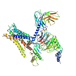
 | | The crystal structure of acyl-acyl carrier protein (acyl-ACP) reductase (AAR) in complex with aldehyde deformylating oxygenase (ADO) | | 分子名称: | Aldehyde decarbonylase, FE (II) ION, HEXADECAN-1-OL, ... | | 著者 | Zhang, H.M, Li, M, Gao, Y. | | 登録日 | 2019-05-03 | | 公開日 | 2020-04-01 | | 最終更新日 | 2024-10-09 | | 実験手法 | X-RAY DIFFRACTION (2.181 Å) | | 主引用文献 | Structural insights into catalytic mechanism and product delivery of cyanobacterial acyl-acyl carrier protein reductase.
Nat Commun, 11, 2020
|
|
6K02
 | | Crystal structure of ceNAP1 core | | 分子名称: | Nucleosome Assembly Protein, ZINC ION | | 著者 | Liu, Y.R. | | 登録日 | 2019-05-05 | | 公開日 | 2019-10-09 | | 最終更新日 | 2024-03-27 | | 実験手法 | X-RAY DIFFRACTION (2.162 Å) | | 主引用文献 | Crystal structure of xlH2A-H2B
Structure, 2019
|
|
6K0J
 | | The co-crystal structure of DYRK2 with a small molecule inhibitor | | 分子名称: | 3-(2,7-dimethoxyacridin-9-yl)sulfanylpropan-1-amine, Dual specificity tyrosine-phosphorylation-regulated kinase 2 | | 著者 | Tiantian, W, Xiao, J. | | 登録日 | 2019-05-06 | | 公開日 | 2019-11-20 | | 最終更新日 | 2019-12-18 | | 実験手法 | X-RAY DIFFRACTION (2.352 Å) | | 主引用文献 | Inhibition of dual-specificity tyrosine phosphorylation-regulated kinase 2 perturbs 26S proteasome-addicted neoplastic progression.
Proc.Natl.Acad.Sci.USA, 116, 2019
|
|
6K0P
 | | Catalytic domain of GH87 alpha-1,3-glucanase D1045A in complex with nigerose | | 分子名称: | ACETIC ACID, Alpha-1,3-glucanase, CALCIUM ION, ... | | 著者 | Itoh, T, Intuy, R, Suyotha, W, Hayashi, J, Yano, S, Makabe, K, Wakayama, M, Hibi, T. | | 登録日 | 2019-05-07 | | 公開日 | 2019-12-25 | | 最終更新日 | 2023-11-22 | | 実験手法 | X-RAY DIFFRACTION (1.424 Å) | | 主引用文献 | Structural insights into substrate recognition and catalysis by glycoside hydrolase family 87 alpha-1,3-glucanase from Paenibacillus glycanilyticus FH11.
Febs J., 287, 2020
|
|
6K1W
 | |
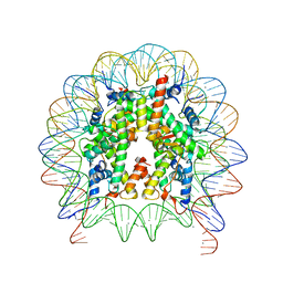
6K20
 | | Structure of Apo YdiU | | 分子名称: | MAGNESIUM ION, Protein adenylyltransferase SelO | | 著者 | Li, B, Yang, Y, Ma, Y. | | 登録日 | 2019-05-13 | | 公開日 | 2020-05-13 | | 最終更新日 | 2024-03-27 | | 実験手法 | X-RAY DIFFRACTION (2.58 Å) | | 主引用文献 | Structure of Apo-YdiU
To Be Published
|
|
6K12
 | |
7W5Z
 | | Cryo-EM structure of Tetrahymena thermophila mitochondrial complex IV, composite dimer model | | 分子名称: | 1,2-DIACYL-SN-GLYCERO-3-PHOSPHOCHOLINE, 2-oxoglutarate/malate carrier protein, CARDIOLIPIN, ... | | 著者 | Zhou, L, Maldonado, M, Padavannil, A, Letts, J. | | 登録日 | 2021-11-30 | | 公開日 | 2022-04-06 | | 最終更新日 | 2022-06-01 | | 実験手法 | ELECTRON MICROSCOPY (3.02 Å) | | 主引用文献 | Structures of Tetrahymena 's respiratory chain reveal the diversity of eukaryotic core metabolism.
Science, 376, 2022
|
|
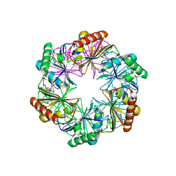
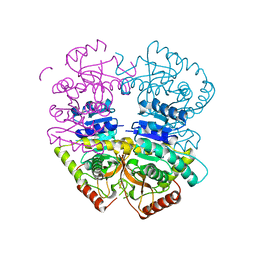
5SUL
 | | Inhibited state structure of yGsy2p | | 分子名称: | Glycogen [starch] synthase isoform 2, URIDINE-5'-DIPHOSPHATE, URIDINE-5'-MONOPHOSPHATE | | 著者 | Mahalingan, K.K, Hurley, T.D. | | 登録日 | 2016-08-03 | | 公開日 | 2017-06-14 | | 最終更新日 | 2023-10-04 | | 実験手法 | X-RAY DIFFRACTION (3.3 Å) | | 主引用文献 | Redox Switch for the Inhibited State of Yeast Glycogen Synthase Mimics Regulation by Phosphorylation.
Biochemistry, 56, 2017
|
|
6K1Z
 | | Crystal structure of farnesylated hGBP1 | | 分子名称: | FARNESYL, Guanylate-binding protein 1 | | 著者 | Du, S, Xiao, J.Y. | | 登録日 | 2019-05-13 | | 公開日 | 2019-06-12 | | 最終更新日 | 2019-12-25 | | 実験手法 | X-RAY DIFFRACTION (2.307 Å) | | 主引用文献 | Structural mechanism for guanylate-binding proteins (GBPs) targeting by the Shigella E3 ligase IpaH9.8.
Plos Pathog., 15, 2019
|
|
6K2G
 | |
6K2J
 | |
6K2Y
 | | Human Galectin-14 | | 分子名称: | Placental protein 13-like | | 著者 | Su, J. | | 登録日 | 2019-05-15 | | 公開日 | 2020-06-17 | | 最終更新日 | 2021-03-10 | | 実験手法 | X-RAY DIFFRACTION (1.57 Å) | | 主引用文献 | Structure-function studies of galectin-14, an important effector molecule in embryology.
Febs J., 288, 2021
|
|
6K35
 | | Crystal structure of GH20 exo beta-N-acetylglucosaminidase from Vibrio harveyi in complex with NAG-thiazoline | | 分子名称: | 3AR,5R,6S,7R,7AR-5-HYDROXYMETHYL-2-METHYL-5,6,7,7A-TETRAHYDRO-3AH-PYRANO[3,2-D]THIAZOLE-6,7-DIOL, Beta-N-acetylglucosaminidase Nag2 | | 著者 | Meekrathok, P, Stubbs, K.A, Bulmer, D.M, van den Berg, B, Suginta, W. | | 登録日 | 2019-05-16 | | 公開日 | 2020-05-13 | | 最終更新日 | 2023-11-22 | | 実験手法 | X-RAY DIFFRACTION (2.36 Å) | | 主引用文献 | NAG-thiazoline is a potent inhibitor of the Vibrio campbellii GH20 beta-N-Acetylglucosaminidase.
Febs J., 287, 2020
|
|
6K42
 | | cryo-EM structure of alpha2BAR-Gi1 complex | | 分子名称: | 4-[(1~{S})-1-(2,3-dimethylphenyl)ethyl]-1~{H}-imidazole, Alpha-2A adrenergic receptor,Endolysin,Alpha-2B adrenergic receptor,Alpha-2B adrenergic receptor, Guanine nucleotide-binding protein G(I)/G(S)/G(O) subunit gamma-2, ... | | 著者 | Yuan, D, Liu, Z, Wang, H.W, Kobilka, B.K. | | 登録日 | 2019-05-23 | | 公開日 | 2020-04-15 | | 最終更新日 | 2020-05-13 | | 実験手法 | ELECTRON MICROSCOPY (4.1 Å) | | 主引用文献 | Activation of the alpha2Badrenoceptor by the sedative sympatholytic dexmedetomidine.
Nat.Chem.Biol., 16, 2020
|
|
5SUR
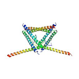
 | | X-ray crystallographic structure of a covalent trimer derived from A-beta 17_36. Synchrotron data set. (ORN)CVF(MEA)CED(ORN)AIIGL(ORN)V. | | 分子名称: | 16mer A-beta peptide: ORN-CYS-VAL-PHE-MEA-CYS-GLU-ASP-ORN-ALA-ILE-ILE-GLY-LEU-ORN-VAL, CHLORIDE ION, HEXANE-1,6-DIOL, ... | | 著者 | Kreutzer, A.G, Yoo, S, Nowick, J.S. | | 登録日 | 2016-08-03 | | 公開日 | 2017-01-11 | | 最終更新日 | 2024-03-27 | | 実験手法 | X-RAY DIFFRACTION (1.801 Å) | | 主引用文献 | Stabilization, Assembly, and Toxicity of Trimers Derived from A beta.
J.Am.Chem.Soc., 139, 2017
|
|
6K2S
 | |
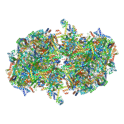
6K33
 | | Structure of PSI-isiA supercomplex from Thermosynechococcus vulcanus | | 分子名称: | 1,2-DIPALMITOYL-PHOSPHATIDYL-GLYCEROLE, 1,2-DISTEAROYL-MONOGALACTOSYL-DIGLYCERIDE, BETA-CAROTENE, ... | | 著者 | Akita, F, Nagao, R, Kato, K, Shen, J.R, Miyazaki, N. | | 登録日 | 2019-05-16 | | 公開日 | 2020-05-20 | | 最終更新日 | 2024-10-16 | | 実験手法 | ELECTRON MICROSCOPY (2.74 Å) | | 主引用文献 | Structure of a cyanobacterial photosystem I surrounded by octadecameric IsiA antenna proteins.
Commun Biol, 3, 2020
|
|
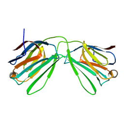
6K7U
 | | Crystal structure of beta-2 microglobulin (beta2m) of Bat (Pteropus Alecto) | | 分子名称: | Bat beta-2-microglobulin | | 著者 | Lu, D, Liu, K.F, Zhang, D, Yue, C, Lu, Q, Cheng, H, Chai, Y, Qi, J.X, Gao, F.G, Liu, W.J. | | 登録日 | 2019-06-08 | | 公開日 | 2019-09-18 | | 最終更新日 | 2019-12-04 | | 実験手法 | X-RAY DIFFRACTION (1.601 Å) | | 主引用文献 | Peptide presentation by bat MHC class I provides new insight into the antiviral immunity of bats.
Plos Biol., 17, 2019
|
|


