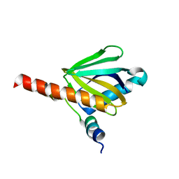
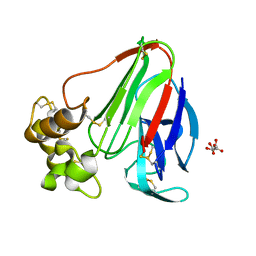
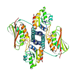
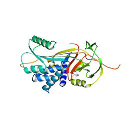
3E9D
 
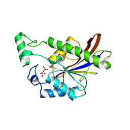 | | Structure of full-length TIGAR from Danio rerio | | 分子名称: | PHOSPHATE ION, POTASSIUM ION, Zgc:56074 | | 著者 | Li, H, Jogl, G. | | 登録日 | 2008-08-21 | | 公開日 | 2008-12-16 | | 最終更新日 | 2017-10-25 | | 実験手法 | X-RAY DIFFRACTION (2 Å) | | 主引用文献 | TIGAR (TP53 induced glycolysis and apoptosis regulator) is a fructose-2,6- and fructose-1,6-bisphosphatase
To be Published
|
|
3EEJ
 | |
3EF0
 | |
3EA7
 | |
3DXE
 | |
3DXL
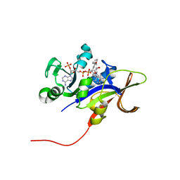
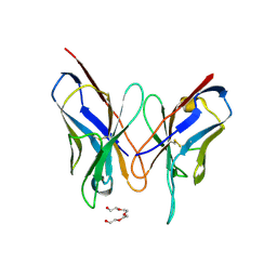
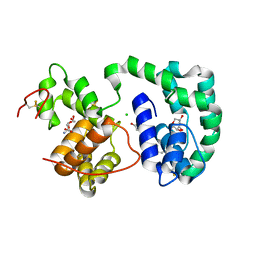 | | Crystal structure of AeD7 from Aedes Aegypti | | 分子名称: | 2-AMINO-2-HYDROXYMETHYL-PROPANE-1,3-DIOL, Allergen Aed a 2, CHLORIDE ION, ... | | 著者 | Andersen, J.F, Calvo, E, Mans, B.J, Ribeiro, J.M. | | 登録日 | 2008-07-24 | | 公開日 | 2009-02-03 | | 最終更新日 | 2021-03-31 | | 実験手法 | X-RAY DIFFRACTION (1.3 Å) | | 主引用文献 | Multifunctionality and mechanism of ligand binding in a mosquito antiinflammatory protein
Proc.Natl.Acad.Sci.USA, 106, 2009
|
|
3DY9
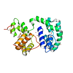 | | Crystal structure of AeD7 potassium bromide soak | | 分子名称: | 2-AMINO-2-HYDROXYMETHYL-PROPANE-1,3-DIOL, BROMIDE ION, D7 protein, ... | | 著者 | Andersen, J.F, Calvo, E, Mans, B.J, Ribeiro, J.M. | | 登録日 | 2008-07-25 | | 公開日 | 2009-02-03 | | 最終更新日 | 2021-03-31 | | 実験手法 | X-RAY DIFFRACTION (1.7 Å) | | 主引用文献 | Multifunctionality and mechanism of ligand binding in a mosquito antiinflammatory protein
Proc.Natl.Acad.Sci.USA, 106, 2009
|
|
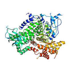
3DJ4
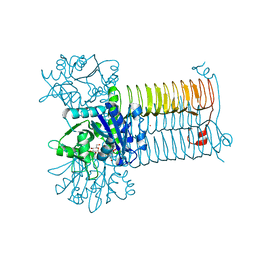 | | Crystal Structure of GlmU from Mycobacterium tuberculosis in complex with URIDINE-DIPHOSPHATE-N-ACETYLGLUCOSAMINE. | | 分子名称: | Bifunctional protein glmU, COBALT (II) ION, MAGNESIUM ION, ... | | 著者 | Verma, S.K, Prakash, B. | | 登録日 | 2008-06-22 | | 公開日 | 2009-05-19 | | 最終更新日 | 2024-03-20 | | 実験手法 | X-RAY DIFFRACTION (2.38 Å) | | 主引用文献 | PknB-mediated phosphorylation of a novel substrate, N-acetylglucosamine-1-phosphate uridyltransferase, modulates its acetyltransferase activity.
J.Mol.Biol., 386, 2009
|
|
3DZR
 | |
3DKC
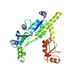
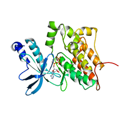 | | Structure of MET receptor tyrosine kinase in complex with ATP | | 分子名称: | ADENOSINE-5'-TRIPHOSPHATE, CHLORIDE ION, Hepatocyte growth factor receptor, ... | | 著者 | Hendle, J. | | 登録日 | 2008-06-24 | | 公開日 | 2009-07-07 | | 最終更新日 | 2024-02-21 | | 実験手法 | X-RAY DIFFRACTION (1.52 Å) | | 主引用文献 | SGX523 is an exquisitely selective, ATP-competitive inhibitor of the MET receptor tyrosine kinase with antitumor activity in vivo.
Mol.Cancer Ther., 8, 2009
|
|
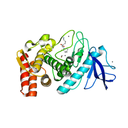
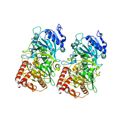
3DL0
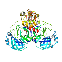
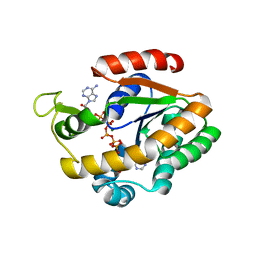 | | Crystal structure of adenylate kinase variant AKlse3 | | 分子名称: | Adenylate kinase, BIS(ADENOSINE)-5'-PENTAPHOSPHATE, MAGNESIUM ION, ... | | 著者 | Bannen, R.M, Bianchetti, C.M, Bingman, C.A, McCoy, J.G. | | 登録日 | 2008-06-26 | | 公開日 | 2009-06-09 | | 最終更新日 | 2023-08-30 | | 実験手法 | X-RAY DIFFRACTION (1.58 Å) | | 主引用文献 | Effectiveness and limitations of local structural entropy optimization in the thermal stabilization of mesophilic and thermophilic adenylate kinases.
Proteins, 82, 2014
|
|
3E1G
 | |
3DGE
 | |
3DO2
 | |
3DOZ
 | | Crystal structure of (3R)-Hydroxyacyl-Acyl Carrier Protein Dehydratase (FabZ) from Helicobacter pylori in complex with compound 3k | | 分子名称: | (3R)-hydroxymyristoyl-acyl carrier protein dehydratase, 3-bromo-N'-[(1E)-(3,5-dibromo-2,4-dihydroxyphenyl)methylidene]benzohydrazide, BENZAMIDINE, ... | | 著者 | Zhang, L, He, L, Liu, X, Liu, H, Shen, X, Jiang, H. | | 登録日 | 2008-07-07 | | 公開日 | 2009-05-05 | | 最終更新日 | 2023-11-01 | | 実験手法 | X-RAY DIFFRACTION (2.5 Å) | | 主引用文献 | Discovering potent inhibitors against the beta-hydroxyacyl-acyl carrier protein dehydratase (FabZ) of Helicobacter pylori: structure-based design, synthesis, bioassay, and crystal structure determination.
J.Med.Chem., 52, 2009
|
|
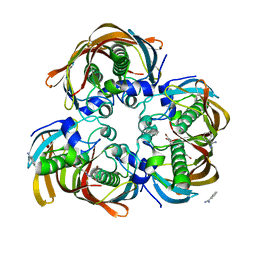
3DQ0
 | | Maize cytokinin oxidase/dehydrogenase complexed with N6-(3-methoxy-phenyl)adenine | | 分子名称: | 2-acetamido-2-deoxy-beta-D-glucopyranose, 2-acetamido-2-deoxy-beta-D-glucopyranose-(1-4)-2-acetamido-2-deoxy-beta-D-glucopyranose, Cytokinin dehydrogenase 1, ... | | 著者 | Kopecny, D, Briozzo, P. | | 登録日 | 2008-07-09 | | 公開日 | 2009-07-14 | | 最終更新日 | 2023-11-01 | | 実験手法 | X-RAY DIFFRACTION (1.9 Å) | | 主引用文献 | Characterization and biological activity of novel purine-derived inhibitor of cytokinin oxidase/dehydrogenase and its potential use for in vivo studies
To be Published
|
|
3DQJ
 | |
3DRU
 | | Crystal Structure of Gly117Phe Alpha1-Antitrypsin | | 分子名称: | Alpha-1-antitrypsin | | 著者 | Gooptu, B, Nobeli, I, Purkiss, A, Phillips, R.L, Mallya, M, Lomas, D.A, Barrett, T.E. | | 登録日 | 2008-07-11 | | 公開日 | 2009-03-31 | | 最終更新日 | 2024-02-21 | | 実験手法 | X-RAY DIFFRACTION (3.2 Å) | | 主引用文献 | Crystallographic and cellular characterisation of two mechanisms stabilising the native fold of alpha1-antitrypsin: implications for disease and drug design.
J.Mol.Biol., 387, 2009
|
|
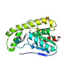
3DPD
 | | Achieving multi-isoform PI3K inhibition in a series of substituted 3,4-Dihydro-2H-benzo[1,4]oxazines | | 分子名称: | 5,5-dimethyl-2-morpholin-4-yl-5,6-dihydro-1,3-benzothiazol-7(4H)-one, Phosphatidylinositol-4,5-bisphosphate 3-kinase catalytic subunit gamma isoform | | 著者 | Ceska, T.A. | | 登録日 | 2008-07-08 | | 公開日 | 2008-08-26 | | 最終更新日 | 2023-11-01 | | 実験手法 | X-RAY DIFFRACTION (2.85 Å) | | 主引用文献 | Achieving multi-isoform PI3K inhibition in a series of substituted 3,4-dihydro-2H-benzo[1,4]oxazines
Bioorg.Med.Chem.Lett., 18, 2008
|
|
3DT4
 | |
3DUS
 | | Crystal structure of SAG506-01, orthorhombic, twinned, crystal 1 | | 分子名称: | 3-deoxy-alpha-D-manno-oct-2-ulopyranosonic acid, Ig-like protein, MAGNESIUM ION, ... | | 著者 | Brooks, C.L, Blackler, R.J, Gerstenbruch, S, Kosma, P, Muller-Loennies, S, Brade, H, Evans, S.V. | | 登録日 | 2008-07-17 | | 公開日 | 2008-12-02 | | 最終更新日 | 2020-07-29 | | 実験手法 | X-RAY DIFFRACTION (1.95 Å) | | 主引用文献 | Pseudo-symmetry and twinning in crystals of homologous antibody Fv fragments.
Acta Crystallogr.,Sect.D, 64, 2008
|
|
3DQI
 | |
3DWS
 | |
3DQU
 | |
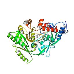
3DX3
 | | Golgi alpha-Mannosidase II in complex with Mannostatin analog (1R,2R,3S,4R,5R)-5-aminocyclopentane-1,2,3,4-tetraol | | 分子名称: | (1R,2R,3S,4R,5R)-5-aminocyclopentane-1,2,3,4-tetrol, (4R)-2-METHYLPENTANE-2,4-DIOL, 2-acetamido-2-deoxy-beta-D-glucopyranose, ... | | 著者 | Kuntz, D.A, Rose, D.R. | | 登録日 | 2008-07-23 | | 公開日 | 2009-07-07 | | 最終更新日 | 2023-08-30 | | 実験手法 | X-RAY DIFFRACTION (1.42 Å) | | 主引用文献 | The molecular basis of inhibition of Golgi alpha-mannosidase II by mannostatin A.
Chembiochem, 10, 2009
|
|


