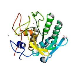
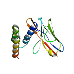
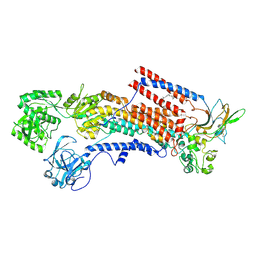
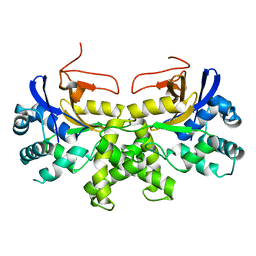
6K58
 
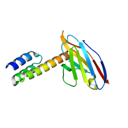
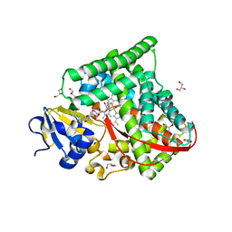 | | Structure of the CYP102A1 Haem Domain with N-Enanthyl-L-Prolyl-L-Phenylalanine | | 分子名称: | (2S)-2-[[(2S)-1-heptylpyrrolidin-2-yl]carbonylamino]-3-phenyl-propanoic acid, Bifunctional cytochrome P450/NADPH--P450 reductase, DIMETHYL SULFOXIDE, ... | | 著者 | Stanfield, J.K, Kasai, C, Sugimoto, H, Shiro, Y, Watanabe, Y, Shoji, O. | | 登録日 | 2019-05-28 | | 公開日 | 2020-03-18 | | 最終更新日 | 2023-11-22 | | 実験手法 | X-RAY DIFFRACTION (1.41 Å) | | 主引用文献 | Crystals in Minutes: Instant On-Site Microcrystallisation of Various Flavours of the CYP102A1 (P450BM3) Haem Domain.
Angew.Chem.Int.Ed.Engl., 59, 2020
|
|
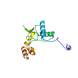
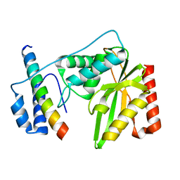
6K1O
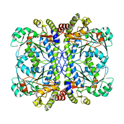 | | Apo form of a putative cystathionine gamma-lyase | | 分子名称: | Cystathionine gamma-lyase | | 著者 | Chen, S, Wang, Y. | | 登録日 | 2019-05-10 | | 公開日 | 2020-05-13 | | 最終更新日 | 2023-11-22 | | 実験手法 | X-RAY DIFFRACTION (2.033 Å) | | 主引用文献 | Structural characterization of cystathionine gamma-lyase smCSE enables aqueous metal quantum dot biosynthesis.
Int.J.Biol.Macromol., 174, 2021
|
|
6K26
 | |
6K2P
 | |
6K2W
 | |
6K36
 | |
6K5S
 | |
6K65
 | | Application of anti-helix antibodies in protein structure determination (9014-1P4B) | | 分子名称: | 1P4B variable heavy chain, 1P4B variable light chain, Immunoglobulin G-binding protein A | | 著者 | Lee, J.O, Jin, M.S, Kim, J.W, Kim, S, Lee, H, Cho, G.Y. | | 登録日 | 2019-06-01 | | 公開日 | 2019-08-14 | | 最終更新日 | 2024-10-16 | | 実験手法 | X-RAY DIFFRACTION (1.65 Å) | | 主引用文献 | Application of antihelix antibodies in protein structure determination.
Proc.Natl.Acad.Sci.USA, 116, 2019
|
|
6K6B
 | | Application of anti-helix antibodies in protein structure determination (8496-3LRH) | | 分子名称: | 3LRH intrabody, Protein A | | 著者 | Lee, J.O, Jin, M.S, Kim, J.W, Kim, S, Lee, H, Cho, G.Y. | | 登録日 | 2019-06-02 | | 公開日 | 2019-08-14 | | 最終更新日 | 2023-11-22 | | 実験手法 | X-RAY DIFFRACTION (2.06 Å) | | 主引用文献 | Application of antihelix antibodies in protein structure determination.
Proc.Natl.Acad.Sci.USA, 116, 2019
|
|
6K6N
 | | Crystal structure of SIVmac239 Nef protein | | 分子名称: | Protein Nef | | 著者 | Hirao, K, Andrews, S, Kuroki, K, Kusaka, H, Tadokoro, T, Kita, S, Ose, T, Rowland-Jones, S, Maenaka, K. | | 登録日 | 2019-06-04 | | 公開日 | 2020-03-25 | | 最終更新日 | 2023-11-22 | | 実験手法 | X-RAY DIFFRACTION (2.0002 Å) | | 主引用文献 | Structure of HIV-2 Nef Reveals Features Distinct from HIV-1 Involved in Immune Regulation.
Iscience, 23, 2020
|
|
3DBK
 | |
6K74
 | |
6K3M
 | | Application of anti-helix antibodies in protein structure determination (8189-3LRH) | | 分子名称: | 3LRH intrabody, SpA IgG-binding domain protein,Protein A | | 著者 | Lee, J.O, Jin, M.S, Kim, J.W, Kim, S, Lee, H, Cho, G.Y. | | 登録日 | 2019-05-20 | | 公開日 | 2019-08-14 | | 最終更新日 | 2023-11-22 | | 実験手法 | X-RAY DIFFRACTION (1.8 Å) | | 主引用文献 | Application of antihelix antibodies in protein structure determination.
Proc.Natl.Acad.Sci.USA, 116, 2019
|
|
6K3Z
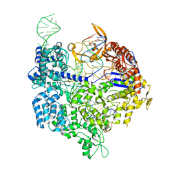 | | Crystal structure of dCas9 in complex with sgRNA and DNA (TGA PAM) | | 分子名称: | CRISPR-associated endonuclease Cas9, DNA (28-MER), DNA (5'-D(*AP*AP*AP*TP*GP*AP*TP*AP*TP*TP*G)-3'), ... | | 著者 | Chen, W, Zhang, H, Zhang, Y, Wang, Y, Gan, J, Ji, Q. | | 登録日 | 2019-05-22 | | 公開日 | 2019-09-25 | | 最終更新日 | 2023-11-22 | | 実験手法 | X-RAY DIFFRACTION (3.2 Å) | | 主引用文献 | Molecular basis for the PAM expansion and fidelity enhancement of an evolved Cas9 nuclease.
Plos Biol., 17, 2019
|
|
6K4P
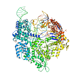 | | Crystal structure of xCas9 in complex with sgRNA and DNA (TGG PAM) | | 分子名称: | CRISPR-associated endonuclease Cas9/Csn1, DNA (28-MER), PHOSPHATE ION, ... | | 著者 | Chen, W, Zhang, H, Zhang, Y, Wang, Y, Gan, J, Ji, Q. | | 登録日 | 2019-05-25 | | 公開日 | 2019-09-25 | | 最終更新日 | 2023-11-22 | | 実験手法 | X-RAY DIFFRACTION (2.9 Å) | | 主引用文献 | Molecular basis for the PAM expansion and fidelity enhancement of an evolved Cas9 nuclease.
Plos Biol., 17, 2019
|
|
6K7C
 | | Dimeric Shewanella violacea cytochrome c5 | | 分子名称: | HEME C, NITRATE ION, Soluble cytochrome cA | | 著者 | Yang, H, Yamanaka, M, Nagao, S, Yasuhara, K, Shibata, N, Higuchi, Y, Hirota, S. | | 登録日 | 2019-06-07 | | 公開日 | 2019-09-04 | | 最終更新日 | 2023-11-22 | | 実験手法 | X-RAY DIFFRACTION (1.15 Å) | | 主引用文献 | Protein surface charge effect on 3D domain swapping in cells for c-type cytochromes.
Biochim Biophys Acta Proteins Proteom, 1867, 2019
|
|
6K7H
 | | Cryo-EM structure of the human P4-type flippase ATP8A1-CDC50 (E1 state class2) | | 分子名称: | 2-acetamido-2-deoxy-beta-D-glucopyranose, CHOLESTEROL HEMISUCCINATE, Cell cycle control protein 50A, ... | | 著者 | Hiraizumi, M, Yamashita, K, Nishizawa, T, Nureki, O. | | 登録日 | 2019-06-07 | | 公開日 | 2019-08-28 | | 最終更新日 | 2021-02-10 | | 実験手法 | ELECTRON MICROSCOPY (3.22 Å) | | 主引用文献 | Cryo-EM structures capture the transport cycle of the P4-ATPase flippase.
Science, 365, 2019
|
|
6K7T
 | | Crystal structure of bat (Pteropus Alecto) MHC class I Ptal-N*01:01 in complex with Hendra virus-derived peptide HeV1--human beta-2 microglobulin | | 分子名称: | Beta-2-microglobulin, HeV1, MHC class I antigen | | 著者 | Lu, D, Liu, K.F, Zhang, D, Yue, C, Lu, Q, Cheng, H, Chai, Y, Qi, J.X, Gao, F.G, Liu, W.J. | | 登録日 | 2019-06-08 | | 公開日 | 2019-11-06 | | 最終更新日 | 2023-11-22 | | 実験手法 | X-RAY DIFFRACTION (1.6 Å) | | 主引用文献 | Peptide presentation by bat MHC class I provides new insight into the antiviral immunity of bats.
Plos Biol., 17, 2019
|
|
6K5F
 | |
6K5T
 | |
6K6L
 | | YGL082W-catalytic domain | | 分子名称: | pseudo deubiquitinase | | 著者 | Lu, L.N, Wang, F. | | 登録日 | 2019-06-03 | | 公開日 | 2019-07-03 | | 最終更新日 | 2023-11-22 | | 実験手法 | X-RAY DIFFRACTION (1.77 Å) | | 主引用文献 | Inactivity of YGL082W in vitro due to impairment of conformational change in the catalytic center loop
Sci China Chem, 2019
|
|
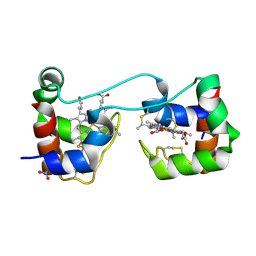
6K81
 | | Crystal structure of human VASH1-SVBP complex | | 分子名称: | Small vasohibin-binding protein, Tubulinyl-Tyr carboxypeptidase 1 | | 著者 | Liu, X, Wang, H, Zhang, Y, Feng, Y. | | 登録日 | 2019-06-11 | | 公開日 | 2020-02-19 | | 最終更新日 | 2024-03-27 | | 実験手法 | X-RAY DIFFRACTION (2.28 Å) | | 主引用文献 | Structural insights into tubulin detyrosination by vasohibins-SVBP complex.
Cell Discov, 5, 2019
|
|
4K5O
 | |
6K87
 | |
6K8G
 | | H/D exchanged Hen egg-white lysozyme | | 分子名称: | CHLORIDE ION, Lysozyme C, SODIUM ION | | 著者 | Kita, A, Morimoto, Y. | | 登録日 | 2019-06-11 | | 公開日 | 2020-06-17 | | 最終更新日 | 2024-04-03 | | 実験手法 | NEUTRON DIFFRACTION (2 Å), X-RAY DIFFRACTION | | 主引用文献 | Hydrogen/deuterium exchange behavior in tetragonal hen egg-white lysozyme crystals affected by solution state.
J.Appl.Crystallogr., 53, 2020
|
|


