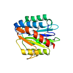
6L64
 
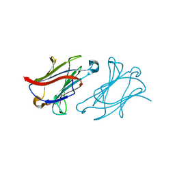 | | X-ray structure of human galectin-10 in complex with D-glucose | | 分子名称: | Galectin-10, beta-D-glucopyranose | | 著者 | Kamitori, S. | | 登録日 | 2019-10-28 | | 公開日 | 2020-03-04 | | 最終更新日 | 2023-11-22 | | 実験手法 | X-RAY DIFFRACTION (2.08 Å) | | 主引用文献 | Structures of human galectin-10/monosaccharide complexes demonstrate potential of monosaccharides as effectors in forming Charcot-Leyden crystals.
Biochem.Biophys.Res.Commun., 2020
|
|
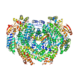
4GA4
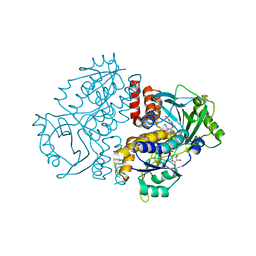
 | | Crystal structure of AMP phosphorylase N-terminal deletion mutant | | 分子名称: | PHOSPHATE ION, Putative thymidine phosphorylase | | 著者 | Nishitani, Y, Aono, R, Nakamura, A, Sato, T, Atomi, H, Imanaka, T, Miki, K. | | 登録日 | 2012-07-25 | | 公開日 | 2013-05-15 | | 最終更新日 | 2023-11-08 | | 実験手法 | X-RAY DIFFRACTION (3.51 Å) | | 主引用文献 | Structure analysis of archaeal AMP phosphorylase reveals two unique modes of dimerization
J.Mol.Biol., 425, 2013
|
|
6L6A
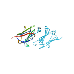 | | X-ray structure of human galectin-10 in complex with D-mannose | | 分子名称: | Galectin-10, beta-D-mannopyranose | | 著者 | Kamitori, S. | | 登録日 | 2019-10-28 | | 公開日 | 2020-03-04 | | 最終更新日 | 2023-11-22 | | 実験手法 | X-RAY DIFFRACTION (1.81 Å) | | 主引用文献 | Structures of human galectin-10/monosaccharide complexes demonstrate potential of monosaccharides as effectors in forming Charcot-Leyden crystals.
Biochem.Biophys.Res.Commun., 2020
|
|
4GAM
 | | Complex structure of Methane monooxygenase hydroxylase and regulatory subunit | | 分子名称: | FE (III) ION, Methane monooxygenase component A alpha chain, Methane monooxygenase component A beta chain, ... | | 著者 | Lee, S.J, Lippard, S.J, Cho, U.-S. | | 登録日 | 2012-07-25 | | 公開日 | 2013-02-06 | | 最終更新日 | 2023-09-13 | | 実験手法 | X-RAY DIFFRACTION (2.902 Å) | | 主引用文献 | Control of substrate access to the active site in methane monooxygenase.
Nature, 494, 2013
|
|
2G83
 | | Structure of activated G-alpha-i1 bound to a nucleotide-state-selective peptide: Minimal determinants for recognizing the active form of a G protein alpha subunit | | 分子名称: | GUANOSINE-5'-DIPHOSPHATE, Guanine nucleotide-binding protein G(i), alpha-1 subunit, ... | | 著者 | Johnston, C.A, Ramer, J.K, Blaesius, R, Kuhlman, B, Arshavsky, V.Y, Siderovski, D.P. | | 登録日 | 2006-03-01 | | 公開日 | 2006-10-10 | | 最終更新日 | 2023-08-30 | | 実験手法 | X-RAY DIFFRACTION (2.8 Å) | | 主引用文献 | Minimal Determinants for Binding Activated Galpha from the Structure of a Galpha(i1)-Peptide Dimer.
Biochemistry, 45, 2006
|
|
7Z97
 | | Crystal structure of the F191M variant of Variovorax paradoxus indole monooxygenase (VpIndA1) in complex with 6-bromoindole | | 分子名称: | 1,2-ETHANEDIOL, 6-bromo-1H-indole, FLAVIN-ADENINE DINUCLEOTIDE, ... | | 著者 | Kratky, J, Weisse, R, Strater, N. | | 登録日 | 2022-03-20 | | 公開日 | 2023-02-22 | | 最終更新日 | 2024-02-07 | | 実験手法 | X-RAY DIFFRACTION (1.46 Å) | | 主引用文献 | Structural and Mechanistic Studies on Substrate and Stereoselectivity of the Indole Monooxygenase VpIndA1: New Avenues for Biocatalytic Epoxidations and Sulfoxidations.
Angew.Chem.Int.Ed.Engl., 62, 2023
|
|
5HV4
 | |
4M9Y
 | | Crystal structure of CED-4 bound CED-3 fragment | | 分子名称: | ADENOSINE-5'-TRIPHOSPHATE, CED-3 fragment, Cell death protein 4, ... | | 著者 | Huang, W.J, Jinag, T.Y, Choi, W.Y, Wang, J.W, Shi, Y.G. | | 登録日 | 2013-08-15 | | 公開日 | 2013-10-23 | | 最終更新日 | 2023-12-06 | | 実験手法 | X-RAY DIFFRACTION (4.2 Å) | | 主引用文献 | Mechanistic insights into CED-4-mediated activation of CED-3.
Genes Dev., 27, 2013
|
|
5HVI
 | |
4HQL
 | | Crystal structure of magnesium-loaded Plasmodium vivax TRAP protein | | 分子名称: | CHLORIDE ION, MAGNESIUM ION, Sporozoite surface protein 2, ... | | 著者 | Song, G, Koksal, A.C, Lu, C, Springer, T.A. | | 登録日 | 2012-10-25 | | 公開日 | 2012-12-26 | | 最終更新日 | 2023-09-20 | | 実験手法 | X-RAY DIFFRACTION (2.241 Å) | | 主引用文献 | Shape change in the receptor for gliding motility in Plasmodium sporozoites.
Proc.Natl.Acad.Sci.USA, 109, 2012
|
|
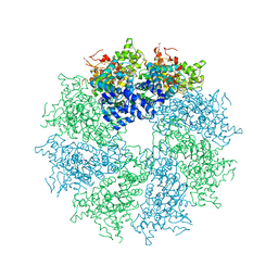
6L3H
 | | Cryo-EM structure of dimeric quinol dependent Nitric Oxide Reductase (qNOR) from the pathogen Neisseria meninigitidis | | 分子名称: | CALCIUM ION, FE (III) ION, Nitric-oxide reductase, ... | | 著者 | Jamali, M.M.A, Gopalasingam, C.C, Johnson, R.M, Tosha, T, Muench, S.P, Muramoto, K, Antonyuk, S.V, Shiro, Y, Hasnain, S.S. | | 登録日 | 2019-10-11 | | 公開日 | 2020-04-01 | | 最終更新日 | 2024-03-27 | | 実験手法 | ELECTRON MICROSCOPY (3.06 Å) | | 主引用文献 | The active form of quinol-dependent nitric oxide reductase fromNeisseria meningitidisis a dimer.
Iucrj, 7, 2020
|
|
4G3U
 | |
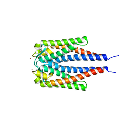
5HNW
 | | Structural basis of backwards motion in kinesin-14: minus-end directed nKn664 in the AMPPNP state | | 分子名称: | GUANOSINE-5'-DIPHOSPHATE, GUANOSINE-5'-TRIPHOSPHATE, MAGNESIUM ION, ... | | 著者 | Shigematsu, H, Yokoyama, T, Kikkawa, M, Shirouzu, M, Nitta, R. | | 登録日 | 2016-01-19 | | 公開日 | 2016-08-10 | | 最終更新日 | 2018-07-25 | | 実験手法 | ELECTRON MICROSCOPY (6.6 Å) | | 主引用文献 | Structural Basis of Backwards Motion in Kinesin-1-Kinesin-14 Chimera: Implication for Kinesin-14 Motility
Structure, 24, 2016
|
|
2G7I
 | |
4HS8
 | |
5HP4
 | | Crystal structure bacteriohage T5 D15 flap endonuclease (D155K) pseudo-enzyme-product complex with DNA and metal ions | | 分子名称: | CALCIUM ION, DNA (5'-D(*GP*AP*TP*CP*TP*AP*TP*AP*TP*GP*CP*CP*AP*TP*CP*GP*G)-3'), Exodeoxyribonuclease, ... | | 著者 | Almalki, F.A, Zhang, J, Sedelnikova, S.E, Rafferty, J.B, Sayers, J.R, Artymiuk, P.A. | | 登録日 | 2016-01-20 | | 公開日 | 2016-06-01 | | 最終更新日 | 2024-01-10 | | 実験手法 | X-RAY DIFFRACTION (1.86 Å) | | 主引用文献 | Direct observation of DNA threading in flap endonuclease complexes.
Nat.Struct.Mol.Biol., 23, 2016
|
|
7CXT
 | |
7Z5F
 | | VP2-only capsid of MVM D263A mutant | | 分子名称: | Capsid protein VP1 | | 著者 | Luque, D, Ortega-Esteban, A, Valbuena, A, Vilas, J.L, Rodriguez-Huete, A, Mateu, M.G, Caston, J.R. | | 登録日 | 2022-03-09 | | 公開日 | 2023-03-08 | | 最終更新日 | 2024-07-17 | | 実験手法 | ELECTRON MICROSCOPY (3.22 Å) | | 主引用文献 | Equilibrium Dynamics of a Biomolecular Complex Analyzed at Single-amino Acid Resolution by Cryo-electron Microscopy.
J.Mol.Biol., 435, 2023
|
|
6LJK
 | |
7Z5E
 | | VP2-only capsid of MVM D263A mutant | | 分子名称: | Capsid protein VP1 | | 著者 | Luque, D, Ortega-Esteban, A, Valbuena, A, Vilas, J.L, Rodriguez-Huete, A, Mateu, M.G, Caston, J.R. | | 登録日 | 2022-03-09 | | 公開日 | 2023-03-08 | | 最終更新日 | 2024-07-17 | | 実験手法 | ELECTRON MICROSCOPY (3.32 Å) | | 主引用文献 | Equilibrium Dynamics of a Biomolecular Complex Analyzed at Single-amino Acid Resolution by Cryo-electron Microscopy.
J.Mol.Biol., 435, 2023
|
|
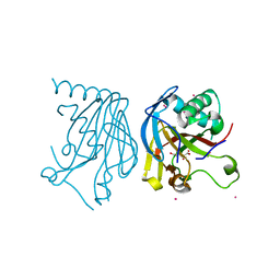
4G8V
 | | Crystal structure of Ribonuclease A in complex with 5a | | 分子名称: | 1-{[1-(alpha-L-arabinofuranosyl)-1H-1,2,3-triazol-4-yl]methyl}-2,4-dioxo-1,2,3,4-tetrahydropyrimidine, Ribonuclease pancreatic | | 著者 | Chatzileontiadou, D.S.M, Kantsadi, A.L, Leonidas, D.D. | | 登録日 | 2012-07-23 | | 公開日 | 2012-11-21 | | 最終更新日 | 2023-09-13 | | 実験手法 | X-RAY DIFFRACTION (1.7 Å) | | 主引用文献 | Triazole pyrimidine nucleosides as inhibitors of Ribonuclease A. Synthesis, biochemical, and structural evaluation.
Bioorg.Med.Chem., 20, 2012
|
|
4HQK
 | | Crystal structure of Plasmodium falciparum TRAP, P4212 form | | 分子名称: | SULFATE ION, Thrombospondin-related anonymous protein, TRAP | | 著者 | Song, G, Koksal, A.C, Lu, C, Springer, T.A. | | 登録日 | 2012-10-25 | | 公開日 | 2012-12-26 | | 最終更新日 | 2023-09-20 | | 実験手法 | X-RAY DIFFRACTION (2.249 Å) | | 主引用文献 | Shape change in the receptor for gliding motility in Plasmodium sporozoites.
Proc.Natl.Acad.Sci.USA, 109, 2012
|
|
2GA0
 | | Variable Small Protein 1 of Borrelia turicatae (VspA or Vsp1) | | 分子名称: | NICKEL (II) ION, surface protein VspA | | 著者 | Lawson, C.L, Yung, B.H, Barbour, A.G, Zuckert, W.R. | | 登録日 | 2006-03-07 | | 公開日 | 2006-06-27 | | 最終更新日 | 2023-08-30 | | 実験手法 | X-RAY DIFFRACTION (2.7 Å) | | 主引用文献 | Crystal structure of neurotropism-associated variable surface protein 1 (Vsp1) of Borrelia turicatae.
J.Bacteriol., 188, 2006
|
|
6LA0
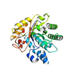 | | Crystal structure of AoRut | | 分子名称: | 2-acetamido-2-deoxy-beta-D-glucopyranose, Glycoside hydrolase family 5 | | 著者 | Koseki, T, Makabe, K. | | 登録日 | 2019-11-11 | | 公開日 | 2020-11-11 | | 最終更新日 | 2023-11-22 | | 実験手法 | X-RAY DIFFRACTION (1.75 Å) | | 主引用文献 | Aspergillus oryzae Rutinosidase: Biochemical and Structural Investigation.
Appl.Environ.Microbiol., 87, 2021
|
|
4HQX
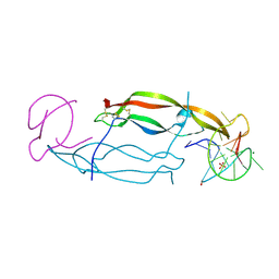 | | CRYSTAL STRUCTURE OF HUMAN PDGF-BB IN COMPLEX WITH A Modified nucleotide aptamer (SOMAmer SL4) | | 分子名称: | MAGNESIUM ION, Platelet-derived growth factor subunit B, SODIUM ION, ... | | 著者 | Davies, D.R, Edwards, T.E, Janjic, N, Gelinas, A.D, Zhang, C, Jarvis, T.C. | | 登録日 | 2012-10-26 | | 公開日 | 2012-11-21 | | 最終更新日 | 2023-09-20 | | 実験手法 | X-RAY DIFFRACTION (2.3 Å) | | 主引用文献 | Unique motifs and hydrophobic interactions shape the binding of modified DNA ligands to protein targets.
Proc.Natl.Acad.Sci.USA, 109, 2012
|
|


