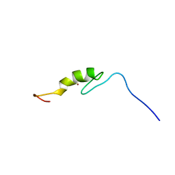
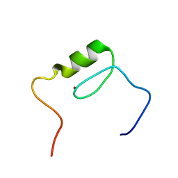
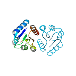
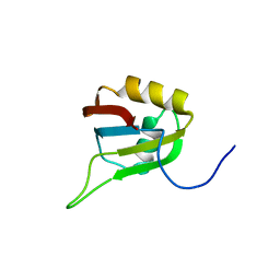
2EOR
 
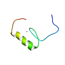 | | Solution structure of the C2H2 type zinc finger (region 255-287) of human Zinc finger protein 224 | | 分子名称: | ZINC ION, Zinc finger protein 224 | | 著者 | Tochio, N, Tomizawa, T, Abe, H, Saito, K, Li, H, Sato, M, Koshiba, S, Kobayashi, N, Kigawa, T, Yokoyama, S, RIKEN Structural Genomics/Proteomics Initiative (RSGI) | | 登録日 | 2007-03-29 | | 公開日 | 2007-10-02 | | 最終更新日 | 2024-05-29 | | 実験手法 | SOLUTION NMR | | 主引用文献 | Solution structure of the C2H2 type zinc finger (region 255-287) of human Zinc finger protein 224
To be Published
|
|
1Z5M
 | |
1Z8O
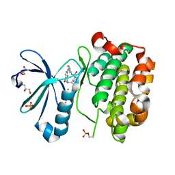
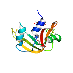
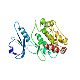
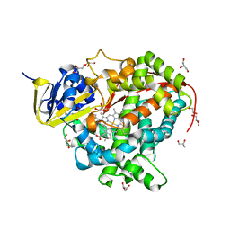
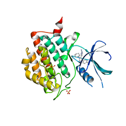
 | | Ferrous dioxygen complex of the wild-type cytochrome P450eryF | | 分子名称: | 6-DEOXYERYTHRONOLIDE B, 6-deoxyerythronolide B hydroxylase, OXYGEN MOLECULE, ... | | 著者 | Nagano, S, Cupp-Vickery, J.R, Poulos, T.L. | | 登録日 | 2005-03-31 | | 公開日 | 2005-04-12 | | 最終更新日 | 2023-08-23 | | 実験手法 | X-RAY DIFFRACTION (1.7 Å) | | 主引用文献 | Crystal structures of the ferrous dioxygen complex of wild-type cytochrome P450eryF and its mutants, A245S and A245T: investigation of the proton transfer system in P450eryF.
J.Biol.Chem., 280, 2005
|
|
2EL5
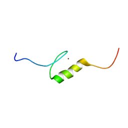 | |
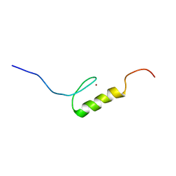
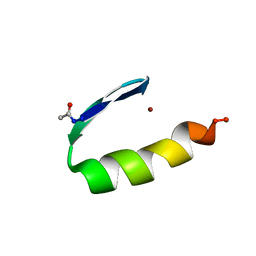
2EN4
 | | Solution structure of the C2H2 type zinc finger (region 284-316) of human Zinc finger protein 347 | | 分子名称: | ZINC ION, Zinc finger protein 347 | | 著者 | Tochio, N, Tomizawa, T, Abe, H, Saito, K, Li, H, Sato, M, Koshiba, S, Kobayashi, N, Kigawa, T, Yokoyama, S, RIKEN Structural Genomics/Proteomics Initiative (RSGI) | | 登録日 | 2007-03-28 | | 公開日 | 2007-10-02 | | 最終更新日 | 2024-05-29 | | 実験手法 | SOLUTION NMR | | 主引用文献 | Solution structure of the C2H2 type zinc finger (region 284-316) of human Zinc finger protein 347
To be Published
|
|
1Z6D
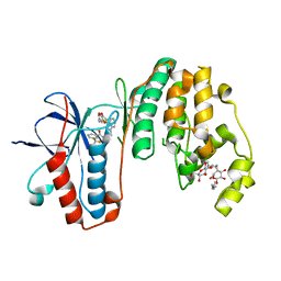
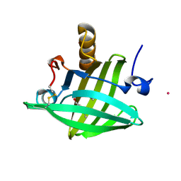
 | | Ribonuclease A- IMP complex | | 分子名称: | INOSINIC ACID, Ribonuclease pancreatic | | 著者 | Hatzopoulos, G.N, Leonidas, D.D, Kardakaris, R, Kobe, J, Oikonomakos, N.G. | | 登録日 | 2005-03-22 | | 公開日 | 2005-08-16 | | 最終更新日 | 2023-10-25 | | 実験手法 | X-RAY DIFFRACTION (1.54 Å) | | 主引用文献 | The binding of IMP to Ribonuclease A
Febs J., 272, 2005
|
|
2EOO
 | | Solution structure of the C2H2 type zinc finger (region 425-457) of human Zinc finger protein 95 homolog | | 分子名称: | ZINC ION, Zinc finger protein 95 homolog | | 著者 | Tochio, N, Tomizawa, T, Abe, H, Saito, K, Li, H, Sato, M, Koshiba, S, Kobayashi, N, Kigawa, T, Yokoyama, S, RIKEN Structural Genomics/Proteomics Initiative (RSGI) | | 登録日 | 2007-03-29 | | 公開日 | 2007-10-02 | | 最終更新日 | 2024-05-29 | | 実験手法 | SOLUTION NMR | | 主引用文献 | Solution structure of the C2H2 type zinc finger (region 425-457) of human Zinc finger protein 95 homolog
To be Published
|
|
3IW5
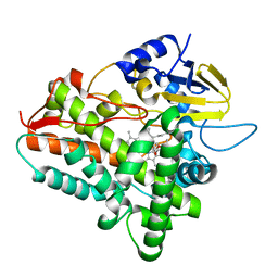
 | | Human p38 MAP Kinase in Complex with an Indole Derivative | | 分子名称: | Mitogen-activated protein kinase 14, N-[2-(3-{[2-(2,3-dihydro-1,4-benzodioxin-6-ylamino)-2-oxoethyl]sulfanyl}-1H-indol-1-yl)ethyl]-3-(trifluoromethyl)benzamide, octyl beta-D-glucopyranoside | | 著者 | Gruetter, C, Simard, J.R, Rauh, D. | | 登録日 | 2009-09-02 | | 公開日 | 2009-11-17 | | 最終更新日 | 2023-11-01 | | 実験手法 | X-RAY DIFFRACTION (2.5 Å) | | 主引用文献 | High-Throughput Screening To Identify Inhibitors Which Stabilize Inactive Kinase Conformations in p38alpha
J.Am.Chem.Soc., 131, 2009
|
|
2EP0
 | | Solution structure of the C2H2 type zinc finger (region 528-560) of human Zinc finger protein 28 homolog | | 分子名称: | ZINC ION, Zinc finger protein 28 homolog | | 著者 | Tochio, N, Tomizawa, T, Abe, H, Saito, K, Li, H, Sato, M, Koshiba, S, Kobayashi, N, Kigawa, T, Yokoyama, S, RIKEN Structural Genomics/Proteomics Initiative (RSGI) | | 登録日 | 2007-03-29 | | 公開日 | 2007-10-02 | | 最終更新日 | 2024-05-29 | | 実験手法 | SOLUTION NMR | | 主引用文献 | Solution structure of the C2H2 type zinc finger (region 528-560) of human Zinc finger protein 28 homolog
To be Published
|
|
1Z9X
 | | Human DRP-1 kinase, W305S S308A D40 mutant, crystal form with 3 monomers in the asymmetric unit | | 分子名称: | Death-associated protein kinase 2 | | 著者 | Kursula, P, Lehmann, F, Shani, G, Kimchi, A, Wilmanns, M. | | 登録日 | 2005-04-05 | | 公開日 | 2006-10-24 | | 最終更新日 | 2023-08-23 | | 実験手法 | X-RAY DIFFRACTION (3.93 Å) | | 主引用文献 | A structural insight into the double-locking mechanism of the human death-associated DRP-1 kinase
To be Published
|
|
1Z9P
 | |
1V2O
 | | Trypsin inhibitor in complex with bovine trypsin variant X(SSYI)bT.B4 | | 分子名称: | CALCIUM ION, METHYL N-[(4-METHYLPHENYL)SULFONYL]GLYCYL-3-[AMINO(IMINO)METHYL]-D-PHENYLALANINATE, SULFATE ION, ... | | 著者 | Rauh, D, Klebe, G, Stubbs, M.T. | | 登録日 | 2003-10-17 | | 公開日 | 2004-06-01 | | 最終更新日 | 2023-12-27 | | 実験手法 | X-RAY DIFFRACTION (1.62 Å) | | 主引用文献 | Understanding protein-ligand interactions: the price of protein flexibility
J.Mol.Biol., 335, 2004
|
|
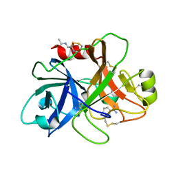
2E0J
 | | Mutant Human Ribonuclease 1 (R31L, R32L) | | 分子名称: | Ribonuclease | | 著者 | Yamada, H, Tamada, T, Kosaka, M, Kuroki, R. | | 登録日 | 2006-10-10 | | 公開日 | 2007-08-28 | | 最終更新日 | 2023-10-25 | | 実験手法 | X-RAY DIFFRACTION (1.6 Å) | | 主引用文献 | 'Crystal lattice engineering,' an approach to engineer protein crystal contacts by creating intermolecular symmetry: crystallization and structure determination of a mutant human RNase 1 with a hydrophobic interface of leucines
Protein Sci., 16, 2007
|
|
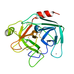
1ZES
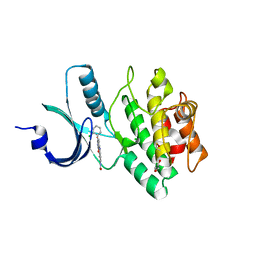
 | | BeF3- activated PhoB receiver domain | | 分子名称: | BERYLLIUM TRIFLUORIDE ION, MAGNESIUM ION, Phosphate regulon transcriptional regulatory protein phoB | | 著者 | Bachhawat, P, Montelione, G.T, Stock, A.M. | | 登録日 | 2005-04-19 | | 公開日 | 2005-09-20 | | 最終更新日 | 2024-02-14 | | 実験手法 | X-RAY DIFFRACTION (1.9 Å) | | 主引用文献 | Mechanism of Activation for Transcription Factor PhoB Suggested by Different Modes of Dimerization in the Inactive and Active States.
Structure, 13, 2005
|
|
1ZHM
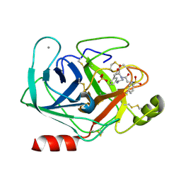
 | | Crystal Structure of the Catalytic Domain of the Coagulation Factor XIa in Complex with Benzamidine (S434A-T475A-K437 Mutant) | | 分子名称: | BENZAMIDINE, GLUTATHIONE, coagulation factor XI | | 著者 | Jin, L, Pandey, P, Babine, R.E, Weaver, D.T, Abdel-Meguid, S.S, Strickler, J.E. | | 登録日 | 2005-04-26 | | 公開日 | 2005-09-20 | | 最終更新日 | 2023-08-23 | | 実験手法 | X-RAY DIFFRACTION (1.96 Å) | | 主引用文献 | Mutation of surface residues to promote crystallization of activated factor XI as a complex with benzamidine: an essential step for the iterative structure-based design of factor XI inhibitors.
Acta Crystallogr.,Sect.D, 61, 2005
|
|
1UTM
 | | Trypsin specificity as elucidated by LIE calculations, X-ray structures and association constant measurements | | 分子名称: | 2-PHENYLETHYLAMINE, CALCIUM ION, TRYPSIN I | | 著者 | Leiros, H.-K.S, Brandsdal, B.O, Andersen, O.A, Os, V, Leiros, I, Helland, R, Otlewski, J, Willassen, N.P, Smalas, A.O. | | 登録日 | 2003-12-09 | | 公開日 | 2004-01-09 | | 最終更新日 | 2019-07-24 | | 実験手法 | X-RAY DIFFRACTION (1.5 Å) | | 主引用文献 | Trypsin Specificity as Elucidated by Lie Calculations, X-Ray Structures, and Association Constant Measurements
Protein Sci., 13, 2004
|
|
1ZH4
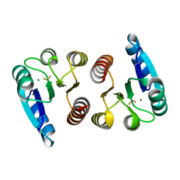 | |
2E5H
 | | Solution structure of RNA binding domain in Zinc finger CCHC-type and RNA binding motif 1 | | 分子名称: | Zinc finger CCHC-type and RNA-binding motif-containing protein 1 | | 著者 | Iibuchi, H, Tsuda, K, Muto, Y, Inoue, M, Kigawa, T, Terada, T, Shirouzu, M, Yokoyama, S, RIKEN Structural Genomics/Proteomics Initiative (RSGI) | | 登録日 | 2006-12-21 | | 公開日 | 2007-06-26 | | 最終更新日 | 2024-05-29 | | 実験手法 | SOLUTION NMR | | 主引用文献 | Solution structure of RNA binding domain in Zinc finger CCHC-type and RNA binding motif 1
To be Published
|
|
1ZNF
 | | THREE-DIMENSIONAL SOLUTION STRUCTURE OF A SINGLE ZINC FINGER DNA-BINDING DOMAIN | | 分子名称: | 31ST ZINC FINGER FROM XFIN, ZINC ION | | 著者 | Lee, M.S, Gippert, G.P, Soman, K.V, Case, D.A, Wright, P.E. | | 登録日 | 1989-09-25 | | 公開日 | 1991-07-15 | | 最終更新日 | 2017-11-29 | | 実験手法 | SOLUTION NMR | | 主引用文献 | Three-dimensional solution structure of a single zinc finger DNA-binding domain.
Science, 245, 1989
|
|
1ZND
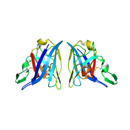
 | | Strong Solute-Solute Dispersive Interactions in a Protein-Ligand Complex | | 分子名称: | CADMIUM ION, Major Urinary Protein, PENTAN-1-OL | | 著者 | Malham, R, Johnstone, S, Bingham, R.J, Barratt, E, Phillips, S.E, Laughton, C.A, Homans, S.W. | | 登録日 | 2005-05-11 | | 公開日 | 2005-12-20 | | 最終更新日 | 2023-08-23 | | 実験手法 | X-RAY DIFFRACTION (1.6 Å) | | 主引用文献 | Strong Solute-Solute Dispersive Interactions in a Protein-Ligand Complex.
J.Am.Chem.Soc., 127, 2005
|
|
1ZNK
 | | Strong Solute-Solute Dispersive Interactions in a Protein-Ligand Complex | | 分子名称: | CADMIUM ION, Major Urinary Protein, NONAN-1-OL | | 著者 | Malham, R, Johnstone, S, Bingham, R.J, Barratt, E, Phillips, S.E, Laughton, C.A, Homans, S.W. | | 登録日 | 2005-05-11 | | 公開日 | 2005-12-20 | | 最終更新日 | 2023-08-23 | | 実験手法 | X-RAY DIFFRACTION (1.6 Å) | | 主引用文献 | Strong Solute-Solute Dispersive Interactions in a Protein-Ligand Complex.
J.Am.Chem.Soc., 127, 2005
|
|
1ZO9
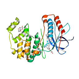
 | | Crystal Structure Of The Wild Type Heme Domain Of P450BM-3 with N-palmitoylmethionine | | 分子名称: | 2-(N-MORPHOLINO)-ETHANESULFONIC ACID, Bifunctional P-450:NADPH-P450 reductase, GLYCEROL, ... | | 著者 | Hegda, A, Chen, B, Tomchick, D.R, Bondlela, M, Haines, D.C, Schaffer, N, Machius, M, Graham, S.E, Peterson, J.A. | | 登録日 | 2005-05-12 | | 公開日 | 2006-08-01 | | 最終更新日 | 2023-08-23 | | 実験手法 | X-RAY DIFFRACTION (1.7 Å) | | 主引用文献 | Interactions of substrates at the surface of P450s can greatly enhance substrate potency.
Biochemistry, 46, 2007
|
|
3UYT
 | | crystal structure of ck1d with PF670462 from P1 crystal form | | 分子名称: | 4-[1-cyclohexyl-4-(4-fluorophenyl)-1H-imidazol-5-yl]pyrimidin-2-amine, Casein kinase I isoform delta, SULFATE ION | | 著者 | Huang, X. | | 登録日 | 2011-12-06 | | 公開日 | 2012-01-11 | | 最終更新日 | 2024-02-28 | | 実験手法 | X-RAY DIFFRACTION (2 Å) | | 主引用文献 | Structural basis for the interaction between casein kinase 1 delta and a potent and selective inhibitor.
J.Med.Chem., 55, 2012
|
|
3UVQ
 | | Human p38 MAP Kinase in Complex with a Dibenzosuberone Derivative | | 分子名称: | Mitogen-activated protein kinase 14, N-{5-[(7-{[(2R)-2,3-dihydroxypropyl]oxy}-5-oxo-10,11-dihydro-5H-dibenzo[a,d][7]annulen-2-yl)amino]-2-fluorophenyl}benzamide, octyl beta-D-glucopyranoside | | 著者 | Mayer-Wrangowski, S.C, Richters, A, Gruetter, C, Rauh, D. | | 登録日 | 2011-11-30 | | 公開日 | 2012-12-05 | | 最終更新日 | 2023-11-08 | | 実験手法 | X-RAY DIFFRACTION (2.2 Å) | | 主引用文献 | Dibenzosuberones as p38 mitogen-activated protein kinase inhibitors with low ATP competitiveness and outstanding whole blood activity.
J.Med.Chem., 56, 2013
|
|
2Z2W
 | | Human Wee1 kinase complexed with inhibitor PF0335770 | | 分子名称: | CHLORIDE ION, GLYCEROL, N-[4-(2-CHLOROPHENYL)-1,3-DIOXO-1,2,3,6-TETRAHYDROPYRROLO[3,4-C]CARBAZOL-9-YL]FORMAMIDE, ... | | 著者 | Squire, C.J, Baker, E.N. | | 登録日 | 2007-05-29 | | 公開日 | 2008-05-06 | | 最終更新日 | 2023-11-15 | | 実験手法 | X-RAY DIFFRACTION (2.22 Å) | | 主引用文献 | Synthesis and Structure-Activity Relationships of 9-Amino-4-(2-chlorophenyl)pyrrolo[3,4-c]carbazole-1,3(2H,6H)-diones and Related Formamides as Inhibitors of the Wee1 and Chk1 Checkpoint Kinases
To be Published
|
|


