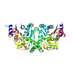
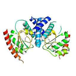
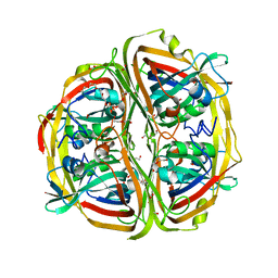
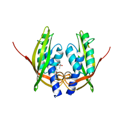
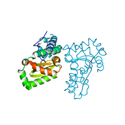
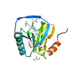
3G4U
 
 | | Ligand migration and cavities within scapharca dimeric hemoglobin: wild type with co bound to heme and dichloropropane bound to the XE4 cavity | | 分子名称: | 1,3-dichloropropane, CARBON MONOXIDE, GLOBIN-1, ... | | 著者 | Knapp, J.E, Pahl, R, Cohen, J, Nichols, J.C, Schulten, K, Gibson, Q.H, Srajer, V, Royer Jr, W.E. | | 登録日 | 2009-02-04 | | 公開日 | 2009-12-01 | | 最終更新日 | 2023-09-06 | | 実験手法 | X-RAY DIFFRACTION (2.1 Å) | | 主引用文献 | Ligand migration and cavities within Scapharca Dimeric HbI: studies by time-resolved crystallo-graphy, Xe binding, and computational analysis.
Structure, 17, 2009
|
|
4L51
 | |
4L5J
 | |
5NL0
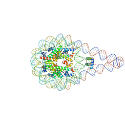 | | Crystal structure of a 197-bp palindromic 601L nucleosome in complex with linker histone H1 | | 分子名称: | DNA (197-MER), Histone H1.0-B, Histone H2A type 1, ... | | 著者 | Garcia-Saez, I, Petosa, C, Dimitrov, S. | | 登録日 | 2017-04-03 | | 公開日 | 2017-05-17 | | 最終更新日 | 2024-01-17 | | 実験手法 | X-RAY DIFFRACTION (5.4 Å) | | 主引用文献 | Structure and Dynamics of a 197 bp Nucleosome in Complex with Linker Histone H1.
Mol. Cell, 66, 2017
|
|
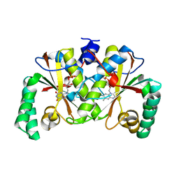
5YPY
 | | Crystal structure of IlvN. Val-1c | | 分子名称: | Acetolactate synthase isozyme 1 small subunit, VALINE | | 著者 | Sarma, S.P, Bansal, A, Schindelin, H, Demeler, B. | | 登録日 | 2017-11-04 | | 公開日 | 2018-09-19 | | 最終更新日 | 2023-11-22 | | 実験手法 | X-RAY DIFFRACTION (1.966 Å) | | 主引用文献 | Crystallographic Structures of IlvN·Val/Ile Complexes: Conformational Selectivity for Feedback Inhibition of Aceto Hydroxy Acid Synthases.
Biochemistry, 58, 2019
|
|
3T38
 | | Corynebacterium glutamicum thioredoxin-dependent arsenate reductase Cg_ArsC1' | | 分子名称: | (4S,5S)-1,2-DITHIANE-4,5-DIOL, Arsenate Reductase | | 著者 | Messens, J, Wahni, K, Dufe, T.V. | | 登録日 | 2011-07-25 | | 公開日 | 2011-11-23 | | 最終更新日 | 2011-11-30 | | 実験手法 | X-RAY DIFFRACTION (2.2 Å) | | 主引用文献 | Corynebacterium glutamicum survives arsenic stress with arsenate reductases coupled to two distinct redox mechanisms.
Mol.Microbiol., 82, 2011
|
|
3BS6
 | |
5WTF
 | | Cryo-EM structure for Hepatitis A virus empty particle | | 分子名称: | VP0, VP1, VP3 | | 著者 | Wang, X, Zhu, L, Dang, M, Hu, Z, Gao, Q, Yuan, S, Sun, Y, Zhang, B, Ren, J, Walter, T.S, Wang, J, Fry, E.E, Stuart, D.I, Rao, Z. | | 登録日 | 2016-12-11 | | 公開日 | 2017-01-25 | | 最終更新日 | 2024-03-27 | | 実験手法 | ELECTRON MICROSCOPY (3.9 Å) | | 主引用文献 | Potent neutralization of hepatitis A virus reveals a receptor mimic mechanism and the receptor recognition site
Proc. Natl. Acad. Sci. U.S.A., 114, 2017
|
|
3BXF
 | | Crystal structure of effector binding domain of central glycolytic gene regulator (CggR) from Bacillus subtilis in complex with effector fructose-1,6-bisphosphate | | 分子名称: | 1,3-DIHYDROXYACETONEPHOSPHATE, 1,6-di-O-phosphono-beta-D-fructofuranose, CHLORIDE ION, ... | | 著者 | Rezacova, P, Otwinowski, Z. | | 登録日 | 2008-01-13 | | 公開日 | 2008-07-01 | | 最終更新日 | 2023-08-30 | | 実験手法 | X-RAY DIFFRACTION (1.7 Å) | | 主引用文献 | Crystal structures of the effector-binding domain of repressor Central glycolytic gene Regulator from Bacillus subtilis reveal ligand-induced structural changes upon binding of several glycolytic intermediates.
Mol.Microbiol., 69, 2008
|
|
2CJ5
 | |
4ZBO
 | | Streptomyces bingchenggensis acetoacetate decarboxylase in non-covalent complex with potassium formate | | 分子名称: | 2-{2-[2-(2-{2-[2-(2-ETHOXY-ETHOXY)-ETHOXY]-ETHOXY}-ETHOXY)-ETHOXY]-ETHOXY}-ETHANOL, 2-{2-[2-2-(METHOXY-ETHOXY)-ETHOXY]-ETHOXY}-ETHANOL, Acetoacetate decarboxylase, ... | | 著者 | Mydy, L.S, Silvaggi, N.R. | | 登録日 | 2015-04-15 | | 公開日 | 2015-06-17 | | 最終更新日 | 2024-05-22 | | 実験手法 | X-RAY DIFFRACTION (1.4 Å) | | 主引用文献 | Sbi00515, a Protein of Unknown Function from Streptomyces bingchenggensis, Highlights the Functional Versatility of the Acetoacetate Decarboxylase Scaffold.
Biochemistry, 54, 2015
|
|
2OR3
 | |
2HY7
 | |
4ZGL
 | | Hit Like Protein | | 分子名称: | ADENOSINE MONOPHOSPHATE, Uncharacterized HIT-like protein HP_0404 | | 著者 | Tarique, K.F, Devi, S, Abdul Rehman, S.A, Gourinath, S. | | 登録日 | 2015-04-23 | | 公開日 | 2015-05-27 | | 最終更新日 | 2023-11-08 | | 実験手法 | X-RAY DIFFRACTION (2.95 Å) | | 主引用文献 | Crystal structure of HINT from Helicobacter pylori.
Acta Crystallogr.,Sect.F, 72, 2016
|
|
2CC3
 | | Structure of Agrobacterium tumefaciens VirB8 protein | | 分子名称: | (4S)-2-METHYL-2,4-PENTANEDIOL, PROTEIN VIRB8 | | 著者 | Bailey, S, Ward, D, Middleton, R, Grossmann, G, Zambryski, P.C. | | 登録日 | 2006-01-11 | | 公開日 | 2006-01-30 | | 最終更新日 | 2023-12-13 | | 実験手法 | X-RAY DIFFRACTION (2.2 Å) | | 主引用文献 | Agrobacterium Tumefaciens Virb8 Structure Reveals Potential Protein-Protein Interactions Sites.
Proc.Natl.Acad.Sci.USA, 103, 2006
|
|
5WP6
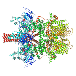 | | Cryo-EM structure of a human TRPM4 channel in complex with calcium and decavanadate | | 分子名称: | DECAVANADATE, Transient receptor potential cation channel subfamily M member 4 | | 著者 | Winkler, P.A, Huang, Y, Sun, W, Du, J, Lu, W. | | 登録日 | 2017-08-03 | | 公開日 | 2017-12-13 | | 最終更新日 | 2024-05-15 | | 実験手法 | ELECTRON MICROSCOPY (3.8 Å) | | 主引用文献 | Electron cryo-microscopy structure of a human TRPM4 channel.
Nature, 552, 2017
|
|
3BM1
 | |
4LI1
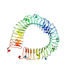 | |
5X2J
 | | Crystal structure of a recombinant hybrid manganese superoxide dismutase from Staphylococcus equorum and Staphylococcus saprophyticus | | 分子名称: | MANGANESE (II) ION, manganese superoxide dismutase | | 著者 | Retnoningrum, D.S, Yoshida, H, Arumsari, S, Kamitori, S, Ismaya, W.T. | | 登録日 | 2017-01-31 | | 公開日 | 2018-01-31 | | 最終更新日 | 2023-11-22 | | 実験手法 | X-RAY DIFFRACTION (1.4 Å) | | 主引用文献 | The first crystal structure of manganese superoxide dismutase from the genus Staphylococcus
Acta Crystallogr F Struct Biol Commun, 74, 2018
|
|
2ZNW
 | | Crystal Structure of ScFv10 in Complex with Hen Egg Lysozyme | | 分子名称: | 2-(2-{2-[2-(2-METHOXY-ETHOXY)-ETHOXY]-ETHOXY}-ETHOXY)-ETHANOL, Lysozyme C, ScFv10 | | 著者 | DeSantis, M.E, Acchione, M, Li, M, Walter, R.L, Wlodawer, A, Smith-Gill, S. | | 登録日 | 2008-05-02 | | 公開日 | 2009-01-27 | | 最終更新日 | 2017-10-11 | | 実験手法 | X-RAY DIFFRACTION (2.71 Å) | | 主引用文献 | Specific fluorine labeling of the HyHEL10 antibody affects antigen binding and dynamics
Biochemistry, 51, 2012
|
|
3TGO
 | | Crystal structure of the E. coli BamCD complex | | 分子名称: | CHLORIDE ION, GLYCEROL, Lipoprotein 34, ... | | 著者 | Paetzel, M, Kim, K.H, Aulakh, S. | | 登録日 | 2011-08-17 | | 公開日 | 2011-09-28 | | 最終更新日 | 2023-09-13 | | 実験手法 | X-RAY DIFFRACTION (2.9 Å) | | 主引用文献 | Crystal structure of the beta-barrel assembly machinery BamCD protein complex
J.Biol.Chem., 286, 2011
|
|
2CJ4
 | |
2CB3
 | | Crystal structure of peptidoglycan recognition protein-LE in complex with tracheal cytotoxin (monomeric diaminopimelic acid-type peptidoglycan) | | 分子名称: | GLCNAC(BETA1-4)-MURNAC(1,6-ANHYDRO)-L-ALA-GAMMA-D-GLU-MESO-A2PM-D-ALA, GLYCEROL, PEPTIDOGLYCAN-RECOGNITION PROTEIN-LE | | 著者 | Lim, J.-H, Kim, M.-S, Oh, B.-H. | | 登録日 | 2005-12-29 | | 公開日 | 2006-01-26 | | 最終更新日 | 2023-12-13 | | 実験手法 | X-RAY DIFFRACTION (2.4 Å) | | 主引用文献 | Structural Basis for Preferential Recognition of Diaminopimelic Acid-Type Peptidoglycan by a Subset of Peptidoglycan Recognition Proteins
J.Biol.Chem., 281, 2006
|
|
6MVS
 | | Structure of a bacterial ALDH16 complexed with NAD | | 分子名称: | Aldehyde dehydrogenase, GLYCEROL, NICOTINAMIDE-ADENINE-DINUCLEOTIDE, ... | | 著者 | Tanner, J.J, Liu, L. | | 登録日 | 2018-10-28 | | 公開日 | 2018-12-26 | | 最終更新日 | 2023-10-11 | | 実験手法 | X-RAY DIFFRACTION (1.65 Å) | | 主引用文献 | Crystal Structure of Aldehyde Dehydrogenase 16 Reveals Trans-Hierarchical Structural Similarity and a New Dimer.
J. Mol. Biol., 431, 2019
|
|
4NSE
 | | BOVINE ENDOTHELIAL NITRIC OXIDE SYNTHASE, H4B-FREE, L-ARG COMPLEX | | 分子名称: | ACETATE ION, ARGININE, CACODYLATE ION, ... | | 著者 | Raman, C.S, Li, H, Martasek, P, Kral, V, Masters, B.S.S, Poulos, T.L. | | 登録日 | 1998-10-07 | | 公開日 | 1999-05-18 | | 最終更新日 | 2024-02-28 | | 実験手法 | X-RAY DIFFRACTION (1.95 Å) | | 主引用文献 | Crystal structure of constitutive endothelial nitric oxide synthase: a paradigm for pterin function involving a novel metal center.
Cell(Cambridge,Mass.), 95, 1998
|
|


