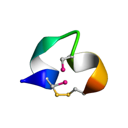
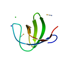
2N07
 
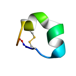 | | Design of a Highly Stable Disulfide-Deleted Mutant of Analgesic Cyclic alpha-Conotoxin Vc1.1 | | 分子名称: | Alpha-conotoxin Vc1A | | 著者 | Yu, R, Seymour, V, Berecki, G, Jia, X, Akcan, M, Adams, D, Kaas, Q, Craik, D. | | 登録日 | 2015-03-04 | | 公開日 | 2016-04-13 | | 実験手法 | SOLUTION NMR | | 主引用文献 | Design of a Highly Stable Disulfide-Deleted Mutant of Analgesic Cyclic alpha-Conotoxin Vc1.1.
To be Published
|
|
1HJE
 | |
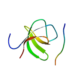
2BC8
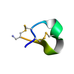
 | | [Sec2,3,8,12]-ImI | | 分子名称: | Alpha-conotoxin ImI | | 著者 | Armishaw, C.J. | | 登録日 | 2005-10-18 | | 公開日 | 2006-03-14 | | 最終更新日 | 2024-05-01 | | 実験手法 | SOLUTION NMR | | 主引用文献 | {alpha}-Selenoconotoxins, a New Class of Potent {alpha}7 Neuronal Nicotinic Receptor Antagonists.
J.Biol.Chem., 281, 2006
|
|
3ZMH
 | | Structure of E.coli rhomboid protease GlpG in complex with monobactam L62 | | 分子名称: | CHLORIDE ION, CYCLOPENTYL 2-OXO-4-PHENYLAZETIDINE-1-CARBOXYLATE, RHOMBOID PROTEASE GLPG, ... | | 著者 | Vinothkumar, K.R, Pierrat, O.A, Large, J.M, Freeman, M. | | 登録日 | 2013-02-11 | | 公開日 | 2013-05-22 | | 最終更新日 | 2024-10-16 | | 実験手法 | X-RAY DIFFRACTION (2.3 Å) | | 主引用文献 | Structure of rhomboid protease in complex with beta-lactam inhibitors defines the S2' cavity.
Structure, 21, 2013
|
|
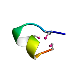
2BC7
 | | Solution structure of [Sec2,8]-ImI | | 分子名称: | Alpha-conotoxin ImI | | 著者 | Armishaw, C.J. | | 登録日 | 2005-10-18 | | 公開日 | 2006-03-14 | | 最終更新日 | 2014-08-06 | | 実験手法 | SOLUTION NMR | | 主引用文献 | {alpha}-Selenoconotoxins, a New Class of Potent {alpha}7 Neuronal Nicotinic Receptor Antagonists.
J.Biol.Chem., 281, 2006
|
|
1IXT
 | | Structure of a Novel P-Superfamily Spasmodic Conotoxin Reveals an Inhibitory Cystine Knot Motif | | 分子名称: | spasmodic protein tx9a-like protein | | 著者 | Miles, L.A, Dy, C.Y, Nielsen, J, Barnham, K.J, Hinds, M.G, Olivera, B.M, Bulaj, G, Norton, R.S. | | 登録日 | 2002-07-04 | | 公開日 | 2003-01-28 | | 最終更新日 | 2023-12-27 | | 実験手法 | SOLUTION NMR | | 主引用文献 | Structure of a Novel P-Superfamily Spasmodic Conotoxin Reveals an Inhibitory Cystine Knot Motif
J.Biol.Chem., 277, 2002
|
|
3IOJ
 | | Crystal structure of the Fucosylgalactoside alpha N-acetylgalactosaminyltransferase (GTA, cisAB mutant L266G, G268A) in complex with UDP | | 分子名称: | GLYCEROL, Histo-blood group ABO system transferase, MANGANESE (II) ION, ... | | 著者 | Pesnot, T, Jorgensen, R, Palcic, M.M, Wagner, G.K. | | 登録日 | 2009-08-14 | | 公開日 | 2010-04-07 | | 最終更新日 | 2023-09-06 | | 実験手法 | X-RAY DIFFRACTION (1.651 Å) | | 主引用文献 | Structural and mechanistic basis for a new mode of glycosyltransferase inhibition.
Nat.Chem.Biol., 6, 2010
|
|
3IOI
 | | Crystal structure of the Fucosylgalactoside alpha N-acetylgalactosaminyltransferase (GTA, cisAB mutant L266G, G268A) in complex with a novel UDP-Gal derived inhibitor (1GW) | | 分子名称: | 5-(2-FORMYLTHIEN-5-YL)-URIDINE-5'-DIPHOSPHATE-ALPHA-D-GALACTOSE, Histo-blood group ABO system transferase, MANGANESE (II) ION, ... | | 著者 | Pesnot, T, Jorgensen, R, Palcic, M.M, Wagner, G.K. | | 登録日 | 2009-08-14 | | 公開日 | 2010-04-07 | | 最終更新日 | 2023-09-06 | | 実験手法 | X-RAY DIFFRACTION (1.45 Å) | | 主引用文献 | Structural and mechanistic basis for a new mode of glycosyltransferase inhibition.
Nat.Chem.Biol., 6, 2010
|
|
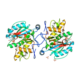
3IOH
 | | Crystal structure of the Fucosylgalactoside alpha N-acetylgalactosaminyltransferase (GTA, cisAB mutant L266G, G268A) | | 分子名称: | GLYCEROL, Histo-blood group ABO system transferase | | 著者 | Pesnot, T, Jorgensen, R, Palcic, M.M, Wagner, G.K. | | 登録日 | 2009-08-14 | | 公開日 | 2010-04-07 | | 最終更新日 | 2023-09-06 | | 実験手法 | X-RAY DIFFRACTION (1.25 Å) | | 主引用文献 | Structural and mechanistic basis for a new mode of glycosyltransferase inhibition.
Nat.Chem.Biol., 6, 2010
|
|
2XOW
 | | Structure of GlpG in complex with a mechanism-based isocoumarin inhibitor | | 分子名称: | 5-AMINO-2-(2-METHOXY-2-OXOETHYL)BENZOIC ACID, RHOMBOID PROTEASE GLPG, nonyl beta-D-glucopyranoside | | 著者 | Vinothkumar, K.R, Strisovsky, K, Andreeva, A, Christova, Y, Verhelst, S, Freeman, M. | | 登録日 | 2010-08-24 | | 公開日 | 2010-10-13 | | 最終更新日 | 2023-12-20 | | 実験手法 | X-RAY DIFFRACTION (2.09 Å) | | 主引用文献 | The Structural Basis for Catalysis and Substrate Specificity of a Rhomboid Protease
Embo J., 29, 2010
|
|
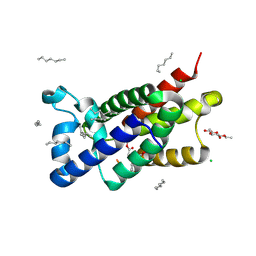
2XT3
 | | HUMAN KIF7, A KINESIN INVOLVED IN HEDGEHOG SIGNALLING | | 分子名称: | ADENOSINE-5'-DIPHOSPHATE, KINESIN-LIKE PROTEIN KIF7, MAGNESIUM ION | | 著者 | Klejnot, M, Kozielski, F. | | 登録日 | 2010-10-04 | | 公開日 | 2011-12-14 | | 最終更新日 | 2023-12-20 | | 実験手法 | X-RAY DIFFRACTION (1.882 Å) | | 主引用文献 | Structural Insights Into Human Kif7, a Kinesin Involved in Hedgehog Signalling.
Acta Crystallogr.,Sect.D, 68, 2012
|
|
2XTU
 | |
2XOV
 | | Crystal Structure of E.coli rhomboid protease GlpG, native enzyme | | 分子名称: | GLYCEROL, RHOMBOID PROTEASE GLPG, nonyl beta-D-glucopyranoside | | 著者 | Vinothkumar, K.R, Strisovsky, K, Andreeva, A, Christova, Y, Verhelst, S, Freeman, M. | | 登録日 | 2010-08-24 | | 公開日 | 2010-10-13 | | 最終更新日 | 2023-12-20 | | 実験手法 | X-RAY DIFFRACTION (1.65 Å) | | 主引用文献 | The structural basis for catalysis and substrate specificity of a rhomboid protease.
EMBO J., 29, 2010
|
|
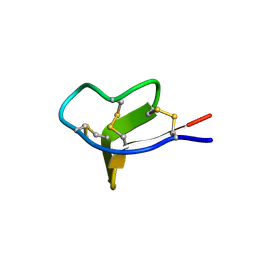
2LO9
 | | NMR solution structure of Mu-contoxin BuIIIB | | 分子名称: | Mu-conotoxin BuIIIB | | 著者 | Kuang, Z. | | 登録日 | 2012-01-20 | | 公開日 | 2013-01-23 | | 最終更新日 | 2023-06-14 | | 実験手法 | SOLUTION NMR | | 主引用文献 | Mammalian neuronal sodium channel blocker mu-conotoxin BuIIIB has a structured N-terminus that influences potency.
Acs Chem.Biol., 8, 2013
|
|
3ZMJ
 | | Structure of E.coli rhomboid protease GlpG in complex with monobactam L61 | | 分子名称: | 2-methylpropyl N-[(1R)-3-oxidanylidene-1-phenyl-propyl]carbamate, CHLORIDE ION, RHOMBOID PROTEASE GLPG, ... | | 著者 | Vinothkumar, K.R, Pierrat, O, Large, J.M, Freeman, M. | | 登録日 | 2013-02-11 | | 公開日 | 2013-05-22 | | 最終更新日 | 2024-10-16 | | 実験手法 | X-RAY DIFFRACTION (2.3 Å) | | 主引用文献 | Structure of Rhomboid Protease in Complex with Beta-Lactam Inhibitors Defines the S2' Cavity.
Structure, 21, 2013
|
|
3ZMI
 | | Structure of E.coli rhomboid protease GlpG in complex with monobactam L29 | | 分子名称: | RHOMBOID PROTEASE GLPG, nonyl beta-D-glucopyranoside, phenyl N-[(1R)-3-oxidanylidene-1-phenyl-propyl]carbamate | | 著者 | Vinothkumar, K.R, Pierrat, O.A, Large, J.M, Freeman, M. | | 登録日 | 2013-02-11 | | 公開日 | 2013-05-22 | | 最終更新日 | 2023-12-20 | | 実験手法 | X-RAY DIFFRACTION (2.2 Å) | | 主引用文献 | Structure of Rhomboid Protease in Complex with Beta-Lactam Inhibitors Defines the S2' Cavity.
Structure, 21, 2013
|
|
3ZOT
 | | Structure of E.coli rhomboid protease GlpG in complex with monobactam L29 (data set 2) | | 分子名称: | CHLORIDE ION, RHOMBOID PROTEASE GLPG, nonyl beta-D-glucopyranoside, ... | | 著者 | Vinothkumar, K.R, Pierrat, O.A, Large, J.M, Freeman, M. | | 登録日 | 2013-02-24 | | 公開日 | 2013-05-22 | | 最終更新日 | 2023-12-20 | | 実験手法 | X-RAY DIFFRACTION (2.399 Å) | | 主引用文献 | Structure of Rhomboid Protease in Complex with Beta-Lactam Inhibitors Defines the S2' Cavity.
Structure, 21, 2013
|
|
4A14
 | | HUMAN KIF7, A KINESIN INVOLVED IN HEDGEHOG SIGNALLING | | 分子名称: | ADENOSINE-5'-DIPHOSPHATE, KINESIN-LIKE PROTEIN KIF7, MAGNESIUM ION | | 著者 | Klejnot, M, Kozielski, F. | | 登録日 | 2011-09-14 | | 公開日 | 2012-01-25 | | 最終更新日 | 2023-12-20 | | 実験手法 | X-RAY DIFFRACTION (1.6 Å) | | 主引用文献 | Structural Insights Into Human Kif7, a Kinesin Involved in Hedgehog Signalling.
Acta Crystallogr.,Sect.D, 68, 2012
|
|
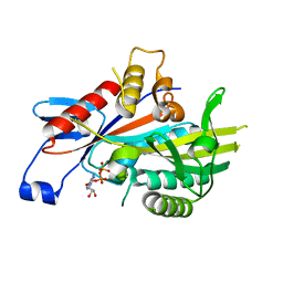
2R47
 | | Crystal structure of MTH_862 protein of unknown function from Methanothermobacter thermautotrophicus | | 分子名称: | SULFATE ION, Uncharacterized protein MTH_862 | | 著者 | Osipiuk, J, Evdokimova, E, Kudritska, M, Savchenko, A, Edwards, A, Joachimiak, A, Midwest Center for Structural Genomics (MCSG) | | 登録日 | 2007-08-30 | | 公開日 | 2007-09-11 | | 最終更新日 | 2017-10-25 | | 実験手法 | X-RAY DIFFRACTION (1.88 Å) | | 主引用文献 | Crystal structure of MTH_862 protein of unknown function from Methanothermobacter thermautotrophicus.
To be Published
|
|
1K3R
 | | Crystal Structure of the Methyltransferase with a Knot from Methanobacterium thermoautotrophicum | | 分子名称: | conserved protein MT0001 | | 著者 | Zarembinski, T.I, Kim, Y, Peterson, K, Christendat, D, Dharamsi, A, Arrowsmith, C.H, Edwards, A.M, Joachimiak, A, Midwest Center for Structural Genomics (MCSG) | | 登録日 | 2001-10-03 | | 公開日 | 2002-05-15 | | 最終更新日 | 2024-02-07 | | 実験手法 | X-RAY DIFFRACTION (2.3 Å) | | 主引用文献 | Deep trefoil knot implicated in RNA binding found in an archaebacterial protein.
Proteins, 50, 2003
|
|
2KCY
 | | SOLUTION STRUCTURE OF RIBOSOMAL PROTEIN S8E FROM Methanothermobacter thermautotrophicus, NORTHEASTSTRUCTURAL GENOMICS CONSORTIUM (NESG) TARGET TR71D | | 分子名称: | 30S ribosomal protein S8e | | 著者 | Liu, G, Rossi, P, Wang, D, Nwosu, C, Owens, L, Xiao, R, Liu, J, Baran, M.C, Swapna, G, Acton, T.B, Rost, B.C, Montelione, G.T, Northeast Structural Genomics Consortium (NESG) | | 登録日 | 2008-12-30 | | 公開日 | 2009-01-20 | | 最終更新日 | 2024-05-22 | | 実験手法 | SOLUTION NMR | | 主引用文献 | SOLUTION STRUCTURE OF RIBOSOMAL PROTEIN S8E FROM Methanothermobacter thermautotrophicus, NORTHEASTSTRUCTURAL GENOMICS CONSORTIUM (NESG) TARGET TR71D
To be Published
|
|
2O9S
 | |
2O9V
 | |
1KES
 | | CONFORMATION OF KERATAN SULPHATE | | 分子名称: | 2-acetamido-2-deoxy-6-O-sulfo-beta-D-glucopyranose-(1-3)-6-O-sulfo-beta-D-galactopyranose-(1-4)-2-acetamido-2-deoxy-6-O-sulfo-beta-D-glucopyranose-(1-3)-6-O-sulfo-beta-D-galactopyranose | | 著者 | Arnott, S. | | 登録日 | 1978-05-23 | | 公開日 | 1980-03-28 | | 最終更新日 | 2024-02-07 | | 実験手法 | FIBER DIFFRACTION (3 Å) | | 主引用文献 | Conformation of keratan sulphate.
J.Mol.Biol., 88, 1974
|
|
2R15
 | |


