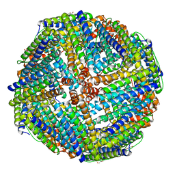
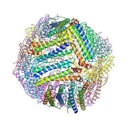
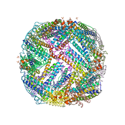
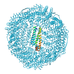
5D8Y
 
 | | 2.05A resolution structure of iron bound BfrB (L68A E81A) from Pseudomonas aeruginosa | | 分子名称: | ACETATE ION, FE (II) ION, Ferroxidase, ... | | 著者 | Lovell, S, Battaile, K.P, Wang, Y, Yao, H, Rivera, M. | | 登録日 | 2015-08-18 | | 公開日 | 2015-09-23 | | 最終更新日 | 2023-09-27 | | 実験手法 | X-RAY DIFFRACTION (2.05 Å) | | 主引用文献 | Characterization of the Bacterioferritin/Bacterioferritin Associated Ferredoxin Protein-Protein Interaction in Solution and Determination of Binding Energy Hot Spots.
Biochemistry, 54, 2015
|
|
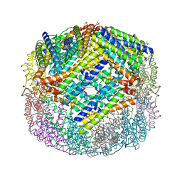
3KWO
 | | Crystal Structure of Putative Bacterioferritin from Campylobacter jejuni | | 分子名称: | 1,4-BUTANEDIOL, ACETIC ACID, GLYCEROL, ... | | 著者 | Kim, Y, Gu, M, Papazisi, L, Anderson, W, Joachimiak, A, Center for Structural Genomics of Infectious Diseases (CSGID) | | 登録日 | 2009-12-01 | | 公開日 | 2010-01-19 | | 最終更新日 | 2024-11-20 | | 実験手法 | X-RAY DIFFRACTION (1.985 Å) | | 主引用文献 | Crystal Structure of Putative Bacterioferritin from Campylobacter jejuni
To be Published
|
|
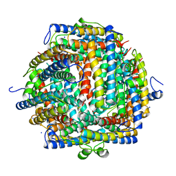
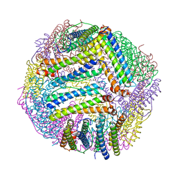
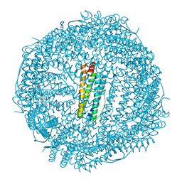
5D8P
 | | 2.35A resolution structure of iron bound BfrB (wild-type, C2221 form) from Pseudomonas aeruginosa | | 分子名称: | ACETATE ION, FE (II) ION, Ferroxidase, ... | | 著者 | Lovell, S, Battaile, K.P, Wang, Y, Yao, H, Rivera, M. | | 登録日 | 2015-08-17 | | 公開日 | 2015-09-23 | | 最終更新日 | 2023-09-27 | | 実験手法 | X-RAY DIFFRACTION (2.35 Å) | | 主引用文献 | Characterization of the Bacterioferritin/Bacterioferritin Associated Ferredoxin Protein-Protein Interaction in Solution and Determination of Binding Energy Hot Spots.
Biochemistry, 54, 2015
|
|
4XGS
 | |
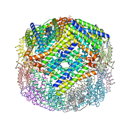
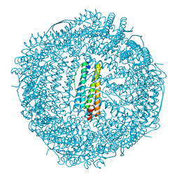
5D8S
 | | 2.55A resolution structure of BfrB (E85A) from Pseudomonas aeruginosa | | 分子名称: | Ferroxidase, POTASSIUM ION, PROTOPORPHYRIN IX CONTAINING FE | | 著者 | Lovell, S, Battaile, K.P, Wang, Y, Yao, H, Rivera, M. | | 登録日 | 2015-08-17 | | 公開日 | 2015-09-23 | | 最終更新日 | 2023-09-27 | | 実験手法 | X-RAY DIFFRACTION (2.55 Å) | | 主引用文献 | Characterization of the Bacterioferritin/Bacterioferritin Associated Ferredoxin Protein-Protein Interaction in Solution and Determination of Binding Energy Hot Spots.
Biochemistry, 54, 2015
|
|
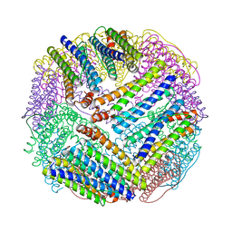
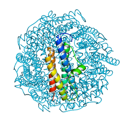
5D8R
 | | 2.50A resolution structure of BfrB (E81A) from Pseudomonas aeruginosa | | 分子名称: | Ferroxidase, POTASSIUM ION, PROTOPORPHYRIN IX CONTAINING FE | | 著者 | Lovell, S, Battaile, K.P, Wang, Y, Yao, H, Rivera, M. | | 登録日 | 2015-08-17 | | 公開日 | 2015-09-23 | | 最終更新日 | 2023-09-27 | | 実験手法 | X-RAY DIFFRACTION (2.5 Å) | | 主引用文献 | Characterization of the Bacterioferritin/Bacterioferritin Associated Ferredoxin Protein-Protein Interaction in Solution and Determination of Binding Energy Hot Spots.
Biochemistry, 54, 2015
|
|
3KX9
 | |
4XKT
 | | E coli BFR variant Y149F | | 分子名称: | Bacterioferritin, SULFATE ION | | 著者 | Bradley, J.M, Hemmings, A.M, Le Brun, N.E. | | 登録日 | 2015-01-12 | | 公開日 | 2015-12-16 | | 最終更新日 | 2024-01-10 | | 実験手法 | X-RAY DIFFRACTION (1.82 Å) | | 主引用文献 | Three Aromatic Residues are Required for Electron Transfer during Iron Mineralization in Bacterioferritin.
Angew.Chem.Int.Ed.Engl., 54, 2015
|
|
8PP5
 | |
1DPS
 | | THE CRYSTAL STRUCTURE OF DPS, A FERRITIN HOMOLOG THAT BINDS AND PROTECTS DNA | | 分子名称: | DPS, SODIUM ION | | 著者 | Grant, R.A, Filman, D.J, Finkel, S.E, Kolter, R, Hogle, J.M. | | 登録日 | 1998-02-23 | | 公開日 | 1998-09-16 | | 最終更新日 | 2024-02-07 | | 実験手法 | X-RAY DIFFRACTION (1.6 Å) | | 主引用文献 | The crystal structure of Dps, a ferritin homolog that binds and protects DNA.
Nat.Struct.Biol., 5, 1998
|
|
8PP4
 | |
8PP2
 | |
8PP3
 | |
4XKU
 | | E coli BFR variant Y114F | | 分子名称: | Bacterioferritin, SULFATE ION | | 著者 | Hemmings, A.M, Le Brun, N.E, Bradley, J.M. | | 登録日 | 2015-01-12 | | 公開日 | 2015-12-16 | | 最終更新日 | 2024-01-10 | | 実験手法 | X-RAY DIFFRACTION (1.78 Å) | | 主引用文献 | Three Aromatic Residues are Required for Electron Transfer during Iron Mineralization in Bacterioferritin.
Angew.Chem.Int.Ed.Engl., 54, 2015
|
|
1EUM
 | | CRYSTAL STRUCTURE OF THE E.COLI FERRITIN ECFTNA | | 分子名称: | FERRITIN 1 | | 著者 | Stillman, T.J, Hempstead, P.D, Artymiuk, P.J, Andrews, S.C, Hudson, A.J, Treffry, A, Guest, J.R, Harrison, P.M. | | 登録日 | 2000-04-17 | | 公開日 | 2001-03-28 | | 最終更新日 | 2024-02-07 | | 実験手法 | X-RAY DIFFRACTION (2.05 Å) | | 主引用文献 | The high-resolution X-ray crystallographic structure of the ferritin (EcFtnA) of Escherichia coli; comparison with human H ferritin (HuHF) and the structures of the Fe(3+) and Zn(2+) derivatives.
J.Mol.Biol., 307, 2001
|
|
4XKS
 | | E. coli BFR variant Y45F | | 分子名称: | Bacterioferritin, SULFATE ION | | 著者 | Hemmings, A.M, Le Brun, N.E, Bradley, J.M. | | 登録日 | 2015-01-12 | | 公開日 | 2015-12-16 | | 最終更新日 | 2024-01-10 | | 実験手法 | X-RAY DIFFRACTION (1.57 Å) | | 主引用文献 | Three Aromatic Residues are Required for Electron Transfer during Iron Mineralization in Bacterioferritin.
Angew.Chem.Int.Ed.Engl., 54, 2015
|
|
4Y08
 | | ONE MINUTE IRON LOADED HUMAN H FERRITIN | | 分子名称: | CHLORIDE ION, FE (II) ION, Ferritin heavy chain, ... | | 著者 | Pozzi, C, Di Pisa, F, Mangani, S. | | 登録日 | 2015-02-05 | | 公開日 | 2015-09-23 | | 最終更新日 | 2024-01-10 | | 実験手法 | X-RAY DIFFRACTION (1.34 Å) | | 主引用文献 | Iron binding to human heavy-chain ferritin.
Acta Crystallogr.,Sect.D, 71, 2015
|
|
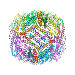
8PVC
 | | Structure of mouse heavy-chain apoferritin determined by cryoEM at 100 keV | | 分子名称: | FE (III) ION, Ferritin heavy chain, ZINC ION | | 著者 | McMullan, G, Naydenova, K, Mihaylov, D, Peet, M.J, Wilson, H, Yamashita, K, Dickerson, J.L, Chen, S, Cannone, G, Lee, Y, Hutchings, K.A, Gittins, O, Sobhy, M, Wells, T, El-Gomati, M.M, Dalby, J, Meffert, M, Schulze-Briese, C, Henderson, R, Russo, C.J. | | 登録日 | 2023-07-17 | | 公開日 | 2023-11-29 | | 最終更新日 | 2023-12-06 | | 実験手法 | ELECTRON MICROSCOPY (2.6 Å) | | 主引用文献 | Structure determination by cryoEM at 100 keV.
Proc.Natl.Acad.Sci.USA, 120, 2023
|
|
8PV9
 | | Structure of DPS determined by cryoEM at 100 keV | | 分子名称: | DNA protection during starvation protein | | 著者 | McMullan, G, Naydenova, K, Mihaylov, D, Peet, M.J, Wilson, H, Yamashita, K, Dickerson, J.L, Chen, S, Cannone, G, Lee, Y, Hutchings, K.A, Gittins, O, Sobhy, M, Wells, T, El-Gomati, M.M, Dalby, J, Meffert, M, Schulze-Briese, C, Henderson, R, Russo, C.J. | | 登録日 | 2023-07-17 | | 公開日 | 2023-11-29 | | 最終更新日 | 2023-12-06 | | 実験手法 | ELECTRON MICROSCOPY (2.7 Å) | | 主引用文献 | Structure determination by cryoEM at 100 keV.
Proc.Natl.Acad.Sci.USA, 120, 2023
|
|
1AEW
 | | L-CHAIN HORSE APOFERRITIN | | 分子名称: | CADMIUM ION, FERRITIN | | 著者 | Hempstead, P.D, Yewdall, S.J, Lawson, D.M, Harrison, P.M, Artymiuk, P.J. | | 登録日 | 1997-02-26 | | 公開日 | 1997-09-04 | | 最終更新日 | 2024-04-03 | | 実験手法 | X-RAY DIFFRACTION (1.95 Å) | | 主引用文献 | Comparison of the three-dimensional structures of recombinant human H and horse L ferritins at high resolution.
J.Mol.Biol., 268, 1997
|
|
1BFR
 | | IRON STORAGE AND ELECTRON TRANSPORT | | 分子名称: | BACTERIOFERRITIN, MANGANESE (II) ION, PROTOPORPHYRIN IX CONTAINING FE | | 著者 | Dautant, A, Yariv, J, Meyer, J.B, Precigoux, G, Sweet, R.M, Frolow, F, Kalb(Gilboa), A.J. | | 登録日 | 1994-12-16 | | 公開日 | 1996-06-20 | | 最終更新日 | 2024-02-07 | | 実験手法 | X-RAY DIFFRACTION (2.94 Å) | | 主引用文献 | Structure of a monoclinic crystal from of cyctochrome b1 (Bacterioferritin) from E. coli.
Acta Crystallogr.,Sect.D, 54, 1998
|
|
4YKH
 | | Thirty minutes iron loaded human H ferritin | | 分子名称: | BICINE, CHLORIDE ION, FE (II) ION, ... | | 著者 | Pozzi, C, Di Pisa, F, Mangani, S. | | 登録日 | 2015-03-04 | | 公開日 | 2015-09-09 | | 最終更新日 | 2024-01-10 | | 実験手法 | X-RAY DIFFRACTION (1.52 Å) | | 主引用文献 | Iron binding to human heavy-chain ferritin.
Acta Crystallogr.,Sect.D, 71, 2015
|
|
1BCF
 | | THE STRUCTURE OF A UNIQUE, TWO-FOLD SYMMETRIC, HAEM-BINDING SITE | | 分子名称: | BACTERIOFERRITIN, MANGANESE (II) ION, PROTOPORPHYRIN IX CONTAINING FE | | 著者 | Frolow, F, Kalb(Gilboa), A.J, Yariv, J. | | 登録日 | 1993-12-06 | | 公開日 | 1994-12-20 | | 最終更新日 | 2024-02-07 | | 実験手法 | X-RAY DIFFRACTION (2.9 Å) | | 主引用文献 | Structure of a unique twofold symmetric haem-binding site.
Nat.Struct.Biol., 1, 1994
|
|
3KXU
 | | Crystal structure of human ferritin FTL498InsTC pathogenic mutant | | 分子名称: | CADMIUM ION, Ferritin, SULFATE ION | | 著者 | Granier, T, Gallois, B, Langlois d'Estaintot, B, Arosio, P. | | 登録日 | 2009-12-04 | | 公開日 | 2010-02-09 | | 最終更新日 | 2023-11-01 | | 実験手法 | X-RAY DIFFRACTION (1.85 Å) | | 主引用文献 | Mutant ferritin L-chains that cause neurodegeneration act in a dominant-negative manner to reduce ferritin iron incorporation.
J.Biol.Chem., 285, 2010
|
|
1BG7
 | | LOCALIZED UNFOLDING AT THE JUNCTION OF THREE FERRITIN SUBUNITS. A MECHANISM FOR IRON RELEASE? | | 分子名称: | CALCIUM ION, FERRITIN | | 著者 | Takagi, H, Shi, D, Ha, Y, Allewell, N.M, Theil, E.C. | | 登録日 | 1998-06-05 | | 公開日 | 1999-01-13 | | 最終更新日 | 2024-05-22 | | 実験手法 | X-RAY DIFFRACTION (1.85 Å) | | 主引用文献 | Localized unfolding at the junction of three ferritin subunits. A mechanism for iron release?
J.Biol.Chem., 273, 1998
|
|


