Loading
PDBj
メニューPDBj@FacebookPDBj@TwitterPDBj@YouTubewwPDB FoundationwwPDB
RCSB PDBPDBeBMRBAdv. SearchSearch help

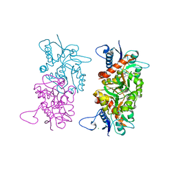
3C31
DownloadVisualize
BU of 3c31 by Molmil
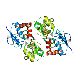
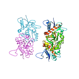
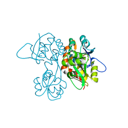
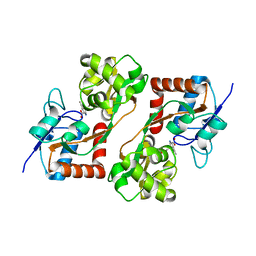
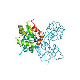
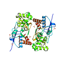
Crystal structure of GluR5 ligand-binding core in complex with lithium at 1.49 Angstrom resolution
分子名称: 3-(CARBOXYMETHYL)-4-ISOPROPENYLPROLINE, CHLORIDE ION, GLUTAMATE RECEPTOR, ...
著者Mayer, M.L.
登録日2008-01-27
公開日2008-06-17
最終更新日2023-08-30
実験手法X-RAY DIFFRACTION (1.49 Å)
主引用文献Molecular basis of kainate receptor modulation by sodium.
Neuron, 58, 2008
1S9T
DownloadVisualize
BU of 1s9t by Molmil
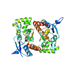
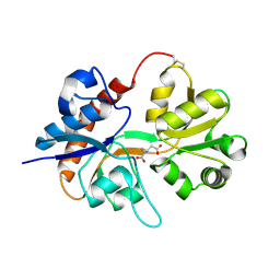
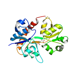
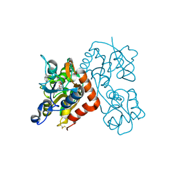
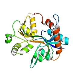
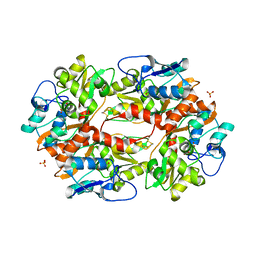
Crystal structure of the GLUR6 ligand binding core in complex with quisqualate at 1.8A resolution
分子名称: (S)-2-AMINO-3-(3,5-DIOXO-[1,2,4]OXADIAZOLIDIN-2-YL)-PROPIONIC ACID, CHLORIDE ION, Glutamate receptor, ...
著者Mayer, M.L.
登録日2004-02-05
公開日2005-02-08
最終更新日2023-10-25
実験手法X-RAY DIFFRACTION (1.8 Å)
主引用文献Crystal structures of the GluR5 and GluR6 ligand binding cores: molecular mechanisms underlying kainate receptor selectivity.
Neuron, 45, 2005
1S7Y
DownloadVisualize
BU of 1s7y by Molmil
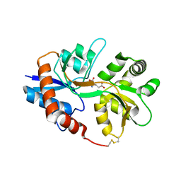
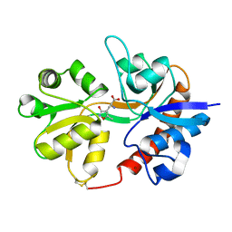
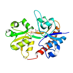
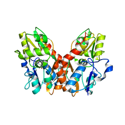
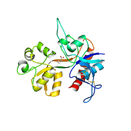
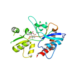
Crystal structure of the GluR6 ligand binding core in complex with glutamate at 1.75 A resolution orthorhombic form
分子名称: GLUTAMIC ACID, Glutamate receptor, ionotropic kainate 2 precursor
著者Mayer, M.L.
登録日2004-01-30
公開日2005-02-08
最終更新日2024-02-14
実験手法X-RAY DIFFRACTION (1.75 Å)
主引用文献Crystal structures of the GluR5 and GluR6 ligand binding cores: Molecular mechanisms underlying kainate receptor selectivity
Neuron, 45, 2005
2I0C
DownloadVisualize
BU of 2i0c by Molmil
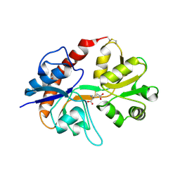
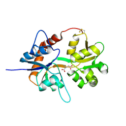
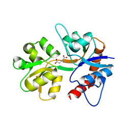
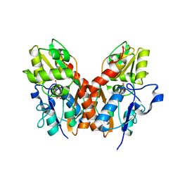
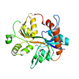
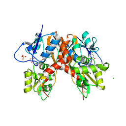
Crystal structure of the GluR6 ligand binding core dimer crosslinked by disulfide bonds between Y490C and L752C at 2.25 Angstroms Resolution
分子名称: GLUTAMIC ACID, Glutamate receptor, ionotropic kainate 2
著者Mayer, M.L.
登録日2006-08-10
公開日2006-11-21
最終更新日2024-04-03
実験手法X-RAY DIFFRACTION (2.25 Å)
主引用文献Conformational restriction blocks glutamate receptor desensitization.
Nat.Struct.Mol.Biol., 13, 2006
2I0B
DownloadVisualize
BU of 2i0b by Molmil
Crystal structure of the GluR6 ligand binding core ELKQ mutant dimer at 1.96 Angstroms Resolution
分子名称: GLUTAMIC ACID, Glutamate receptor, ionotropic kainate 2, ...
著者Mayer, M.L.
登録日2006-08-10
公開日2006-11-21
最終更新日2024-04-03
実験手法X-RAY DIFFRACTION (1.96 Å)
主引用文献Conformational restriction blocks glutamate receptor desensitization.
Nat.Struct.Mol.Biol., 13, 2006
2I3W
DownloadVisualize
BU of 2i3w by Molmil
Measurement of conformational changes accompanying desensitization in an ionotropic glutamate receptor: Structure of S729C mutant
分子名称: GLUTAMATE RECEPTOR SUBUNIT 2, GLUTAMIC ACID
著者Armstrong, N, Jasti, J, Beich-Frandsen, M, Gouaux, E.
登録日2006-08-21
公開日2006-10-17
最終更新日2023-08-30
実験手法X-RAY DIFFRACTION (2.3 Å)
主引用文献Measurement of Conformational Changes accompanying Desensitization in an Ionotropic Glutamate Receptor.
Cell(Cambridge,Mass.), 127, 2006
1SD3
DownloadVisualize
BU of 1sd3 by Molmil
Crystal structure of the GLUR6 ligand binding core in complex with 2S,4R-4-methylglutamate at 1.8 Angstrom resolution
分子名称: 2S,4R-4-METHYLGLUTAMATE, Glutamate receptor, ionotropic kainate 2
著者Mayer, M.L.
登録日2004-02-12
公開日2005-02-15
最終更新日2023-10-25
実験手法X-RAY DIFFRACTION (1.8 Å)
主引用文献Crystal structures of the GluR5 and GluR6 ligand binding cores: molecular mechanisms underlying kainate receptor selectivity.
Neuron, 45, 2005
2I3V
DownloadVisualize
BU of 2i3v by Molmil
Measurement of conformational changes accompanying desensitization in an ionotropic glutamate receptor: Structure of G725C mutant
分子名称: GLUTAMIC ACID, Glutamate receptor 2, ZINC ION
著者Armstrong, N, Jasti, J, Beich-Frandsen, M, Gouaux, E.
登録日2006-08-21
公開日2006-10-17
最終更新日2023-08-30
実験手法X-RAY DIFFRACTION (2.4 Å)
主引用文献Measurement of Conformational Changes accompanying Desensitization in an Ionotropic Glutamate Receptor.
Cell(Cambridge,Mass.), 127, 2006
3DP4
DownloadVisualize
BU of 3dp4 by Molmil
Crystal structure of the binding domain of the AMPA subunit GluR3 bound to AMPA
分子名称: (S)-ALPHA-AMINO-3-HYDROXY-5-METHYL-4-ISOXAZOLEPROPIONIC ACID, Glutamate receptor 3, ZINC ION
著者Ahmed, A.H, Wang, Q, Sondermann, H, Oswald, R.E.
登録日2008-07-07
公開日2008-11-25
最終更新日2023-08-30
実験手法X-RAY DIFFRACTION (2.11 Å)
主引用文献Structure of the S1S2 glutamate binding domain of GLuR3.
Proteins, 75, 2008
1S50
DownloadVisualize
BU of 1s50 by Molmil
X-ray structure of the GluR6 ligand binding core (S1S2A) in complex with glutamate at 1.65 A resolution
分子名称: GLUTAMIC ACID, Glutamate Receptor 6
著者Mayer, M.L.
登録日2004-01-19
公開日2005-02-08
最終更新日2023-08-23
実験手法X-RAY DIFFRACTION (1.65 Å)
主引用文献Crystal structures of the GluR5 and GluR6 ligand binding cores: Molecular mechanisms underlying kainate receptor selectivity
Neuron, 45, 2005
1VSO
DownloadVisualize
BU of 1vso by Molmil
Crystal Structure of the Ligand-Binding Core of iGluR5 in Complex With the Antagonist (S)-ATPO at 1.85 A resolution
分子名称: (S)-2-AMINO-3-(5-TERT-BUTYL-3-(PHOSPHONOMETHOXY)-4-ISOXAZOLYL)PROPIONIC ACID, GLYCEROL, Glutamate receptor, ...
著者Hald, H, Naur, P, Gajhede, M, Kastrup, J.S.
登録日2007-03-29
公開日2007-07-03
最終更新日2023-08-23
実験手法X-RAY DIFFRACTION (1.85 Å)
主引用文献Partial agonism and antagonism of the ionotropic glutamate receptor iGLuR5: structures of the ligand-binding core in complex with domoic acid and 2-amino-3-[5-tert-butyl-3-(phosphonomethoxy)-4-isoxazolyl]propionic acid.
J.Biol.Chem., 282, 2007
3DP6
DownloadVisualize
BU of 3dp6 by Molmil
Crystal structure of the binding domain of the AMPA subunit GluR2 bound to glutamate
分子名称: GLUTAMIC ACID, Glutamate receptor 2, ZINC ION
著者Ahmed, A.H, Wang, Q, Sondermann, H, Oswald, R.E.
登録日2008-07-07
公開日2008-11-25
最終更新日2023-08-30
実験手法X-RAY DIFFRACTION (1.55 Å)
主引用文献Structure of the S1S2 glutamate binding domain of GLuR3.
Proteins, 75, 2008
3DLN
DownloadVisualize
BU of 3dln by Molmil
Crystal structure of the binding domain of the AMPA subunit GluR3 bound to glutamate
分子名称: GLUTAMIC ACID, Glutamate receptor 3, ZINC ION
著者Ahmed, A.H, Wang, Q, Sondermann, H, Oswald, R.E.
登録日2008-06-27
公開日2008-11-25
最終更新日2023-08-30
実験手法X-RAY DIFFRACTION (1.91 Å)
主引用文献Structure of the S1S2 glutamate binding domain of GLuR3.
Proteins, 75, 2008
1TXF
DownloadVisualize
BU of 1txf by Molmil
CRYSTAL STRUCTURE OF THE GLUR5 LIGAND BINDING CORE IN COMPLEX WITH GLUTAMATE AT 2.1 ANGSTROM RESOLUTION
分子名称: GLUTAMIC ACID, Glutamate receptor, ionotropic kainate 1
著者Mayer, M.L.
登録日2004-07-04
公開日2005-02-08
最終更新日2023-08-23
実験手法X-RAY DIFFRACTION (2.1 Å)
主引用文献Crystal structures of the GluR5 and GluR6 ligand binding cores: Molecular mechanisms underlying kainate receptor selectivity
Neuron, 45, 2005
1TT1
DownloadVisualize
BU of 1tt1 by Molmil
CRYSTAL STRUCTURE OF THE GLUR6 LIGAND BINDING CORE IN COMPLEX WITH KAINATE 1.93 A RESOLUTION
分子名称: 3-(CARBOXYMETHYL)-4-ISOPROPENYLPROLINE, Glutamate receptor, ionotropic kainate 2
著者Mayer, M.L.
登録日2004-06-21
公開日2005-02-08
最終更新日2023-08-23
実験手法X-RAY DIFFRACTION (1.93 Å)
主引用文献Crystal structures of the GluR5 and GluR6 ligand binding cores: Molecular mechanisms underlying kainate receptor selectivity
Neuron, 45, 2005
3EN3
DownloadVisualize
BU of 3en3 by Molmil
Crystal Structure of the GluR4 Ligand-Binding domain in complex with kainate
分子名称: 3-(CARBOXYMETHYL)-4-ISOPROPENYLPROLINE, Glutamate receptor 4,Glutamate receptor
著者Gill, A, Madden, D.R.
登録日2008-09-25
公開日2009-05-19
最終更新日2017-07-26
実験手法X-RAY DIFFRACTION (2.43 Å)
主引用文献Correlating AMPA receptor activation and cleft closure across subunits: crystal structures of the GluR4 ligand-binding domain in complex with full and partial agonists
Biochemistry, 47, 2008
3EPE
DownloadVisualize
BU of 3epe by Molmil
Crystal Structure of the GluR4 Ligand-Binding domain in complex with glutamate
分子名称: GLUTAMIC ACID, Glutamate receptor 4,Glutamate receptor
著者Gill, A, Madden, D.R.
登録日2008-09-29
公開日2009-05-19
最終更新日2023-09-06
実験手法X-RAY DIFFRACTION (1.85 Å)
主引用文献Correlating AMPA receptor activation and cleft closure across subunits: crystal structures of the GluR4 ligand-binding domain in complex with full and partial agonists
Biochemistry, 47, 2008
1SYI
DownloadVisualize
BU of 1syi by Molmil
X-RAY STRUCTURE OF THE Y702F MUTANT OF THE GLUR2 LIGAND-BINDING CORE (S1S2J) IN COMPLEX WITH (S)-CPW399 AT 2.1 A RESOLUTION.
分子名称: (S)-2-AMINO-3-(1,3,5,7-PENTAHYDRO-2,4-DIOXO-CYCLOPENTA[E]PYRIMIDIN-1-YL) PROIONIC ACID, Glutamate receptor 2
著者Frandsen, A, Pickering, D.S, Vestergaard, B, Kasper, C, Nielsen, B.B, Greenwood, J.R, Campiani, G, Gajhede, M, Schousboe, A, Kastrup, J.S.
登録日2004-04-01
公開日2005-03-22
最終更新日2023-08-23
実験手法X-RAY DIFFRACTION (2.1 Å)
主引用文献Tyr702 Is an Important Determinant of Agonist Binding and Domain Closure of the Ligand-Binding Core of GluR2.
Mol.Pharmacol., 67, 2005
1XHY
DownloadVisualize
BU of 1xhy by Molmil
X-ray structure of the Y702F mutant of the GluR2 ligand-binding core (S1S2J) in complex with kainate at 1.85 A resolution
分子名称: 3-(CARBOXYMETHYL)-4-ISOPROPENYLPROLINE, Glutamate receptor, SULFATE ION
著者Frandsen, A, Pickering, D.S, Vestergaard, B, Kasper, C, Nielsen, B.B, Greenwood, J.R, Campiani, G, Gajhede, M, Schousboe, A, Kastrup, J.S.
登録日2004-09-21
公開日2005-03-22
最終更新日2023-08-23
実験手法X-RAY DIFFRACTION (1.85 Å)
主引用文献Tyr702 Is an Important Determinant of Agonist Binding and Domain Closure of the Ligand-Binding Core of GluR2.
Mol.Pharmacol., 67, 2005
1Y20
DownloadVisualize
BU of 1y20 by Molmil
Crystal structure of the NR1 ligand-binding core in complex with ACPC
分子名称: 1-AMINOCYCLOPROPANECARBOXYLIC ACID, Glutamate [NMDA] receptor subunit zeta 1
著者Inanobe, A, Gouaux, E.
登録日2004-11-19
公開日2005-07-12
最終更新日2017-07-26
実験手法X-RAY DIFFRACTION (1.4 Å)
主引用文献Mechanism of Partial Agonist Action at the NR1 Subunit of NMDA Receptors.
Neuron, 47, 2005
1Y1M
DownloadVisualize
BU of 1y1m by Molmil
Crystal structure of the NR1 ligand binding core in complex with cycloleucine
分子名称: 1-AMINOCYCLOPENTANECARBOXYLIC ACID, Glutamate [NMDA] receptor subunit zeta 1
著者Inanobe, A, Gouaux, E.
登録日2004-11-18
公開日2005-07-12
最終更新日2017-07-26
実験手法X-RAY DIFFRACTION (1.8 Å)
主引用文献Mechanism of Partial Agonist Action at the NR1 Subunit of NMDA Receptors.
Neuron, 47, 2005
1Y1Z
DownloadVisualize
BU of 1y1z by Molmil
Crystal structure of the NR1 ligand binding core in complex with ACBC
分子名称: 1-AMINOCYCLOBUTANECARBOXLIC ACID, Glutamate [NMDA] receptor subunit zeta 1
著者Inanobe, A, Gouaux, E.
登録日2004-11-19
公開日2005-07-12
最終更新日2017-07-26
実験手法X-RAY DIFFRACTION (1.5 Å)
主引用文献Mechanism of Partial Agonist Action at the NR1 Subunit of NMDA Receptors.
Neuron, 47, 2005
1YCJ
DownloadVisualize
BU of 1ycj by Molmil
Crystal structure of the kainate receptor GluR5 ligand-binding core in complex with (S)-glutamate
分子名称: GLUTAMIC ACID, Ionotropic glutamate receptor 5, SULFATE ION
著者Naur, P, Vestergaard, B, Skov, L.K, Egebjerg, J, Gajhede, M, Kastrup, J.S.
登録日2004-12-22
公開日2005-02-01
最終更新日2023-10-25
実験手法X-RAY DIFFRACTION (1.95 Å)
主引用文献Crystal structure of the kainate receptor GluR5 ligand-binding core in complex with (S)-glutamate
Febs Lett., 579, 2005
2F36
DownloadVisualize
BU of 2f36 by Molmil
Crystal Structure of the GluR5 Ligand Binding Core Dimer with Glutamate At 2.1 Angstroms Resolution
分子名称: GLUTAMATE RECEPTOR, IONOTROPIC KAINATE 1, GLUTAMIC ACID, ...
著者Mayer, M.L.
登録日2005-11-18
公開日2006-04-04
最終更新日2023-08-23
実験手法X-RAY DIFFRACTION (2.11 Å)
主引用文献Crystal structures of the kainate receptor GluR5 ligand binding core dimer with novel GluR5-selective antagonists.
J.Neurosci., 26, 2006
1YAE
DownloadVisualize
BU of 1yae by Molmil
Structure of the Kainate Receptor Subunit GluR6 Agonist Binding Domain Complexed with Domoic Acid
分子名称: (2S,3S,4S)-2-CARBOXY-4-[(1Z,3E,5R)-5-CARBOXY-1-METHYL-1,3-HEXADIENYL]-3-PYRROLIDINEACETIC ACID, 2-acetamido-2-deoxy-beta-D-glucopyranose, Glutamate receptor, ...
著者Nanao, M.H, Green, T, Stern-Bach, Y, Heinemann, S.F, Choe, S.
登録日2004-12-17
公開日2005-02-01
最終更新日2023-08-23
実験手法X-RAY DIFFRACTION (3.11 Å)
主引用文献Structure of the kainate receptor subunit GluR6 agonist-binding domain complexed with domoic acid.
Proc.Natl.Acad.Sci.USA, 102, 2005

223532

件を2024-08-07に公開中

PDB statisticsPDBj update infoContact PDBjnumon