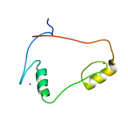
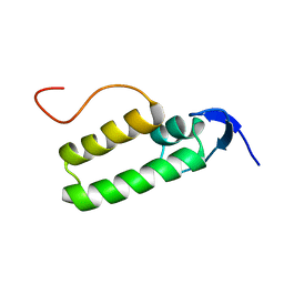
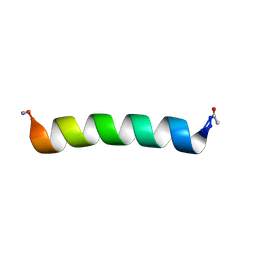
1WJT
 
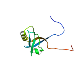
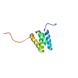 | | Solution structure of the N-terminal Domain I of mouse transcription elongation factor S-II protein 3 | | 分子名称: | Transcription elongation factor S-II protein 3 | | 著者 | Yoneyama, M, Tochio, N, Koshiba, S, Inoue, M, Kigawa, T, Yokoyama, S, RIKEN Structural Genomics/Proteomics Initiative (RSGI) | | 登録日 | 2004-05-29 | | 公開日 | 2004-11-29 | | 最終更新日 | 2024-05-29 | | 実験手法 | SOLUTION NMR | | 主引用文献 | Solution structure of the N-terminal Domain I of mouse transcription elongation factor S-II protein 3
To be Published
|
|
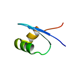
2MXF
 | |
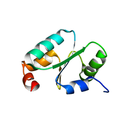
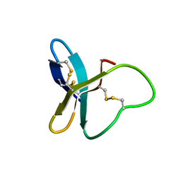
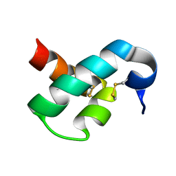
2MZ9
 | | Solution structure of oxidized triheme cytochrome PpcA from Geobacter sulfurreducens | | 分子名称: | HEME C, PpcA | | 著者 | Morgado, L, Bruix, M, Pokkuluri, R, Salgueiro, C.A, Turner, D.L. | | 登録日 | 2015-02-08 | | 公開日 | 2016-02-10 | | 最終更新日 | 2024-05-01 | | 実験手法 | SOLUTION NMR | | 主引用文献 | Redox- and pH-linked conformational changes in triheme cytochrome PpcA from Geobacter sulfurreducens.
Biochem. J., 474, 2017
|
|
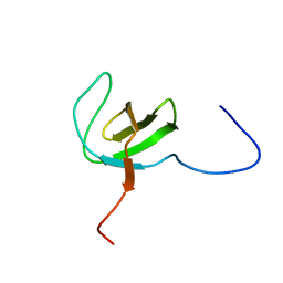
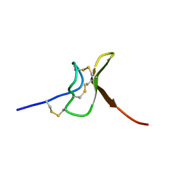
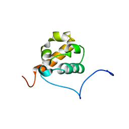
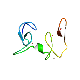
1X6B
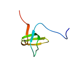 | | Solution structures of the SH3 domain of human rho guanine exchange factor (GEF) 16 | | 分子名称: | Rho guanine exchange factor (GEF) 16 | | 著者 | Sato, M, Saito, K, Koshiba, S, Inoue, M, Kigawa, T, Yokoyama, S, RIKEN Structural Genomics/Proteomics Initiative (RSGI) | | 登録日 | 2005-05-17 | | 公開日 | 2005-11-17 | | 最終更新日 | 2024-05-29 | | 実験手法 | SOLUTION NMR | | 主引用文献 | Solution structures of the SH3 domain of human rho guanine exchange factor (GEF) 16
To be Published
|
|
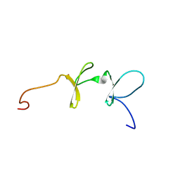
1X6H
 | | Solution structures of the C2H2 type zinc finger domain of human Transcriptional repressor CTCF | | 分子名称: | Transcriptional repressor CTCF, ZINC ION | | 著者 | Sato, M, Saito, K, Koshiba, S, Inoue, M, Kigawa, T, Yokoyama, S, RIKEN Structural Genomics/Proteomics Initiative (RSGI) | | 登録日 | 2005-05-17 | | 公開日 | 2005-11-17 | | 最終更新日 | 2024-05-29 | | 実験手法 | SOLUTION NMR | | 主引用文献 | Solution structures of the C2H2 type zinc finger domain of human Transcriptional repressor CTCF
To be Published
|
|
2N52
 | |
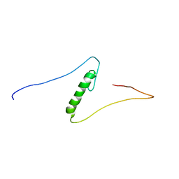
1X6G
 | | Solution structures of the SH3 domain of human megakaryocyte-associated tyrosine-protein kinase. | | 分子名称: | Megakaryocyte-associated tyrosine-protein kinase | | 著者 | Sato, M, Koshiba, S, Inoue, M, Kigawa, T, Yokoyama, S, RIKEN Structural Genomics/Proteomics Initiative (RSGI) | | 登録日 | 2005-05-17 | | 公開日 | 2005-11-17 | | 最終更新日 | 2024-05-29 | | 実験手法 | SOLUTION NMR | | 主引用文献 | Solution structures of the SH3 domain of human megakaryocyte-associated tyrosine-protein kinase.
To be Published
|
|
1X68
 | | Solution structures of the C-terminal LIM domain of human FHL5 protein | | 分子名称: | FHL5 protein, ZINC ION | | 著者 | Nameki, N, Sasagawa, A, Sato, M, Koshiba, S, Inoue, M, Kigawa, T, Yokoyama, S, RIKEN Structural Genomics/Proteomics Initiative (RSGI) | | 登録日 | 2005-05-17 | | 公開日 | 2005-11-17 | | 最終更新日 | 2024-05-29 | | 実験手法 | SOLUTION NMR | | 主引用文献 | Solution structures of the C-terminal LIM domain of human FHL5 protein
To be Published
|
|
1X6F
 | | Solution structures of the C2H2 type zinc finger domain of human Zinc finger protein 462 | | 分子名称: | ZINC ION, Zinc finger protein 462 | | 著者 | Sato, M, Saito, K, Koshiba, S, Inoue, M, Kigawa, T, Yokoyama, S, RIKEN Structural Genomics/Proteomics Initiative (RSGI) | | 登録日 | 2005-05-17 | | 公開日 | 2005-11-17 | | 最終更新日 | 2024-05-29 | | 実験手法 | SOLUTION NMR | | 主引用文献 | Solution structures of the C2H2 type zinc finger domain of human Zinc finger protein 462
To be Published
|
|
1X6D
 | | Solution structures of the PDZ domain of human Interleukin-16 | | 分子名称: | Interleukin-16 | | 著者 | Sato, M, Koshiba, S, Inoue, M, Kigawa, T, Yokoyama, S, RIKEN Structural Genomics/Proteomics Initiative (RSGI) | | 登録日 | 2005-05-17 | | 公開日 | 2005-11-17 | | 最終更新日 | 2024-05-29 | | 実験手法 | SOLUTION NMR | | 主引用文献 | Solution structures of the PDZ domain of human Interleukin-16
To be Published
|
|
2MXE
 | |
2MYG
 | | Solution structure of the dithiolic glutaredoxin 2-C-Grx1 from the pathogen Trypanosoma brucei brucei | | 分子名称: | Dithiol glutaredoxin 1 | | 著者 | Sturlese, M, Stefani, M, Manta, B, Mammi, S, Comini, M, Bellanda, M. | | 登録日 | 2015-01-22 | | 公開日 | 2016-02-10 | | 最終更新日 | 2024-05-01 | | 実験手法 | SOLUTION NMR | | 主引用文献 | Solution structure of the dithiolic glutaredoxin 2-C-Grx1 from the pathogen Trypanosoma brucei brucei
To be Published
|
|
2N1N
 | | Solution structure of VSTx1 | | 分子名称: | Kappa-theraphotoxin-Gr3a | | 著者 | Lau, H.Y, King, G.F, Mobli, M. | | 登録日 | 2015-04-12 | | 公開日 | 2016-03-02 | | 最終更新日 | 2016-10-12 | | 実験手法 | SOLUTION NMR | | 主引用文献 | Molecular basis of the interaction between gating modifier spider toxins and the voltage sensor of voltage-gated ion channels.
Sci Rep, 6
|
|
2MT7
 | |
1X6C
 | | Solution structures of the SH2 domain of human protein-tyrosine phosphatase SHP-1 | | 分子名称: | Tyrosine-protein phosphatase, non-receptor type 6 | | 著者 | Sato, M, Koshiba, S, Inoue, M, Kigawa, T, Yokoyama, S, RIKEN Structural Genomics/Proteomics Initiative (RSGI) | | 登録日 | 2005-05-17 | | 公開日 | 2005-11-17 | | 最終更新日 | 2024-05-29 | | 実験手法 | SOLUTION NMR | | 主引用文献 | Solution structures of the SH2 domain of human protein-tyrosine phosphatase SHP-1
To be Published
|
|
2MS5
 | |
2MPQ
 | |
2MU9
 | | Changing ABRA protein peptide to fit the HLA-DR B1*0301 molecule renders it protection-inducing | | 分子名称: | P101/acidic basic repeat antigen | | 著者 | Salazar, L, Alba, M, Curtidor, H, Bermudez, A, Vargas, L, Rivera, Z, Patarroyo, M. | | 登録日 | 2014-09-04 | | 公開日 | 2015-09-16 | | 最終更新日 | 2024-05-15 | | 実験手法 | SOLUTION NMR | | 主引用文献 | Changing ABRA protein peptide to fit into the HLA-DRbeta1*0301 molecule renders it protection-inducing.
Biochem.Biophys.Res.Commun., 322, 2004
|
|
1XS8
 | |
2MUB
 | |
1X66
 | | Solution structure of the SAM_PNT-domain of the human friend LEUKEMIAINTEGRATION 1 transcription factor | | 分子名称: | Friend leukemia integration 1 transcription factor | | 著者 | Goroncy, A, Kigawa, T, Koshiba, S, Sato, M, Kobayashi, N, Tochio, N, Inoue, M, Yokoyama, S, RIKEN Structural Genomics/Proteomics Initiative (RSGI) | | 登録日 | 2005-05-17 | | 公開日 | 2005-11-17 | | 最終更新日 | 2024-05-29 | | 実験手法 | SOLUTION NMR | | 主引用文献 | Solution structure of the SAM_PNT-domain of the human friend LEUKEMIAINTEGRATION 1 transcription factor
To be Published
|
|
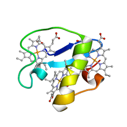
1YNR
 | | Crystal structure of the cytochrome c-552 from Hydrogenobacter thermophilus at 2.0 resolution | | 分子名称: | (4S)-2-METHYL-2,4-PENTANEDIOL, Cytochrome c-552, HEME C, ... | | 著者 | Travaglini-Allocatelli, C, Gianni, S, Dubey, V.K, Borgia, A, Di Matteo, A, Bonivento, D, Cutruzzola, F, Bren, K.L, Brunori, M. | | 登録日 | 2005-01-25 | | 公開日 | 2005-05-17 | | 最終更新日 | 2023-10-25 | | 実験手法 | X-RAY DIFFRACTION (2 Å) | | 主引用文献 | An Obligatory Intermediate in the Folding Pathway of Cytochrome c552 from Hydrogenobacter thermophilus
J.Biol.Chem., 280, 2005
|
|
2MUG
 | | Protective cellular immunity against P. falciparum malaria merozoite is associated with a different P7 and P8 residue orientation in the MHC-peptide-TCR complex | | 分子名称: | Serine-repeat antigen protein | | 著者 | Patarroyo, M, Salazar, L, Cifuentes, G, Lozano, J, Delgado, G, Rivera, Z, Rosas, J, Vargas, L. | | 登録日 | 2014-09-09 | | 公開日 | 2014-10-01 | | 最終更新日 | 2023-06-14 | | 実験手法 | SOLUTION NMR | | 主引用文献 | Protective cellular immunity against P. falciparum malaria merozoites is associated with a different P7 and P8 residue orientation in the MHC-peptide-TCR complex.
Biochimie, 88, 2006
|
|
2MUN
 | |
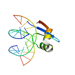
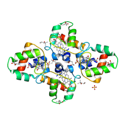
2NAA
 | | NSD1-PHD_5-C5HCH tandem domain structure | | 分子名称: | Histone-lysine N-methyltransferase, H3 lysine-36 and H4 lysine-20 specific, ZINC ION | | 著者 | Berardi, A, Quilici, G, Spiliotopoulos, D, Musco, G. | | 登録日 | 2015-12-22 | | 公開日 | 2016-03-09 | | 最終更新日 | 2024-05-15 | | 実験手法 | SOLUTION NMR | | 主引用文献 | Structural basis for PHDVC5HCHNSD1-C2HRNizp1 interaction: implications for Sotos syndrome.
Nucleic Acids Res., 44, 2016
|
|


