1JRF
 
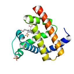
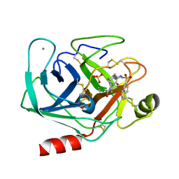
 | | NMR Solution Structure of the Viral Receptor Domain of Tva | | 分子名称: | CALCIUM ION, SUBGROUP A ROUS SARCOMA VIRUS RECEPTORS PG800 AND PG950 | | 著者 | Wang, Q.-Y, Huang, W, Dolmer, K, Gettins, P.G.W, Rong, L. | | 登録日 | 2001-08-13 | | 公開日 | 2002-03-08 | | 最終更新日 | 2022-02-23 | | 実験手法 | SOLUTION NMR | | 主引用文献 | Solution structure of the viral receptor domain of Tva and its implications in viral entry.
J.Virol., 76, 2002
|
|
1VDP
 | | The crystal structure of the monoclinic form of hen egg white lysozyme at 1.7 angstroms resolution in space | | 分子名称: | Lysozyme C | | 著者 | Aibara, S, Suzuki, A, Kidera, A, Shibata, K, Yamane, T, DeLucas, L.J, Hirose, M. | | 登録日 | 2004-03-24 | | 公開日 | 2004-04-13 | | 最終更新日 | 2023-12-27 | | 実験手法 | X-RAY DIFFRACTION (1.7 Å) | | 主引用文献 | The crystal structure of the monoclinic form of hen egg white lysozyme at 1.7 angstroms resolution in space
to be published
|
|
3FY0
 | |
2DMD
 | | Solution structure of the N-terminal C2H2 type zinc-binding domain of the Zinc finger protein 64, isoforms 1 and 2 | | 分子名称: | ZINC ION, Zinc finger protein 64, isoforms 1 and 2 | | 著者 | Yoneyama, M, Tochio, N, Koshiba, S, Inoue, M, Kigawa, T, Yokoyama, S, RIKEN Structural Genomics/Proteomics Initiative (RSGI) | | 登録日 | 2006-04-21 | | 公開日 | 2006-10-21 | | 最終更新日 | 2024-05-29 | | 実験手法 | SOLUTION NMR | | 主引用文献 | Solution structure of the N-terminal C2H2 type zinc-binding domain of the Zinc finger
protein 64, isoforms 1 and 2
To be Published
|
|
2DN0
 | | Solution structure of the second homeobox domain of human zinc fingers and homeoboxes protein 3 | | 分子名称: | Zinc fingers and homeoboxes protein 3 | | 著者 | Seimiya, K, Kurosaki, C, Hayashi, F, Yoshida, M, Yokoyama, S, RIKEN Structural Genomics/Proteomics Initiative (RSGI) | | 登録日 | 2006-04-24 | | 公開日 | 2006-10-24 | | 最終更新日 | 2024-05-29 | | 実験手法 | SOLUTION NMR | | 主引用文献 | Solution structure of the second homeobox domain of human zinc fingers
and homeoboxes protein 3
To be published
|
|
2DNP
 | | Solution structure of RNA binding domain 2 in RNA-binding protein 14 | | 分子名称: | RNA-binding protein 14 | | 著者 | Kusuhara, M, Tsuda, K, Muto, Y, Inoue, M, Kigawa, T, Terada, T, Shirouzu, M, Yokoyama, S, RIKEN Structural Genomics/Proteomics Initiative (RSGI) | | 登録日 | 2006-04-26 | | 公開日 | 2006-10-26 | | 最終更新日 | 2024-05-29 | | 実験手法 | SOLUTION NMR | | 主引用文献 | Solution structure of RNA binding domain 2 in RNA-binding protein 14
To be Published
|
|
1JV4
 | | Crystal structure of recombinant major mouse urinary protein (rmup) at 1.75 A resolution | | 分子名称: | 2-(SEC-BUTYL)THIAZOLE, CADMIUM ION, Major urinary protein 2 | | 著者 | Kuser, P.R, Franzoni, L, Ferrari, E, Spisni, A, Polikarpov, I. | | 登録日 | 2001-08-28 | | 公開日 | 2001-12-05 | | 最終更新日 | 2023-08-16 | | 実験手法 | X-RAY DIFFRACTION (1.75 Å) | | 主引用文献 | The X-ray structure of a recombinant major urinary protein at 1.75 A resolution. A comparative study of X-ray and NMR-derived structures.
Acta Crystallogr.,Sect.D, 57, 2001
|
|
1JWR
 | | Crystal structure of human lysozyme at 100 K | | 分子名称: | lysozyme | | 著者 | Higo, J, Nakasako, M. | | 登録日 | 2001-09-05 | | 公開日 | 2001-09-19 | | 最終更新日 | 2023-10-25 | | 実験手法 | X-RAY DIFFRACTION (1.4 Å) | | 主引用文献 | Hydration structure of human lysozyme investigated by molecular dynamics simulation and cryogenic X-ray crystal structure analyses: on the correlation between crystal water sites, solvent density, and solvent dipole
J.Comput.Chem., 23, 2002
|
|
3FSK
 | | P38 kinase crystal structure in complex with RO6257 | | 分子名称: | 3-(2-chlorophenyl)-7-[(trans-4-hydroxycyclohexyl)amino]-3,4-dihydropyrimido[4,5-d]pyrimidin-2(1H)-one, Mitogen-activated protein kinase 14 | | 著者 | Kuglstatter, A, Bertrand, J, Takahara, P, Villasenor, A. | | 登録日 | 2009-01-09 | | 公開日 | 2009-12-22 | | 最終更新日 | 2023-09-06 | | 実験手法 | X-RAY DIFFRACTION (2 Å) | | 主引用文献 | Mapping Binding Pocket Volume: Potential Applications towards Ligand Design and Selectivity
To be Published
|
|
2DRK
 | |
1JYJ
 | | Crystal Structure of a Double Variant (W67L/W91H) of Recombinant Human Serum Retinol-binding Protein at 2.0 A Resolution | | 分子名称: | GLYCEROL, PLASMA RETINOL-BINDING PROTEIN | | 著者 | Greene, L.H, Chrysina, E.D, Irons, L.I, Papageorgiou, A.C, Acharya, K.R, Brew, K. | | 登録日 | 2001-09-12 | | 公開日 | 2003-07-01 | | 最終更新日 | 2023-08-16 | | 実験手法 | X-RAY DIFFRACTION (2 Å) | | 主引用文献 | Role of Conserved Residues in Structure and Stability: Tryptophans of Human Serum Retinol-Binding Protein, a Model for the Lipocalin Superfamily
Protein Sci., 10, 2001
|
|
1JYQ
 | | Xray Structure of Grb2 SH2 Domain Complexed with a Highly Affine Phospho Peptide | | 分子名称: | GROWTH FACTOR RECEPTOR-BOUND PROTEIN 2, mAZ-pY-(alpha Me)pY-N-NH2 peptide inhibitor | | 著者 | Nioche, P, Liu, W.-Q, Broutin, I, Charbonnier, F, Latreille, M.-T, Vidal, M, Roques, B, Garbay, C, Ducruix, A. | | 登録日 | 2001-09-13 | | 公開日 | 2002-03-13 | | 最終更新日 | 2024-07-10 | | 実験手法 | X-RAY DIFFRACTION (2 Å) | | 主引用文献 | Crystal structures of the SH2 domain of Grb2: highlight on the binding of a new high-affinity inhibitor.
J.Mol.Biol., 315, 2002
|
|
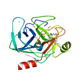
1K1P
 | | BOVINE TRYPSIN-INHIBITOR COMPLEX | | 分子名称: | CALCIUM ION, TRYPSIN, [((1R)-2-{(2S)-2-[({4-[AMINO(IMINO)METHYL]BENZYL}AMINO)CARBONYL]AZETIDINYL}-1-CYCLOHEXYL-2-OXOETHYL)AMINO]ACETIC ACID | | 著者 | Stubbs, M.T. | | 登録日 | 2001-09-25 | | 公開日 | 2001-11-28 | | 最終更新日 | 2011-07-13 | | 実験手法 | X-RAY DIFFRACTION (1.9 Å) | | 主引用文献 | Factorising ligand affinity: a combined thermodynamic and crystallographic study of trypsin and thrombin inhibition.
J.Mol.Biol., 313, 2001
|
|
3A7I
 | | Human MST3 kinase in complex with adenine | | 分子名称: | ADENINE, Serine/threonine kinase 24 (STE20 homolog, yeast) | | 著者 | Ko, T.P, Jeng, W.Y, Liu, C.I, Lai, M.D, Wang, A.H.J. | | 登録日 | 2009-09-26 | | 公開日 | 2010-02-02 | | 最終更新日 | 2023-11-01 | | 実験手法 | X-RAY DIFFRACTION (1.45 Å) | | 主引用文献 | Structures of human MST3 kinase in complex with adenine, ADP and Mn2+.
Acta Crystallogr.,Sect.D, 66, 2010
|
|
3FWQ
 | | Inactive conformation of human protein kinase CK2 catalytic subunit | | 分子名称: | CHLORIDE ION, Casein kinase II subunit alpha, GLYCEROL, ... | | 著者 | Niefind, K, Raaf, J, Issinger, O.G. | | 登録日 | 2009-01-19 | | 公開日 | 2009-02-17 | | 最終更新日 | 2023-11-01 | | 実験手法 | X-RAY DIFFRACTION (2.3 Å) | | 主引用文献 | First inactive conformation of CK2 alpha, the catalytic subunit of protein kinase CK2
J.Mol.Biol., 386, 2009
|
|
1K0Y
 | | X-ray Crystallographic Analyses of Symmetrical Allosteric Effectors of Hemoglobin. Compounds Designed to Link Primary and Secondary Binding Sites | | 分子名称: | 2-{4-[(3{2-[4-(1-CARBOXY-1-METHYL-ETHOXY)-PHENYL]-ACETYLAMINO}-PHENYLCARBAMOYL)-METHYL]-PHENOXY}-2-METHYL-PROPIONIC ACID, PROTOPORPHYRIN IX CONTAINING FE, SULFATE ION, ... | | 著者 | Safo, M.K, Boyiri, T, Burnett, J.C, Danso-Danquah, R, Moure, C.M, Joshi, G.S, Abraham, D.J. | | 登録日 | 2001-09-21 | | 公開日 | 2001-10-03 | | 最終更新日 | 2023-08-16 | | 実験手法 | X-RAY DIFFRACTION (1.87 Å) | | 主引用文献 | X-ray crystallographic analyses of symmetrical allosteric effectors of hemoglobin: compounds designed to link primary and secondary binding sites.
Acta Crystallogr.,Sect.D, 58, 2002
|
|
1JKK
 | | 2.4A X-RAY STRUCTURE OF TERNARY COMPLEX OF A CATALYTIC DOMAIN OF DEATH-ASSOCIATED PROTEIN KINASE WITH ATP ANALOGUE AND MG. | | 分子名称: | DEATH-ASSOCIATED PROTEIN KINASE, MAGNESIUM ION, PHOSPHOAMINOPHOSPHONIC ACID-ADENYLATE ESTER | | 著者 | Tereshko, V, Teplova, M, Brunzelle, J, Watterson, D.M, Egli, M. | | 登録日 | 2001-07-12 | | 公開日 | 2002-04-01 | | 最終更新日 | 2024-02-07 | | 実験手法 | X-RAY DIFFRACTION (2.4 Å) | | 主引用文献 | Crystal structures of the catalytic domain of human protein kinase associated with apoptosis and tumor suppression.
Nat.Struct.Biol., 8, 2001
|
|
1W5X
 | | HIV-1 protease in complex with fluoro substituted diol-based C2- symmetric inhibitor | | 分子名称: | (2R,3R,4R,5R)-2,5-BIS[(2,3-DIFLUOROBENZYL)OXY]-3,4-DIHYDROXY-N,N'-BIS[(1S,2R)-2-HYDROXY-2,3-DIHYDRO-1H-INDEN-1-YL]HEXAN EDIAMIDE, POL POLYPROTEIN | | 著者 | Lindberg, J, Pyring, D, Loewgren, S, Rosenquist, A, Zuccarello, G, Kvarnstroem, I, Zhang, H, Vrang, L, Claesson, B, Hallberg, A, Samuelsson, B, Unge, T. | | 登録日 | 2004-08-10 | | 公開日 | 2004-12-22 | | 最終更新日 | 2024-05-08 | | 実験手法 | X-RAY DIFFRACTION (1.9 Å) | | 主引用文献 | Symmetric Fluoro-Substituted Diol-Based HIV Protease Inhibitors. Ortho-Fluorinated and Meta-Fluorinated P1/P1'-Benzyloxy Side Groups Significantly Improve the Antiviral Activity and Preserve Binding Efficacy
Eur.J.Biochem., 271, 2004
|
|
1JLD
 | |
1RTF
 | |
1K1J
 | | BOVINE TRYPSIN-INHIBITOR COMPLEX | | 分子名称: | CALCIUM ION, N-ALPHA-(2-NAPHTHYLSULFONYL)-N(3-AMIDINO-L-PHENYLALANINYL)ISOPIPECOLINIC ACID METHYL ESTER, SULFATE ION, ... | | 著者 | Stubbs, M.T. | | 登録日 | 2001-09-25 | | 公開日 | 2001-11-28 | | 最終更新日 | 2011-07-13 | | 実験手法 | X-RAY DIFFRACTION (2.2 Å) | | 主引用文献 | Factorising ligand affinity: a combined thermodynamic and crystallographic study of trypsin and thrombin inhibition.
J.Mol.Biol., 313, 2001
|
|
1JP6
 | |
2DC3
 | | Crystal structure of human cytoglobin at 1.68 angstroms resolution | | 分子名称: | ACETIC ACID, Cytoglobin, PROTOPORPHYRIN IX CONTAINING FE | | 著者 | Makino, M, Sugimoto, H, Sawai, H, Kawada, N, Yoshizato, K, Shiro, Y, RIKEN Structural Genomics/Proteomics Initiative (RSGI) | | 登録日 | 2005-12-21 | | 公開日 | 2006-05-23 | | 最終更新日 | 2023-10-25 | | 実験手法 | X-RAY DIFFRACTION (1.68 Å) | | 主引用文献 | High-resolution structure of human cytoglobin: identification of extra N- and C-termini and a new dimerization mode.
Acta Crystallogr.,Sect.D, 62, 2006
|
|
1K1M
 | | BOVINE TRYPSIN-INHIBITOR COMPLEX | | 分子名称: | CALCIUM ION, N-ALPHA-(2-NAPHTHYLSULFONYL)-N(3-AMIDINO-L-PHENYLALANINYL)-4-ACETYL-PIPERAZINE, SULFATE ION, ... | | 著者 | Stubbs, M.T. | | 登録日 | 2001-09-25 | | 公開日 | 2001-11-28 | | 最終更新日 | 2011-07-13 | | 実験手法 | X-RAY DIFFRACTION (2.2 Å) | | 主引用文献 | Factorising ligand affinity: a combined thermodynamic and crystallographic study of trypsin and thrombin inhibition.
J.Mol.Biol., 313, 2001
|
|
1VJY
 | | Crystal Structure of a Naphthyridine Inhibitor of Human TGF-beta Type I Receptor | | 分子名称: | 2-[5-(6-METHYLPYRIDIN-2-YL)-2,3-DIHYDRO-1H-PYRAZOL-4-YL]-1,5-NAPHTHYRIDINE, TGF-beta receptor type I | | 著者 | Gellibert, F, Woolven, J, Fouchet, M.-H, Mathews, N, Goodland, H, Lovegrove, V, Laroze, A, Nguyen, V.-L, Sautet, S, Wang, R, Janson, C, Smith, W, Krysa, G, Boullay, V, de Gouville, A.-C, Huet, S, Hartley, D. | | 登録日 | 2004-04-07 | | 公開日 | 2004-08-31 | | 最終更新日 | 2023-12-27 | | 実験手法 | X-RAY DIFFRACTION (2 Å) | | 主引用文献 | Identification of 1,5-Naphthyridine Derivatives as a Novel Series of Potent and Selective TGF-beta Type I Receptor Inhibitors.
J.Med.Chem., 47, 2004
|
|


