Loading
PDBj
メニューPDBj@FacebookPDBj@TwitterPDBj@YouTubewwPDB FoundationwwPDB
RCSB PDBPDBeBMRBAdv. SearchSearch help

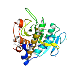
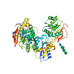
6E7S
DownloadVisualize
BU of 6e7s by Molmil
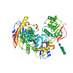
Heterodimer of the GluN1b-GluN2B NMDA receptor amino-terminal domains bound to allosteric inhibitor 93-5
分子名称: 2-acetamido-2-deoxy-beta-D-glucopyranose, 2-acetamido-2-deoxy-beta-D-glucopyranose-(1-4)-2-acetamido-2-deoxy-beta-D-glucopyranose, CHLORIDE ION, ...
著者Regan, M.C, Furukawa, H.
登録日2018-07-27
公開日2019-01-30
最終更新日2023-10-11
実験手法X-RAY DIFFRACTION (2.72 Å)
主引用文献Structural elements of a pH-sensitive inhibitor binding site in NMDA receptors
Nat Commun, 10, 2019
6E7U
DownloadVisualize
BU of 6e7u by Molmil
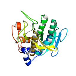
Heterodimer of the GluN1b-GluN2B NMDA receptor amino-terminal domains bound to allosteric inhibitor 93-31
分子名称: 2-acetamido-2-deoxy-beta-D-glucopyranose, CHLORIDE ION, Glutamate receptor ionotropic, ...
著者Regan, M.C, Furukawa, H.
登録日2018-07-27
公開日2019-01-30
最終更新日2023-10-11
実験手法X-RAY DIFFRACTION (2.27 Å)
主引用文献Structural elements of a pH-sensitive inhibitor binding site in NMDA receptors
Nat Commun, 10, 2019
7A36
DownloadVisualize
BU of 7a36 by Molmil
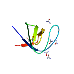
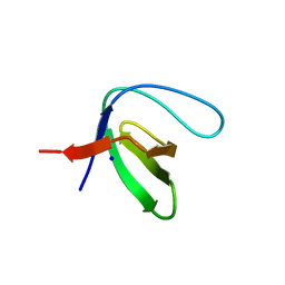
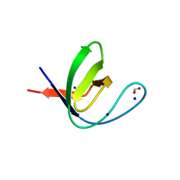
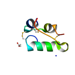
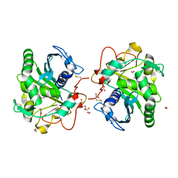
Crystal structure of the c-Src SH3 domain mutant V111L-N113S-T114S in 7 M urea
分子名称: Proto-oncogene tyrosine-protein kinase Src, SODIUM ION, UREA
著者Camara-Artigas, A, Plaza-Garrido, M, Salinas-Garcia, M.C.
登録日2020-08-18
公開日2021-08-25
最終更新日2024-01-31
実験手法X-RAY DIFFRACTION (1.5 Å)
主引用文献Crystal structure of the c-Src SH3 domain mutant V111L-N113S-T114S in 7 M urea
To be published
6E7X
DownloadVisualize
BU of 6e7x by Molmil
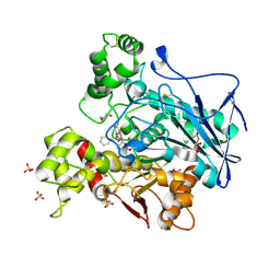
Heterodimer of the GluN1b-GluN2B NMDA receptor amino-terminal domains bound to allosteric inhibitor 93-97
分子名称: 2-acetamido-2-deoxy-beta-D-glucopyranose, CHLORIDE ION, Glutamate receptor ionotropic, ...
著者Regan, M.C, Furukawa, H.
登録日2018-07-27
公開日2019-01-30
最終更新日2023-10-11
実験手法X-RAY DIFFRACTION (2.58 Å)
主引用文献Structural elements of a pH-sensitive inhibitor binding site in NMDA receptors
Nat Commun, 10, 2019
7A2K
DownloadVisualize
BU of 7a2k by Molmil
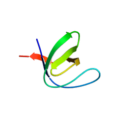
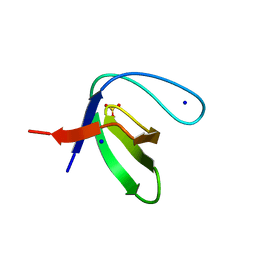
Crystal structure of the Fyn SH3 domain in space group P1 at pH 7.5
分子名称: SODIUM ION, Tyrosine-protein kinase Fyn
著者Camara-Artigas, A, Plaza-Garrido, M, Salinas-Garcia, M.C.
登録日2020-08-18
公開日2021-08-25
最終更新日2024-01-31
実験手法X-RAY DIFFRACTION (1.5 Å)
主引用文献Crystal structure of the Fyn SH3 domain in space group P1 at pH 7.5
To be published
7A0A
DownloadVisualize
BU of 7a0a by Molmil
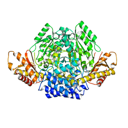
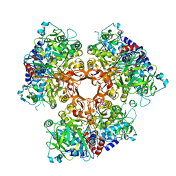
Crystal structure of mouse CSAD in apo form
分子名称: Cysteine sulfinic acid decarboxylase, SODIUM ION, SULFATE ION
著者Mahootchi, E, Raasakka, A, Haavik, J, Kursula, P.
登録日2020-08-07
公開日2021-08-18
最終更新日2024-01-31
実験手法X-RAY DIFFRACTION (2.8 Å)
主引用文献The structure of cysteine sulphinic acid decarboxylase reveals structural determinants for substrate specificity of pyridoxal phosphate-dependent decarboxylases
To be published
6EFD
DownloadVisualize
BU of 6efd by Molmil
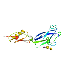
Hsa Siglec and Unique domains in complex with the sialyl T antigen trisaccharide
分子名称: N-acetyl-alpha-neuraminic acid-(2-3)-beta-D-galactopyranose-(1-3)-2-acetamido-2-deoxy-alpha-D-galactopyranose, SODIUM ION, Streptococcal hemagglutinin
著者Iverson, T.M.
登録日2018-08-16
公開日2020-02-19
最終更新日2024-05-22
実験手法X-RAY DIFFRACTION (1.85 Å)
主引用文献Origins of glycan selectivity in streptococcal Siglec-like adhesins suggest mechanisms of receptor adaptation.
Nat Commun, 13, 2022
7A2L
DownloadVisualize
BU of 7a2l by Molmil
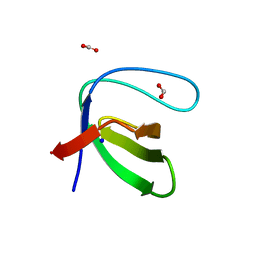
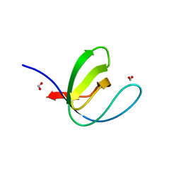
Crystal structure of the Fyn SH3 domain mutant E129Q in space group C21 at pH 4.0
分子名称: FORMIC ACID, SODIUM ION, Tyrosine-protein kinase Fyn
著者Camara-Artigas, A, Plaza-Garrido, M, Salinas-Garcia, M.C.
登録日2020-08-18
公開日2021-08-25
最終更新日2024-01-31
実験手法X-RAY DIFFRACTION (1.9 Å)
主引用文献Crystal structure of the Fyn SH3 domain mutant E129Q in space group C21 at pH 4.0
To be published
6EJ7
DownloadVisualize
BU of 6ej7 by Molmil
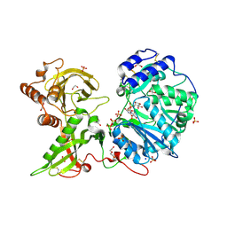
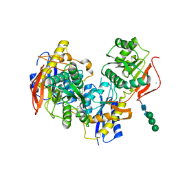
Human Xylosyltransferase 1 in complex with UDP-xylose and peptide QEEEGAGGGQGG
分子名称: 1,2-ETHANEDIOL, 2-acetamido-2-deoxy-beta-D-glucopyranose, DI(HYDROXYETHYL)ETHER, ...
著者Briggs, D.C, Hohenester, E.
登録日2017-09-20
公開日2018-05-02
最終更新日2024-01-17
実験手法X-RAY DIFFRACTION (2 Å)
主引用文献Structural Basis for the Initiation of Glycosaminoglycan Biosynthesis by Human Xylosyltransferase 1.
Structure, 26, 2018
7A2J
DownloadVisualize
BU of 7a2j by Molmil
Crystal structure of the Fyn SH3 domain in space group C21 at pH 7.5
分子名称: SODIUM ION, Tyrosine-protein kinase Fyn
著者Camara-Artigas, A, Plaza-Garrido, M, Salinas-Garcia, M.C.
登録日2020-08-18
公開日2021-08-25
最終更新日2024-01-31
実験手法X-RAY DIFFRACTION (1.5 Å)
主引用文献Crystal structure of the Fyn SH3 domain in space group C21 at pH 7.5
To be published
7A33
DownloadVisualize
BU of 7a33 by Molmil
Crystal structure of the c-Src SH3 domain mutant S94A-T98D-V111L-N113S-T114S at pH 3.0
分子名称: FORMIC ACID, Proto-oncogene tyrosine-protein kinase Src, SODIUM ION
著者Camara-Artigas, A, Plaza-Garrido, M, Salinas-Garcia, M.C.
登録日2020-08-18
公開日2021-08-25
最終更新日2024-01-31
実験手法X-RAY DIFFRACTION (0.96 Å)
主引用文献Crystal structure of the c-Src SH3 domain mutant S94A-T98D-V111L-N113S-T114S at pH 3.0
To be published
7AE7
DownloadVisualize
BU of 7ae7 by Molmil
Structure of Sedimentibacter hydroxybenzoicus vanillic acid decarboxylase (ShVdcCD) in open form, with truncated ShVdcD (V59X)
分子名称: Phenolic acid decarboxylase, Protein ShdD, SODIUM ION, ...
著者Marshall, S.A, Leys, D.
登録日2020-09-17
公開日2021-08-25
最終更新日2024-01-31
実験手法X-RAY DIFFRACTION (2.66 Å)
主引用文献Domain mobility and allosteric activation of UbiD decarboxylases
To Be Published
7A2N
DownloadVisualize
BU of 7a2n by Molmil
Crystal structure of the Fyn SH3 domain A95S-D99T mutant in space group P21
分子名称: FORMIC ACID, SODIUM ION, Tyrosine-protein kinase Fyn
著者Camara-Artigas, A, Plaza-Garrido, M, Salinas-Garcia, M.C.
登録日2020-08-18
公開日2021-08-25
最終更新日2024-01-31
実験手法X-RAY DIFFRACTION (1.4 Å)
主引用文献Crystal structure of the Fyn SH3 domain A95S-D99T mutant in space group P21
To be published
7A2M
DownloadVisualize
BU of 7a2m by Molmil
Crystal structure of the Fyn SH3 domain A95S-D99T mutant in C21 space group
分子名称: FORMIC ACID, SODIUM ION, SULFATE ION, ...
著者Camara-Artigas, A, Plaza-Garrido, M, Salinas-Garcia, M.C.
登録日2020-08-18
公開日2021-08-25
最終更新日2024-01-31
実験手法X-RAY DIFFRACTION (1.5 Å)
主引用文献Crystal structure of the Fyn SH3 domain A95S-D99T mutant in C21 space group
To be published
7AC4
DownloadVisualize
BU of 7ac4 by Molmil
Structure of insulin collected by rotation serial crystallography on a COC membrane at a synchrotron source
分子名称: Insulin, R-1,2-PROPANEDIOL, SODIUM ION
著者Martiel, I, Padeste, C, Karpik, A, Huang, C.Y, Vera, L, Wang, M, Marsh, M.
登録日2020-09-09
公開日2021-09-01
最終更新日2024-01-31
実験手法X-RAY DIFFRACTION (1.46 Å)
主引用文献Versatile microporous polymer-based supports for serial macromolecular crystallography.
Acta Crystallogr D Struct Biol, 77, 2021
7AAW
DownloadVisualize
BU of 7aaw by Molmil
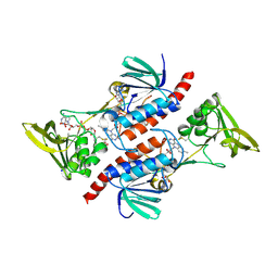
Thioredoxin Reductase from Bacillus cereus
分子名称: ACETATE ION, FLAVIN-ADENINE DINUCLEOTIDE, NADP NICOTINAMIDE-ADENINE-DINUCLEOTIDE PHOSPHATE, ...
著者Shoor, M, Gudim, I, Hersleth, H.-P, Hammerstad, M.
登録日2020-09-04
公開日2021-09-15
最終更新日2024-01-31
実験手法X-RAY DIFFRACTION (2.25 Å)
主引用文献Thioredoxin reductase from Bacillus cereus exhibits distinct reduction and NADPH-binding properties.
Febs Open Bio, 11, 2021
6E7V
DownloadVisualize
BU of 6e7v by Molmil
Heterodimer of the GluN1b-GluN2B NMDA receptor amino-terminal domains bound to allosteric inhibitor 93-88
分子名称: 2-acetamido-2-deoxy-beta-D-glucopyranose, CHLORIDE ION, Glutamate receptor ionotropic, ...
著者Regan, M.C, Furukawa, H.
登録日2018-07-27
公開日2019-01-30
最終更新日2023-10-11
実験手法X-RAY DIFFRACTION (2.6 Å)
主引用文献Structural elements of a pH-sensitive inhibitor binding site in NMDA receptors
Nat Commun, 10, 2019
7AVC
DownloadVisualize
BU of 7avc by Molmil
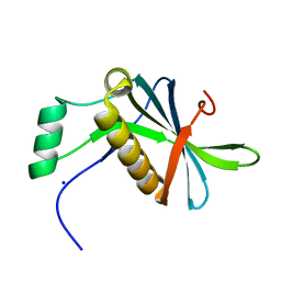
DoBi scaffold based on PIH1D1 N-terminal domain
分子名称: GLYCEROL, PIH1 domain-containing protein 1, SODIUM ION
著者Kolenko, P, Pham, N.P, Pavlicek, J, Mikulecky, P, Schneider, B.
登録日2020-11-05
公開日2021-02-10
最終更新日2024-01-31
実験手法X-RAY DIFFRACTION (1.2 Å)
主引用文献Protein Binder (ProBi) as a New Class of Structurally Robust Non-Antibody Protein Scaffold for Directed Evolution.
Viruses, 13, 2021
7AM8
DownloadVisualize
BU of 7am8 by Molmil
Crystal structure of Omniligase mutant W189F
分子名称: ACRYLIC ACID, CHLORIDE ION, HISTIDINE, ...
著者Rozeboom, H.J, Janssen, D.J.
登録日2020-10-08
公開日2021-02-17
最終更新日2024-05-01
実験手法X-RAY DIFFRACTION (2.04 Å)
主引用文献From thiol-subtilisin to omniligase: Design and structure of a broadly applicable peptide ligase.
Comput Struct Biotechnol J, 19, 2021
7AM5
DownloadVisualize
BU of 7am5 by Molmil
Crystal structure of Peptiligase mutant - L217H/M222P/A225N
分子名称: SODIUM ION, Subtilisin BPN'
著者Rozeboom, H.J, Janssen, D.J.
登録日2020-10-08
公開日2021-02-17
最終更新日2021-03-24
実験手法X-RAY DIFFRACTION (2.3 Å)
主引用文献From thiol-subtilisin to omniligase: Design and structure of a broadly applicable peptide ligase.
Comput Struct Biotechnol J, 19, 2021
7APP
DownloadVisualize
BU of 7app by Molmil
Structure of Lipase TL from capillary grown crystal in the presence of agarose
分子名称: 2-acetamido-2-deoxy-beta-D-glucopyranose, FORMIC ACID, Lipase, ...
著者Gavira, J.A, Martinez-Rodriguez, S, Fernande-Penas, R, Verdugo-Escamilla, C.
登録日2020-10-19
公開日2021-03-03
最終更新日2024-01-31
実験手法X-RAY DIFFRACTION (1.7 Å)
主引用文献Production of Cross-Linked Lipase Crystals at a Preparative Scale.
Cryst.Growth Des., 21, 2021
7AWH
DownloadVisualize
BU of 7awh by Molmil
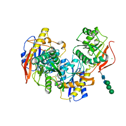
Crystal structure of human butyrylcholinesterase in complex with tert-butyl 3-(((2-((1-(benzenesulfonyl)-1H-indol-4-yl)oxy)ethyl)amino)methyl)piperidine-1-carboxylate
分子名称: 2-(N-MORPHOLINO)-ETHANESULFONIC ACID, 2-acetamido-2-deoxy-beta-D-glucopyranose, 2-acetamido-2-deoxy-beta-D-glucopyranose-(1-4)-[alpha-L-fucopyranose-(1-6)]2-acetamido-2-deoxy-beta-D-glucopyranose, ...
著者Brazzolotto, X, Wichur, T, Wieckowska, A.
登録日2020-11-08
公開日2021-09-29
最終更新日2024-01-31
実験手法X-RAY DIFFRACTION (2.3 Å)
主引用文献Development and crystallography-aided SAR studies of multifunctional BuChE inhibitors and 5-HT 6 R antagonists with beta-amyloid anti-aggregation properties.
Eur.J.Med.Chem., 225, 2021
7AEZ
DownloadVisualize
BU of 7aez by Molmil
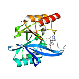
Crystal structure of the metallo-beta-lactamase NDM-7 with 407
分子名称: 1,2-ETHANEDIOL, 2-ETHOXYETHANOL, 7-propan-2-yl-3-[4-(1,2,4-triazol-1-ylmethyl)phenyl]-1~{H}-indole-2-carboxylic acid, ...
著者Brem, J, Schofield, C.J.
登録日2020-09-18
公開日2021-10-06
最終更新日2024-01-31
実験手法X-RAY DIFFRACTION (1.018 Å)
主引用文献Not available yet
To Be Published
7AFY
DownloadVisualize
BU of 7afy by Molmil
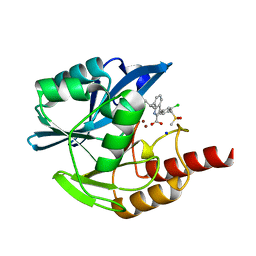
Crystal structure of the metallo-beta-lactamase VIM1 with 1306
分子名称: 3-[3-chloranyl-4-(methylsulfonylmethyl)phenyl]-7-propan-2-yl-1~{H}-indole-2-carboxylic acid, Beta-lactamase VIM-1, SODIUM ION, ...
著者Brem, J, Schofield, C.J.
登録日2020-09-21
公開日2021-10-06
最終更新日2024-01-31
実験手法X-RAY DIFFRACTION (1.105 Å)
主引用文献Crystal structure of the metallo-beta-lactamase VIM2 with 139
To Be Published
7AKT
DownloadVisualize
BU of 7akt by Molmil
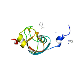
CrPetF variant - A39G_A41V
分子名称: BENZAMIDINE, CHLORIDE ION, FE2/S2 (INORGANIC) CLUSTER, ...
著者Kurisu, G, Ohnishi, Y, Engelbrecht, V, Happe, T.
登録日2020-10-02
公開日2021-10-13
最終更新日2024-01-31
実験手法X-RAY DIFFRACTION (1.11 Å)
主引用文献Ferredoxin 2.0: an electron transfer protein designed into a photosystem I-driven hydrogenase
To Be Published

222624

件を2024-07-17に公開中

PDB statisticsPDBj update infoContact PDBjnumon