1Z0R
 
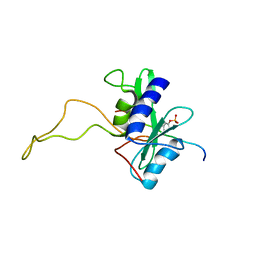
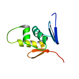
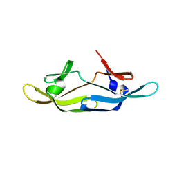
 | | Solution Structure of the N-terminal DNA Recognition Domain of the Bacillus subtilis Transcription-State Regulator AbrB | | 分子名称: | Transition state regulatory protein abrB | | 著者 | Bobay, B.G, Andreeva, A, Mueller, G.A, Cavanagh, J, Murzin, A.G. | | 登録日 | 2005-03-02 | | 公開日 | 2005-03-15 | | 最終更新日 | 2024-05-29 | | 実験手法 | SOLUTION NMR | | 主引用文献 | Revised structure of the AbrB N-terminal domain unifies a diverse superfamily of putative DNA-binding proteins.
Febs Lett., 579, 2005
|
|
1BBI
 | |
1AYI
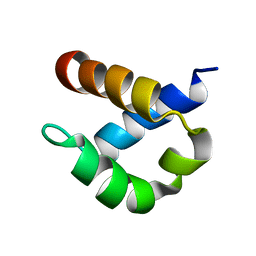
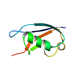
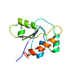
 | | COLICIN E7 IMMUNITY PROTEIN IM7 | | 分子名称: | COLICIN E IMMUNITY PROTEIN 7 | | 著者 | Dennis, C.A, Pauptit, R.A, Wallis, R, James, R, Moore, G.R, Kleanthous, C. | | 登録日 | 1997-11-04 | | 公開日 | 1998-01-28 | | 最終更新日 | 2024-04-03 | | 実験手法 | X-RAY DIFFRACTION (2 Å) | | 主引用文献 | A structural comparison of the colicin immunity proteins Im7 and Im9 gives new insights into the molecular determinants of immunity-protein specificity.
Biochem.J., 333 ( Pt 1), 1998
|
|
5Z31
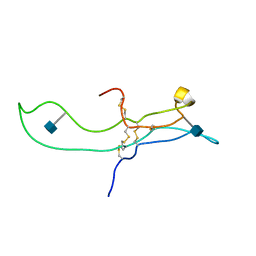
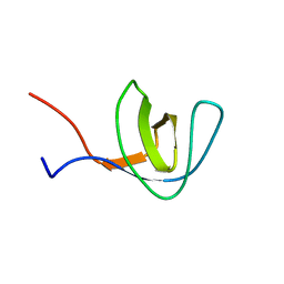
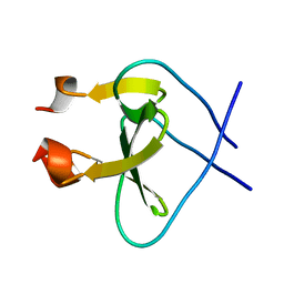
 | | LPS bound solution structure of WS2-KG18 | | 分子名称: | LYS-ASN-LYS-SER-ARG-VAL-ALA-ARG-GLY-TRP-GLY-ARG-LYS-CYS-PRO-LEU-PHE-GLY | | 著者 | Bhunia, A, Mohid, S.A. | | 登録日 | 2018-01-05 | | 公開日 | 2019-02-06 | | 最終更新日 | 2024-05-15 | | 実験手法 | SOLUTION NMR | | 主引用文献 | Application of tungsten disulfide quantum dot-conjugated antimicrobial peptides in bio-imaging and antimicrobial therapy.
Colloids Surf B Biointerfaces, 176, 2019
|
|
1E9J
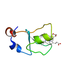
 | | SOLUTION STRUCTURE OF THE A-SUBUNIT OF HUMAN CHORIONIC GONADOTROPIN [INCLUDING A SINGLE GLCNAC RESIDUE AT ASN52 AND ASN78] | | 分子名称: | 2-acetamido-2-deoxy-beta-D-glucopyranose, CHORIONIC GONADOTROPIN | | 著者 | Erbel, P.J.A, Karimi-Nejad, Y, van Kuik, J.A, Boelens, R, Kamerling, J.P, Vliegenthart, J.F.G. | | 登録日 | 2000-10-18 | | 公開日 | 2002-07-25 | | 最終更新日 | 2020-07-29 | | 実験手法 | SOLUTION NMR | | 主引用文献 | Effects of the N-linked glycans on the 3D structure of the free alpha-subunit of human chorionic gonadotropin.
Biochemistry, 39, 2000
|
|
2B7E
 | | First FF domain of Prp40 Yeast Protein | | 分子名称: | Pre-mRNA processing protein PRP40 | | 著者 | Gasch, A, Wiesner, S, Martin-Malpartida, P, Ramirez-Espain, X, Ruiz, L, Macias, M.J. | | 登録日 | 2005-10-04 | | 公開日 | 2005-11-01 | | 最終更新日 | 2024-05-22 | | 実験手法 | SOLUTION NMR | | 主引用文献 | The structure of Prp40 FF1 domain and its interaction with the crn-TPR1 motif of Clf1 gives a new insight into the binding mode of FF domains.
J.Biol.Chem., 281, 2006
|
|
1E8Q
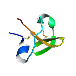 | | Characterisation of the cellulose docking domain from Piromyces equi | | 分子名称: | Endoglucanase 45A | | 著者 | Raghothama, S, Eberhardt, R.Y, White, P, Hazlewood, G.P, Gilbert, H.J, Simpson, P.J, Williamson, M.P. | | 登録日 | 2000-09-28 | | 公開日 | 2001-09-07 | | 最終更新日 | 2018-06-20 | | 実験手法 | SOLUTION NMR | | 主引用文献 | Characterization of a cellulosome dockerin domain from the anaerobic fungus Piromyces equi.
Nat. Struct. Biol., 8, 2001
|
|
1C17
 | |
2BBU
 | |
1ZKH
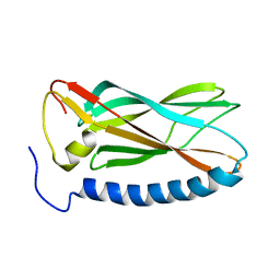
 | | Solution structure of a human ubiquitin-like domain in SF3A1 | | 分子名称: | Splicing factor 3 subunit 1 | | 著者 | Lukin, J.A, Dhe-Paganon, S, Guido, V, Lemak, A, Avvakumov, G.V, Xue, S, Newman, E.M, Mackenzie, F, Sundstrom, M, Edwards, A, Arrowsmith, C.H, Structural Genomics Consortium (SGC) | | 登録日 | 2005-05-02 | | 公開日 | 2005-05-10 | | 最終更新日 | 2024-05-22 | | 実験手法 | SOLUTION NMR | | 主引用文献 | Solution structure of a human ubiquitin-like domain in SF3A1
To be Published
|
|
1YYC
 | |
1Z9Q
 | |
1Z8M
 | |
1BW4
 | |
2AN7
 | | Solution structure of the bacterial antidote ParD | | 分子名称: | Protein parD | | 著者 | Oberer, M, Zangger, K, Gruber, K, Keller, W. | | 登録日 | 2005-08-11 | | 公開日 | 2006-09-05 | | 最終更新日 | 2024-05-15 | | 実験手法 | SOLUTION NMR | | 主引用文献 | The solution structure of ParD, the antidote of the ParDE toxin antitoxin module, provides the structural basis for DNA and toxin binding.
Protein Sci., 16, 2007
|
|
1C15
 | | SOLUTION STRUCTURE OF APAF-1 CARD | | 分子名称: | APOPTOTIC PROTEASE ACTIVATING FACTOR 1 | | 著者 | Zhou, P, Chou, J, Olea, R.S, Yuan, J, Wagner, G. | | 登録日 | 1999-07-20 | | 公開日 | 1999-09-20 | | 最終更新日 | 2024-05-22 | | 実験手法 | SOLUTION NMR | | 主引用文献 | Solution structure of Apaf-1 CARD and its interaction with caspase-9 CARD: a structural basis for specific adaptor/caspase interaction.
Proc.Natl.Acad.Sci.USA, 96, 1999
|
|
1BNX
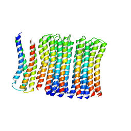
 | | STRUCTURAL STUDIES ON THE EFFECTS OF THE DELETION IN THE RED CELL ANION EXCHANGER (BAND3, AE1) ASSOCIATED WITH SOUTH EAST ASIAN OVALOCYTOSIS. | | 分子名称: | PROTEIN (BAND 3) | | 著者 | Chambers, E.J, Bloomberg, G.B, Ring, S.M, Tanner, M.J.A. | | 登録日 | 1998-07-30 | | 公開日 | 1998-08-05 | | 最終更新日 | 2022-02-16 | | 実験手法 | SOLUTION NMR | | 主引用文献 | Studies on the structure of a transmembrane region and a cytoplasmic loop of the human red cell anion exchanger (band 3, AE1).
Biochem.Soc.Trans., 26, 1998
|
|
1E8P
 | | Characterisation of the cellulose docking domain from Piromyces equi | | 分子名称: | Endoglucanase 45A | | 著者 | Raghothama, S, Eberhardt, R.Y, White, P, Hazlewood, G.P, Gilbert, H.J, Simpson, P.J, Williamson, M.P. | | 登録日 | 2000-09-28 | | 公開日 | 2001-09-07 | | 最終更新日 | 2018-06-20 | | 実験手法 | SOLUTION NMR | | 主引用文献 | Characterization of a cellulosome dockerin domain from the anaerobic fungus Piromyces equi.
Nat. Struct. Biol., 8, 2001
|
|
2A37
 | | Solution structure of the T22G mutant of N-terminal SH3 domain of DRK (DRKN SH3 DOMAIN) | | 分子名称: | Protein E(sev)2B | | 著者 | Bezsonova, I, Singer, A, Choy, W.-Y, Tollinger, M, Forman-Kay, J.D. | | 登録日 | 2005-06-23 | | 公開日 | 2005-12-13 | | 最終更新日 | 2024-05-22 | | 実験手法 | SOLUTION NMR | | 主引用文献 | Structural Comparison of the Unstable drkN SH3 Domain and a Stable Mutant
Biochemistry, 44, 2005
|
|
1BW3
 | |
1JEX
 | |
1P8A
 | |
1RFO
 | | Trimeric Foldon of the T4 phagehead fibritin | | 分子名称: | whisker antigen control protein | | 著者 | Guthe, S, Kapinos, L, Moglich, A, Meier, S, Kiefhaber, T, Grzesiek, S. | | 登録日 | 2003-11-10 | | 公開日 | 2004-03-30 | | 最終更新日 | 2024-05-22 | | 実験手法 | SOLUTION NMR | | 主引用文献 | Very fast folding and association of a trimerization domain from bacteriophage t4 fibritin.
J.Mol.Biol., 337, 2004
|
|
2M87
 | |
2RDK
 | | Five site mutated Cyanovirin-N with Mannose dimer bound | | 分子名称: | Cyanovirin-N, alpha-D-mannopyranose-(1-2)-alpha-D-mannopyranose | | 著者 | Fromme, R, Katiliene, Z, Fromme, P, Ghirlanda, G. | | 登録日 | 2007-09-24 | | 公開日 | 2008-05-06 | | 最終更新日 | 2023-08-30 | | 実験手法 | X-RAY DIFFRACTION (1.35 Å) | | 主引用文献 | Conformational gating of dimannose binding to the antiviral protein cyanovirin revealed from the crystal structure at 1.35 A resolution.
Protein Sci., 17, 2008
|
|


