2QMW
 
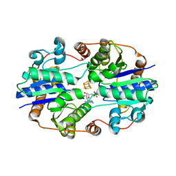
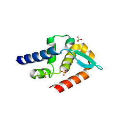
 | | The crystal structure of the prephenate dehydratase (PDT) from Staphylococcus aureus subsp. aureus Mu50 | | 分子名称: | 1,2-ETHANEDIOL, ACETATE ION, DI(HYDROXYETHYL)ETHER, ... | | 著者 | Tan, K, Zhang, R, Li, H, Gu, M, Joachimiak, A, Midwest Center for Structural Genomics (MCSG) | | 登録日 | 2007-07-17 | | 公開日 | 2007-08-07 | | 最終更新日 | 2024-10-16 | | 実験手法 | X-RAY DIFFRACTION (2.3 Å) | | 主引用文献 | Structures of open (R) and close (T) states of prephenate dehydratase (PDT) - implication of allosteric regulation by L-phenylalanine.
J.Struct.Biol., 162, 2008
|
|
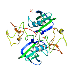
2QJ4
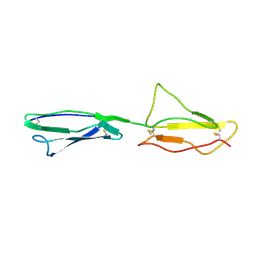
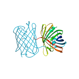
 | | A Mechanistic Basis for Converting a Receptor Tyrosine Kinase Agonist to an Antagonist | | 分子名称: | Hepatocyte growth factor, SULFATE ION | | 著者 | Tolbert, W.D, Daugherty, J, Gao, C.-F, Xe, Q, Miranti, C, Gherardi, E, Vande Woude, G, Xu, H.E. | | 登録日 | 2007-07-06 | | 公開日 | 2007-09-18 | | 最終更新日 | 2023-08-30 | | 実験手法 | X-RAY DIFFRACTION (2.5 Å) | | 主引用文献 | A mechanistic basis for converting a receptor tyrosine kinase agonist to an antagonist
Proc.Natl.Acad.Sci.Usa, 104, 2007
|
|
1PYO
 | |
1NAY
 | | GPP-Foldon:X-ray structure | | 分子名称: | Collagen-like peptide | | 著者 | Stetefeld, J. | | 登録日 | 2002-11-29 | | 公開日 | 2003-03-25 | | 最終更新日 | 2024-03-13 | | 実験手法 | X-RAY DIFFRACTION (2.6 Å) | | 主引用文献 | Collagen Stabilization at Atomic Level. Crystal Structure of Designed (GlyProPro)(10)foldon
Structure, 11, 2003
|
|
1NCN
 | |
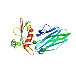
1NDG
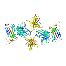
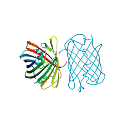
 | | Crystal structure of Fab fragment of antibody HyHEL-8 complexed with its antigen lysozyme | | 分子名称: | ACETIC ACID, Lysozyme C, antibody kappa light chain, ... | | 著者 | Mariuzza, R.A, Li, Y, Li, H, Yang, F, Smith-Gill, S.J. | | 登録日 | 2002-12-09 | | 公開日 | 2003-06-03 | | 最終更新日 | 2024-10-16 | | 実験手法 | X-RAY DIFFRACTION (1.9 Å) | | 主引用文献 | X-ray snapshots of the maturation of an antibody response to a protein antigen
Nat.Struct.Biol., 10, 2003
|
|
2QMX
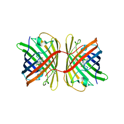
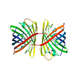
 | | The crystal structure of L-Phe inhibited prephenate dehydratase from Chlorobium tepidum TLS | | 分子名称: | 1,2-ETHANEDIOL, ACETATE ION, PHENYLALANINE, ... | | 著者 | Tan, K, Li, H, Clancy, S, Joachimiak, A, Midwest Center for Structural Genomics (MCSG) | | 登録日 | 2007-07-17 | | 公開日 | 2007-08-07 | | 最終更新日 | 2011-07-13 | | 実験手法 | X-RAY DIFFRACTION (2.3 Å) | | 主引用文献 | Structures of open (R) and close (T) states of prephenate dehydratase (PDT) - implication of allosteric regulation by L-phenylalanine.
J.Struct.Biol., 162, 2008
|
|
1NH9
 | | Crystal Structure of a DNA Binding Protein Mja10b from the hyperthermophile Methanococcus jannaschii | | 分子名称: | DNA-binding protein Alba | | 著者 | Wang, G, Bartlam, M, Guo, R, Yang, H, Xue, H, Liu, Y, Huang, L, Rao, Z. | | 登録日 | 2002-12-19 | | 公開日 | 2003-12-23 | | 最終更新日 | 2023-10-25 | | 実験手法 | X-RAY DIFFRACTION (2 Å) | | 主引用文献 | Crystal structure of a DNA binding protein from the hyperthermophilic euryarchaeon Methanococcus jannaschii
Protein Sci., 12, 2003
|
|
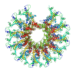
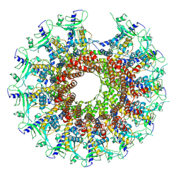
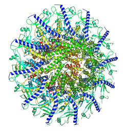
1NIP
 | | CRYSTALLOGRAPHIC STRUCTURE OF THE NITROGENASE IRON PROTEIN FROM AZOTOBACTER VINELANDII | | 分子名称: | ADENOSINE-5'-DIPHOSPHATE, IRON/SULFUR CLUSTER, MAGNESIUM ION, ... | | 著者 | Komiya, H, Georgiadis, M.M, Chakrabarti, P, Woo, D, Kornuc, J.J, Rees, D.C. | | 登録日 | 1992-09-29 | | 公開日 | 1993-10-31 | | 最終更新日 | 2024-02-14 | | 実験手法 | X-RAY DIFFRACTION (2.9 Å) | | 主引用文献 | Crystallographic structure of the nitrogenase iron protein from Azotobacter vinelandii.
Science, 257, 1992
|
|
2QJ2
 | | A Mechanistic Basis for Converting a Receptor Tyrosine Kinase Agonist to an Antagonist | | 分子名称: | Hepatocyte growth factor, SULFATE ION | | 著者 | Tolbert, W.D, Daugherty, J, Gao, C.-F, Xe, Q, Miranti, C, Gherardi, E, Vande Woude, G, Xu, H.E. | | 登録日 | 2007-07-06 | | 公開日 | 2007-09-18 | | 最終更新日 | 2024-10-16 | | 実験手法 | X-RAY DIFFRACTION (1.81 Å) | | 主引用文献 | A mechanistic basis for converting a receptor tyrosine kinase agonist to an antagonist
Proc.Natl.Acad.Sci.Usa, 104, 2007
|
|
1QTS
 | |
1R2M
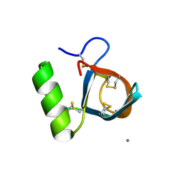 | | Atomic resolution structure of the HFBII hydrophobin: a self-assembling amphiphile | | 分子名称: | Hydrophobin II, MANGANESE (II) ION | | 著者 | Hakanpaa, J, Paananen, A, Askolin, S, Nakari-Setala, T, Parkkinen, T, Penttila, M, Linder, M.B, Rouvinen, J. | | 登録日 | 2003-09-29 | | 公開日 | 2004-01-13 | | 最終更新日 | 2011-07-13 | | 実験手法 | X-RAY DIFFRACTION (1 Å) | | 主引用文献 | Atomic resolution structure of the HFBII hydrophobin, a self-assembling amphiphile.
J.Biol.Chem., 279, 2004
|
|
2RLQ
 | | NMR structure of CCP modules 2-3 of complement factor H | | 分子名称: | Complement factor H | | 著者 | Hocking, H.G, Herbert, A.P, Pangburn, M.K, Kavanagh, D, Barlow, P.N, Uhrin, D. | | 登録日 | 2007-07-29 | | 公開日 | 2008-02-19 | | 最終更新日 | 2024-10-16 | | 実験手法 | SOLUTION NMR | | 主引用文献 | Structure of the N-terminal region of complement factor H and conformational implications of disease-linked sequence variations.
J.Biol.Chem., 283, 2008
|
|
7TE1
 | |
7SZ6
 | | Kinetically trapped Pseudomonas-phage PaP3 portal protein - delta barrel mutant class-3 | | 分子名称: | Portal protein | | 著者 | Hou, C.-F.D, Swanson, N.A, Li, F, Yang, R, Lokareddy, R.K, Cingolani, G. | | 登録日 | 2021-11-25 | | 公開日 | 2022-03-30 | | 最終更新日 | 2024-02-28 | | 実験手法 | ELECTRON MICROSCOPY (6.24 Å) | | 主引用文献 | Cryo-EM Structure of a Kinetically Trapped Dodecameric Portal Protein from the Pseudomonas-phage PaP3.
J.Mol.Biol., 434, 2022
|
|
7SZ4
 | | Kinetically trapped Pseudomonas-phage PaP3 portal protein - delta barrel mutant class-2 | | 分子名称: | Portal protein | | 著者 | Hou, C.-F.D, Swanson, N.A, Li, F, Yang, R, Lokareddy, R.K, Cingolani, G. | | 登録日 | 2021-11-25 | | 公開日 | 2022-03-30 | | 最終更新日 | 2024-02-28 | | 実験手法 | ELECTRON MICROSCOPY (4.8 Å) | | 主引用文献 | Cryo-EM Structure of a Kinetically Trapped Dodecameric Portal Protein from the Pseudomonas-phage PaP3.
J.Mol.Biol., 434, 2022
|
|
7SWS
 | | Crystal structure of the chromoprotein amilCP | | 分子名称: | BROMIDE ION, CHLORIDE ION, Chromoprotein amilCP | | 著者 | Caputo, A.T, Newman, J, Scott, C, Ahmed, H. | | 登録日 | 2021-11-21 | | 公開日 | 2022-04-20 | | 最終更新日 | 2024-10-09 | | 実験手法 | X-RAY DIFFRACTION (1.642 Å) | | 主引用文献 | Over the rainbow: structural characterization of the chromoproteins gfasPurple, amilCP, spisPink and eforRed.
Acta Crystallogr D Struct Biol, 78, 2022
|
|
7SYA
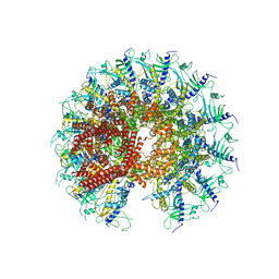
 | | Kinetically trapped Pseudomonas-phage PaP3 portal protein - Full Length | | 分子名称: | Portal protein | | 著者 | Hou, C.F.D, Swanson, N.A, Li, F, Yang, R, Lokareddy, R.K, Cingolani, G. | | 登録日 | 2021-11-24 | | 公開日 | 2022-04-20 | | 最終更新日 | 2024-02-28 | | 実験手法 | ELECTRON MICROSCOPY (3.5 Å) | | 主引用文献 | Cryo-EM Structure of a Kinetically Trapped Dodecameric Portal Protein from the Pseudomonas-phage PaP3.
J.Mol.Biol., 434, 2022
|
|
7SWT
 | | Crystal structure of the chromoprotein eforRED | | 分子名称: | Chromoprotein eforRED | | 著者 | Caputo, A.T, Newman, J, Scott, C, Ahmed, H. | | 登録日 | 2021-11-21 | | 公開日 | 2022-04-20 | | 最終更新日 | 2023-11-15 | | 実験手法 | X-RAY DIFFRACTION (2.005 Å) | | 主引用文献 | Over the rainbow: structural characterization of the chromoproteins gfasPurple, amilCP, spisPink and eforRed.
Acta Crystallogr D Struct Biol, 78, 2022
|
|
7T5H
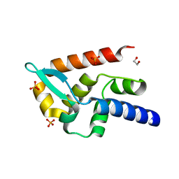 | | Structure of rabies virus phosphoprotein C-terminal domain, wild type | | 分子名称: | 1,2-ETHANEDIOL, PHOSPHATE ION, Phosphoprotein, ... | | 著者 | Zhan, J, Metcalfe, R.D, Gooley, P.R, Griffin, M.D.W. | | 登録日 | 2021-12-12 | | 公開日 | 2022-04-20 | | 最終更新日 | 2023-10-18 | | 実験手法 | X-RAY DIFFRACTION (1.5 Å) | | 主引用文献 | Molecular Basis of Functional Effects of Phosphorylation of the C-Terminal Domain of the Rabies Virus P Protein.
J.Virol., 96, 2022
|
|
7T5G
 | | Structure of rabies virus phosphoprotein C-terminal domain, S210E mutant | | 分子名称: | Phosphoprotein, SULFATE ION | | 著者 | Zhan, J, Metcalfe, R.D, Gooley, P.R, Griffin, M.D.W. | | 登録日 | 2021-12-12 | | 公開日 | 2022-04-20 | | 最終更新日 | 2023-10-18 | | 実験手法 | X-RAY DIFFRACTION (1.7 Å) | | 主引用文献 | Molecular Basis of Functional Effects of Phosphorylation of the C-Terminal Domain of the Rabies Virus P Protein.
J.Virol., 96, 2022
|
|
7SWR
 | | Crystal structure of the chromoprotein gfasPurple | | 分子名称: | CHLORIDE ION, Chromoprotein gfasPurple | | 著者 | Caputo, A.T, Newman, J, Peat, T.S, Scott, C, Ahmed, H. | | 登録日 | 2021-11-21 | | 公開日 | 2022-04-20 | | 最終更新日 | 2023-11-15 | | 実験手法 | X-RAY DIFFRACTION (1.388 Å) | | 主引用文献 | Over the rainbow: structural characterization of the chromoproteins gfasPurple, amilCP, spisPink and eforRed.
Acta Crystallogr D Struct Biol, 78, 2022
|
|
7SXK
 | | Kinetically trapped Pseudomonas-phage PaP3 portal protein - Full Length | | 分子名称: | Portal protein | | 著者 | Hou, C.F.D, Swanson, N.A, Li, F, Yang, R, Lokareddy, R.K, Cingolani, G. | | 登録日 | 2021-11-23 | | 公開日 | 2022-04-20 | | 最終更新日 | 2024-10-09 | | 実験手法 | ELECTRON MICROSCOPY (3.4 Å) | | 主引用文献 | Cryo-EM Structure of a Kinetically Trapped Dodecameric Portal Protein from the Pseudomonas-phage PaP3.
J.Mol.Biol., 434, 2022
|
|
7SWU
 | | Crystal structure of the chromoprotein spisPINK | | 分子名称: | Chromoprotein spisPINK | | 著者 | Caputo, A.T, Newman, J, Scott, C, Ahmed, H. | | 登録日 | 2021-11-21 | | 公開日 | 2022-04-20 | | 最終更新日 | 2024-07-10 | | 実験手法 | X-RAY DIFFRACTION (1.444 Å) | | 主引用文献 | Over the rainbow: structural characterization of the chromoproteins gfasPurple, amilCP, spisPink and eforRed.
Acta Crystallogr D Struct Biol, 78, 2022
|
|
7SQO
 | | Structure of the orexin-2 receptor(OX2R) bound to TAK-925, Gi and scFv16 | | 分子名称: | Guanine nucleotide-binding protein G(I)/G(S)/G(O) subunit gamma-2, Guanine nucleotide-binding protein G(I)/G(S)/G(T) subunit beta-1, Guanine nucleotide-binding protein G(i) subunit alpha-1, ... | | 著者 | McGrath, A.P, Kang, Y, Flinspach, M. | | 登録日 | 2021-11-05 | | 公開日 | 2022-05-25 | | 最終更新日 | 2022-07-06 | | 実験手法 | ELECTRON MICROSCOPY (3.17 Å) | | 主引用文献 | Molecular mechanism of the wake-promoting agent TAK-925.
Nat Commun, 13, 2022
|
|


