2JVJ
 
 | |
2LL3
 | |
2JZQ
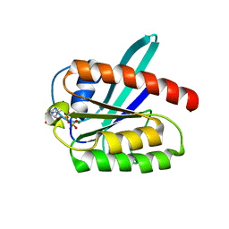
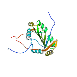
 | | Design of an Active Ultra-Stable Single-Chain Insulin Analog 20 Structures | | 分子名称: | Insulin | | 著者 | Hua, Q.X, Nakarawa, S, Jia, W.H, Huang, K, Philips, N.F, Hu, S.Q, Weiss, M.A. | | 登録日 | 2008-01-11 | | 公開日 | 2008-02-26 | | 最終更新日 | 2021-10-20 | | 実験手法 | SOLUTION NMR | | 主引用文献 | Design of an active ultrastable single-chain insulin analog: synthesis, structure, and therapeutic implications.
J.Biol.Chem., 283, 2008
|
|
2K0G
 | |
2L0X
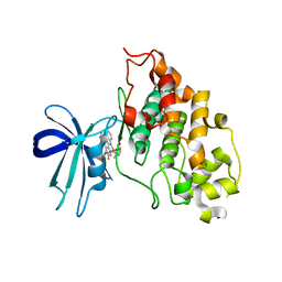
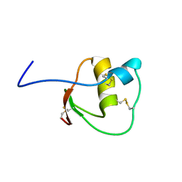
 | | Solution structure of the 21 kDa GTPase RHEB bound to GDP | | 分子名称: | GTP-binding protein Rheb, GUANOSINE-5'-DIPHOSPHATE, MAGNESIUM ION | | 著者 | Stoll, R, Heumann, R, Berghaus, C, Kock, G. | | 登録日 | 2010-07-19 | | 公開日 | 2010-08-04 | | 最終更新日 | 2024-05-01 | | 実験手法 | SOLUTION NMR | | 主引用文献 | Ras homolog enriched in brain (Rheb) enhances apoptotic signaling.
J.Biol.Chem., 285, 2010
|
|
2JLD
 | | Extremely Tight Binding of Ruthenium Complex to Glycogen Synthase Kinase 3 | | 分子名称: | GLYCOGEN SYNTHASE KINASE-3 BETA, PEPTIDE (ALA-GLY-GLY-ALA-ALA-ALA-ALA-ALA), RUTHENIUM PYRIDOCARBAZOLE | | 著者 | Atilla-Gokcumen, G.E, Pagano, N, Streu, C, Maksimoska, J, Filippakopoulos, P, Knapp, S, Meggers, E. | | 登録日 | 2008-09-08 | | 公開日 | 2008-12-09 | | 最終更新日 | 2023-12-13 | | 実験手法 | X-RAY DIFFRACTION (2.35 Å) | | 主引用文献 | Extremely Tight Binding of a Ruthenium Complex to Glycogen Synthase Kinase 3.
Chembiochem, 9, 2008
|
|
2L2M
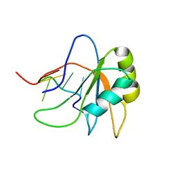
 | | Solution structure of the second dsRBD of HYL1 | | 分子名称: | Hyponastic leave 1 | | 著者 | Rasia, R.M, Mateos, J.L, Bologna, N.G, Burdisso, P, Imbert, L, Palatnik, J.F, Boisbouvier, J. | | 登録日 | 2010-08-22 | | 公開日 | 2010-09-29 | | 最終更新日 | 2024-05-01 | | 実験手法 | SOLUTION NMR | | 主引用文献 | Structure and RNA Interactions of the Plant MicroRNA Processing-Associated Protein HYL1.
Biochemistry, 49, 2010
|
|
2JRS
 | | Solution NMR Structure of CAPER RRM2 Domain. Northeast Structural Genomics Target HR4730A | | 分子名称: | RNA-binding protein 39 | | 著者 | Rossi, P, Zhao, L, Nwosu, C, Cunningham, K, Owens, L, Xiao, R, Liu, J, Baran, M.C, Swapna, G, Acton, T.B, Rost, B, Montelione, G.T, Northeast Structural Genomics Consortium (NESG) | | 登録日 | 2007-06-28 | | 公開日 | 2007-09-04 | | 最終更新日 | 2024-05-01 | | 実験手法 | SOLUTION NMR | | 主引用文献 | Solution NMR Structure of CAPER RRM2 Domain.
To be Published
|
|
2LMV
 | | Androcam at high calcium with three explicit Ca2+ | | 分子名称: | CALCIUM ION, Calmodulin-related protein 97A | | 著者 | Joshi, M.K, Moran, S.T, Beckingham, K.M, Mackenzie, K.R. | | 登録日 | 2011-12-12 | | 公開日 | 2012-08-22 | | 最終更新日 | 2024-05-01 | | 実験手法 | SOLUTION NMR | | 主引用文献 | Structure of androcam supports specialized interactions with myosin VI.
Proc.Natl.Acad.Sci.USA, 109, 2012
|
|
2JLP
 | | Crystal structure of human extracellular copper-zinc superoxide dismutase. | | 分子名称: | COPPER (II) ION, EXTRACELLULAR SUPEROXIDE DISMUTASE (CU-ZN), THIOCYANATE ION, ... | | 著者 | Antonyuk, S.V, Strange, R.W, Marklund, S.L, Hasnain, S.S. | | 登録日 | 2008-09-14 | | 公開日 | 2009-03-17 | | 最終更新日 | 2023-12-13 | | 実験手法 | X-RAY DIFFRACTION (1.7 Å) | | 主引用文献 | The Structure of Human Extracellular Copper-Zinc Superoxide Dismutase at 1.7 A Resolution: Insights Into Heparin and Collagen Binding.
J.Mol.Biol., 388, 2009
|
|
2L7M
 | |
2JVO
 | | Segmental isotope labeling of Npl3 | | 分子名称: | Nucleolar protein 3 | | 著者 | Skrisovska, L, Allain, F.H.-T. | | 登録日 | 2007-09-24 | | 公開日 | 2007-12-18 | | 最終更新日 | 2024-05-01 | | 実験手法 | SOLUTION NMR | | 主引用文献 | Improved segmental isotope labeling methods for the NMR study of multidomain or large proteins: application to the RRMs of Npl3p and hnRNP L
J.Mol.Biol., 375, 2008
|
|
2JWN
 | |
2LEO
 | |
2LEB
 | | Solution structure of human SRSF2 (SC35) RRM in complex with 5'-UCCAGU-3' | | 分子名称: | RNA (5'-R(*UP*CP*CP*AP*GP*U)-3'), Serine/arginine-rich splicing factor 2 | | 著者 | Daubner, G.M, Clery, A, Jayne, S, Stevenin, J, Allain, F.H.-T. | | 登録日 | 2011-06-15 | | 公開日 | 2011-11-23 | | 最終更新日 | 2024-05-08 | | 実験手法 | SOLUTION NMR | | 主引用文献 | A syn-anti conformational difference allows SRSF2 to recognize guanines and cytosines equally well.
Embo J., 31, 2012
|
|
2GEE
 | | Crystal Structure of Human Type III Fibronectin Extradomain B and Domain 8 | | 分子名称: | hypothetical protein | | 著者 | Bencharit, S, Cui, C.B, Siddiqui, A, Howard-Williams, E.L, Aukhil, I. | | 登録日 | 2006-03-19 | | 公開日 | 2006-10-17 | | 最終更新日 | 2023-08-30 | | 実験手法 | X-RAY DIFFRACTION (2.01 Å) | | 主引用文献 | Structural insights into fibronectin type III domain-mediated signaling.
J.Mol.Biol., 367, 2007
|
|
2GHP
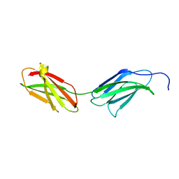
 | | Crystal structure of the N-terminal 3 RNA binding domains of the yeast splicing factor Prp24 | | 分子名称: | U4/U6 snRNA-associated splicing factor PRP24 | | 著者 | Bae, E, Wesenberg, G.E, Phillips Jr, G.N, Bitto, E, Bingman, C.A, Center for Eukaryotic Structural Genomics (CESG) | | 登録日 | 2006-03-27 | | 公開日 | 2006-04-25 | | 最終更新日 | 2017-10-18 | | 実験手法 | X-RAY DIFFRACTION (2.7 Å) | | 主引用文献 | Structure and interactions of the first three RNA recognition motifs of splicing factor prp24.
J.Mol.Biol., 367, 2007
|
|
2GGZ
 | |
2L2P
 | | Folding Intermediate of the Fyn SH3 A39V/N53P/V55L from NMR Relaxation Dispersion Experiments | | 分子名称: | Tyrosine-protein kinase Fyn | | 著者 | Neudecker, P, Robustelli, P, Cavalli, A, Vendruscolo, M, Kay, L.E. | | 登録日 | 2010-08-25 | | 公開日 | 2011-09-14 | | 最終更新日 | 2024-05-01 | | 実験手法 | SOLUTION NMR | | 主引用文献 | Structure of an intermediate state in protein folding and aggregation.
Science, 336, 2012
|
|
2LT5
 | | Zymogen-FLG of the onconase | | 分子名称: | Protein P-30 | | 著者 | Vilanova, M, Callis, M, Laurents, D.V, Ribo, M, Bruix, M, Serrano, S. | | 登録日 | 2012-05-14 | | 公開日 | 2012-10-24 | | 最終更新日 | 2023-06-14 | | 実験手法 | SOLUTION NMR | | 主引用文献 |
|
|
2L3J
 | |
2FMR
 | | KH1 FROM THE FRAGILE X PROTEIN FMR1, NMR, 18 STRUCTURES | | 分子名称: | FMR1 PROTEIN | | 著者 | Musco, G, Kharrat, A, Stier, G, Fraternali, F, Gibson, T.J, Nilges, M, Pastore, A. | | 登録日 | 1997-10-06 | | 公開日 | 1998-04-08 | | 最終更新日 | 2024-05-29 | | 実験手法 | SOLUTION NMR | | 主引用文献 | The solution structure of the first KH domain of FMR1, the protein responsible for the fragile X syndrome.
Nat.Struct.Biol., 4, 1997
|
|
2GF1
 | |
2GBQ
 | | SOLUTION NMR STRUCTURE OF THE GRB2 N-TERMINAL SH3 DOMAIN COMPLEXED WITH A TEN-RESIDUE PEPTIDE DERIVED FROM SOS DIRECT REFINEMENT AGAINST NOES, J-COUPLINGS, AND 1H AND 13C CHEMICAL SHIFTS, 15 STRUCTURES | | 分子名称: | GRB2, SOS-1 | | 著者 | Wittekind, M, Mapelli, C, Lee, V, Goldfarb, V, Friedrichs, M.S, Meyers, C.A, Mueller, L. | | 登録日 | 1996-12-23 | | 公開日 | 1997-09-04 | | 最終更新日 | 2022-03-09 | | 実験手法 | SOLUTION NMR | | 主引用文献 | Solution structure of the Grb2 N-terminal SH3 domain complexed with a ten-residue peptide derived from SOS: direct refinement against NOEs, J-couplings and 1H and 13C chemical shifts.
J.Mol.Biol., 267, 1997
|
|
2GCT
 | |


