Loading
PDBj
メニューPDBj@FacebookPDBj@TwitterPDBj@YouTubewwPDB FoundationwwPDB
RCSB PDBPDBeBMRBAdv. SearchSearch help

8BGL
DownloadVisualize
BU of 8bgl by Molmil
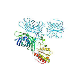
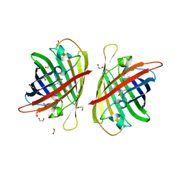
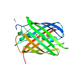
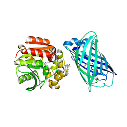
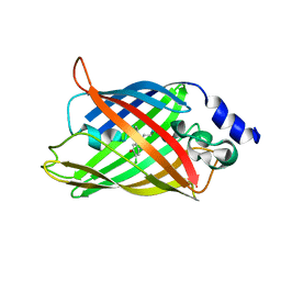
Structure of the dimeric rsCherryRev1.4
分子名称: 1,2-ETHANEDIOL, ALPHA-AMINOBUTYRIC ACID, DI(HYDROXYETHYL)ETHER, ...
著者Bui, T.Y.H, Van Meervelt, L.
登録日2022-10-28
公開日2023-02-15
最終更新日2024-02-07
実験手法X-RAY DIFFRACTION (2 Å)
主引用文献An unusual disulfide-linked dimerization in the fluorescent protein rsCherryRev1.4.
Acta Crystallogr.,Sect.F, 79, 2023
8BBJ
DownloadVisualize
BU of 8bbj by Molmil
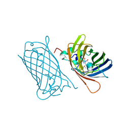
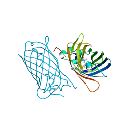
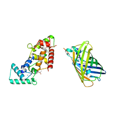
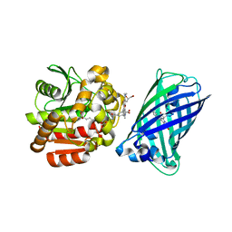
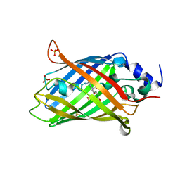
Secretagogin (mouse) in complex with its target peptide from Syntaxin-4
分子名称: CACODYLATE ION, CALCIUM ION, Green fluorescent protein,Syntaxin-4, ...
著者Schnell, R, Szodorai, E.
登録日2022-10-13
公開日2024-04-03
最終更新日2024-04-24
実験手法X-RAY DIFFRACTION (2.65 Å)
主引用文献A hydrophobic groove in secretagogin allows for alternate interactions with SNAP-25 and syntaxin-4 in endocrine tissues.
Proc.Natl.Acad.Sci.USA, 121, 2024
8BAV
DownloadVisualize
BU of 8bav by Molmil
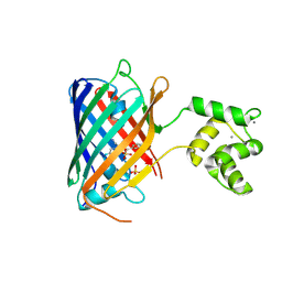
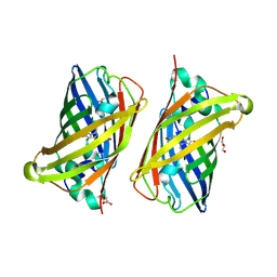
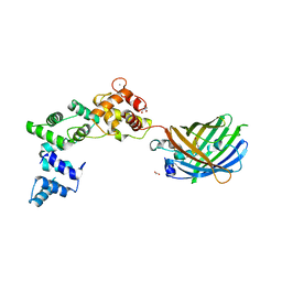
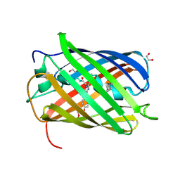
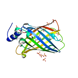
Secretagogin (human) in complex with its target peptide from SNAP-25
分子名称: ACETATE ION, CALCIUM ION, Green fluorescent protein,Synaptosomal-associated protein 25, ...
著者Schnell, R, Szodorai, E.
登録日2022-10-12
公開日2024-04-03
最終更新日2024-04-24
実験手法X-RAY DIFFRACTION (2.3 Å)
主引用文献A hydrophobic groove in secretagogin allows for alternate interactions with SNAP-25 and syntaxin-4 in endocrine tissues.
Proc.Natl.Acad.Sci.USA, 121, 2024
8BAN
DownloadVisualize
BU of 8ban by Molmil
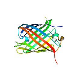
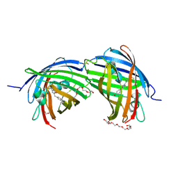
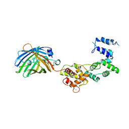
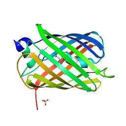
Secretagogin (mouse) in complex with its target peptide from SNAP-25
分子名称: CALCIUM ION, Green fluorescent protein,Synaptosomal-associated protein 25, Secretagogin
著者Schnell, R, Szodorai, E.
登録日2022-10-11
公開日2024-04-03
最終更新日2024-04-24
実験手法X-RAY DIFFRACTION (2.35 Å)
主引用文献A hydrophobic groove in secretagogin allows for alternate interactions with SNAP-25 and syntaxin-4 in endocrine tissues.
Proc.Natl.Acad.Sci.USA, 121, 2024
8B7G
DownloadVisualize
BU of 8b7g by Molmil
Structure of oxygen-degraded rsCherry
分子名称: 1,2-ETHANEDIOL, 2-(N-MORPHOLINO)-ETHANESULFONIC ACID, DI(HYDROXYETHYL)ETHER, ...
著者Bui, T.Y.H, Van Meervelt, L.
登録日2022-09-29
公開日2023-04-05
最終更新日2024-02-07
実験手法X-RAY DIFFRACTION (1.3 Å)
主引用文献Oxygen-induced chromophore degradation in the photoswitchable red fluorescent protein rsCherry.
Int.J.Biol.Macromol., 239, 2023
8B6T
DownloadVisualize
BU of 8b6t by Molmil
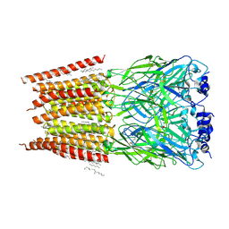
X-ray structure of the interface optimized haloalkane dehalogenase HaloTag7 fusion to the green fluorescent protein GFP (ChemoG5-TMR) labeled with a chloroalkane tetramethylrhodamine fluorophore substrate
分子名称: CHLORIDE ION, Green fluorescent protein,Haloalkane dehalogenase, [9-[2-carboxy-5-[2-[2-(6-chloranylhexoxy)ethoxy]ethylcarbamoyl]phenyl]-6-(dimethylamino)xanthen-3-ylidene]-dimethyl-azanium
著者Tarnawski, M, Hellweg, L, Hiblot, J.
登録日2022-09-27
公開日2023-07-26
最終更新日2023-11-15
実験手法X-RAY DIFFRACTION (2 Å)
主引用文献A general method for the development of multicolor biosensors with large dynamic ranges.
Nat.Chem.Biol., 19, 2023
8B6S
DownloadVisualize
BU of 8b6s by Molmil
X-ray structure of the haloalkane dehalogenase HaloTag7 fusion to the green fluorescent protein GFP (ChemoG1) labeled with a chloroalkane tetramethylrhodamine fluorophore substrate
分子名称: CHLORIDE ION, GLYCEROL, Green fluorescent protein,Haloalkane dehalogenase, ...
著者Tarnawski, M, Hellweg, L, Hiblot, J.
登録日2022-09-27
公開日2023-07-26
最終更新日2023-11-15
実験手法X-RAY DIFFRACTION (1.8 Å)
主引用文献A general method for the development of multicolor biosensors with large dynamic ranges.
Nat.Chem.Biol., 19, 2023
8B65
DownloadVisualize
BU of 8b65 by Molmil
Structure of rsCherry crystallized in anaerobic conditions
分子名称: 1,2-ETHANEDIOL, 2-(N-MORPHOLINO)-ETHANESULFONIC ACID, DI(HYDROXYETHYL)ETHER, ...
著者Bui, T.Y.H, Van Meervelt, L.
登録日2022-09-26
公開日2023-04-05
最終更新日2024-02-07
実験手法X-RAY DIFFRACTION (1.55 Å)
主引用文献Oxygen-induced chromophore degradation in the photoswitchable red fluorescent protein rsCherry.
Int.J.Biol.Macromol., 239, 2023
8ARM
DownloadVisualize
BU of 8arm by Molmil
Crystal structure of LSSmScarlet2
分子名称: LSSmScarlet2, SULFATE ION
著者Samygina, V.R, Subach, O.M, Vlaskina, A.V, Subach, F.V.
登録日2022-08-17
公開日2022-11-02
最終更新日2024-01-31
実験手法X-RAY DIFFRACTION (1.41 Å)
主引用文献LSSmScarlet2 and LSSmScarlet3, Chemically Stable Genetically Encoded Red Fluorescent Proteins with a Large Stokes' Shift.
Int J Mol Sci, 23, 2022
8AM4
DownloadVisualize
BU of 8am4 by Molmil
Cl-rsEGFP2 Long Wavelength Structure
分子名称: Green fluorescent protein
著者Orr, C.M, Fadini, A, van Thor, J.
登録日2022-08-02
公開日2023-08-02
最終更新日2024-01-31
実験手法X-RAY DIFFRACTION (2.02 Å)
主引用文献Serial Femtosecond Crystallography Reveals that Photoactivation in a Fluorescent Protein Proceeds via the Hula Twist Mechanism.
J.Am.Chem.Soc., 2023
8AHB
DownloadVisualize
BU of 8ahb by Molmil
rsEGFP2 photoswitched to its off-state at room temperature and back-switched to its on-state at 100K
分子名称: Green fluorescent protein, SULFATE ION
著者Mantovanelli, A, Adam, V.
登録日2022-07-21
公開日2023-07-19
最終更新日2024-02-07
実験手法X-RAY DIFFRACTION (1.79 Å)
主引用文献Photophysical Studies at Cryogenic Temperature Reveal a Novel Photoswitching Mechanism of rsEGFP2.
J.Am.Chem.Soc., 145, 2023
8AHA
DownloadVisualize
BU of 8aha by Molmil
rsEGFP2 photoswitched to its off-state at 100K
分子名称: Green fluorescent protein, SULFATE ION
著者Mantovanelli, A, Adam, V.
登録日2022-07-21
公開日2023-07-19
最終更新日2024-02-07
実験手法X-RAY DIFFRACTION (2.38 Å)
主引用文献Photophysical Studies at Cryogenic Temperature Reveal a Novel Photoswitching Mechanism of rsEGFP2.
J.Am.Chem.Soc., 145, 2023
8AAB
DownloadVisualize
BU of 8aab by Molmil
S148F mutant of blue-to-red fluorescent timer mRubyFT
分子名称: mRubyFT S148F mutant of blue-to-red fluorescent timer
著者Boyko, K.M, Nikolaeva, A.Y, Vlaskina, A.V, Dorovatovskii, P.V, Subach, O.M, Popov, V.O, Subach, F.V.
登録日2022-06-30
公開日2022-08-17
最終更新日2024-02-07
実験手法X-RAY DIFFRACTION (1.6 Å)
主引用文献mRubyFT/S147I, a mutant of blue-to-red fluorescent timer
Crystallography Reports, 2022
8A83
DownloadVisualize
BU of 8a83 by Molmil
rsEGFP2 with a chlorinated chromophore in the fluorescent ON-state in a crystal dehydrated after illumination
分子名称: Green fluorescent protein, SULFATE ION
著者Fadini, A, van Thor, J, Chang, J.
登録日2022-06-21
公開日2023-07-19
最終更新日2024-02-07
実験手法X-RAY DIFFRACTION (1.81 Å)
主引用文献Serial Femtosecond Crystallography Reveals that Photoactivation in a Fluorescent Protein Proceeds via the Hula Twist Mechanism.
J.Am.Chem.Soc., 145, 2023
8A7V
DownloadVisualize
BU of 8a7v by Molmil
Room temperature rsEGFP2 in its OFF-state obtained with SFX
分子名称: Green fluorescent protein
著者Fadini, A, van Thor, J.
登録日2022-06-21
公開日2023-07-19
最終更新日2024-02-07
実験手法X-RAY DIFFRACTION (1.46 Å)
主引用文献Serial Femtosecond Crystallography Reveals that Photoactivation in a Fluorescent Protein Proceeds via the Hula Twist Mechanism.
J.Am.Chem.Soc., 145, 2023
8A6S
DownloadVisualize
BU of 8a6s by Molmil
Room temperature rsEGFP2 with a chlorinated chromophore 1 microsecond after Photoexcitation
分子名称: Green fluorescent protein
著者Fadini, A, van Thor, J.
登録日2022-06-19
公開日2023-07-19
最終更新日2024-02-07
実験手法X-RAY DIFFRACTION (1.63 Å)
主引用文献Serial Femtosecond Crystallography Reveals that Photoactivation in a Fluorescent Protein Proceeds via the Hula Twist Mechanism.
J.Am.Chem.Soc., 145, 2023
8A6R
DownloadVisualize
BU of 8a6r by Molmil
Room temperature rsEGFP2 with a chlorinated chromophore 100 ps after Photoexcitation
分子名称: Green fluorescent protein
著者Fadini, A, van Thor, J.
登録日2022-06-18
公開日2023-07-19
最終更新日2024-02-07
実験手法X-RAY DIFFRACTION (1.63 Å)
主引用文献Serial Femtosecond Crystallography Reveals that Photoactivation in a Fluorescent Protein Proceeds via the Hula Twist Mechanism.
J.Am.Chem.Soc., 145, 2023
8A6Q
DownloadVisualize
BU of 8a6q by Molmil
Room temperature rsEGFP2 with a chlorinated chromophore 5 ps after photoexcitation
分子名称: Green fluorescent protein
著者Fadini, A, van Thor, J.
登録日2022-06-18
公開日2023-07-19
最終更新日2024-02-07
実験手法X-RAY DIFFRACTION (1.63 Å)
主引用文献Serial Femtosecond Crystallography Reveals that Photoactivation in a Fluorescent Protein Proceeds via the Hula Twist Mechanism.
J.Am.Chem.Soc., 145, 2023
8A6P
DownloadVisualize
BU of 8a6p by Molmil
Room temperature rsEGFP2 with a chlorinated chromophore 900 fs after photoexcitation
分子名称: Green fluorescent protein
著者Fadini, A, van Thor, J.
登録日2022-06-18
公開日2023-07-19
最終更新日2024-02-07
実験手法X-RAY DIFFRACTION (1.63 Å)
主引用文献Serial Femtosecond Crystallography Reveals that Photoactivation in a Fluorescent Protein Proceeds via the Hula Twist Mechanism.
J.Am.Chem.Soc., 145, 2023
8A6O
DownloadVisualize
BU of 8a6o by Molmil
Room temperature rsEGFP2 with a chlorinated chromophore 600 fs after Photoexcitation
分子名称: Green fluorescent protein
著者Fadini, A, van Thor, J.
登録日2022-06-18
公開日2023-07-19
最終更新日2024-02-07
実験手法X-RAY DIFFRACTION (1.63 Å)
主引用文献Serial Femtosecond Crystallography Reveals that Photoactivation in a Fluorescent Protein Proceeds via the Hula Twist Mechanism.
J.Am.Chem.Soc., 145, 2023
8A6N
DownloadVisualize
BU of 8a6n by Molmil
Room temperature rsEGFP2 with a chlorinated chromophore 300 fs after Photoexcitation
分子名称: Green fluorescent protein
著者Fadini, A, van Thor, J.
登録日2022-06-18
公開日2023-07-19
最終更新日2024-02-07
実験手法X-RAY DIFFRACTION (1.63 Å)
主引用文献Serial Femtosecond Crystallography Reveals that Photoactivation in a Fluorescent Protein Proceeds via the Hula Twist Mechanism.
J.Am.Chem.Soc., 145, 2023
8A6G
DownloadVisualize
BU of 8a6g by Molmil
Room temperature rsEGFP2 with a chlorinated chromophore in the non-fluorescent OFF-state
分子名称: Green fluorescent protein
著者Fadini, A, van Thor, J.
登録日2022-06-17
公開日2023-07-19
最終更新日2024-02-07
実験手法X-RAY DIFFRACTION (1.63 Å)
主引用文献Serial Femtosecond Crystallography Reveals that Photoactivation in a Fluorescent Protein Proceeds via the Hula Twist Mechanism.
J.Am.Chem.Soc., 145, 2023
7ZCT
DownloadVisualize
BU of 7zct by Molmil
Structure of the red fluorescent protein mScarlet3 at pH 7.5
分子名称: Red fluorescent protein drFP583
著者Aumonier, S, Dupuy, J, Royant, A.
登録日2022-03-28
公開日2023-04-05
最終更新日2024-02-07
実験手法X-RAY DIFFRACTION (1.33 Å)
主引用文献mScarlet3: a brilliant and fast-maturing red fluorescent protein.
Nat.Methods, 20, 2023
7Z7Q
DownloadVisualize
BU of 7z7q by Molmil
Structure of the T207D single-point mutant of the fluorescent protein NeonCyan1 at pH 6.5
分子名称: NeonCyan1
著者Duarte, K, Dupuy, J, Royant, A.
登録日2022-03-16
公開日2022-05-04
最終更新日2024-01-31
実験手法X-RAY DIFFRACTION (1.6 Å)
主引用文献Cyan fluorescent proteins derived from mNeonGreen.
Protein Eng.Des.Sel., 35, 2022
7Z7P
DownloadVisualize
BU of 7z7p by Molmil
Structure of the fluorescent protein NeonCyan0.95 at pH 5.6
分子名称: NeonCyan0.95, SULFATE ION
著者Depernet, H, Dupuy, J, Royant, A.
登録日2022-03-16
公開日2022-05-04
最終更新日2024-01-31
実験手法X-RAY DIFFRACTION (1.95 Å)
主引用文献Cyan fluorescent proteins derived from mNeonGreen.
Protein Eng.Des.Sel., 35, 2022

221051

件を2024-06-12に公開中

PDB statisticsPDBj update infoContact PDBjnumon