6GQB
 
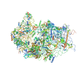
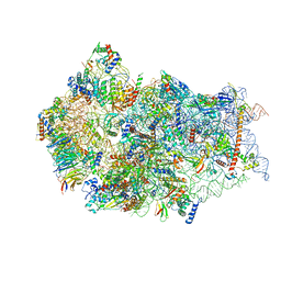
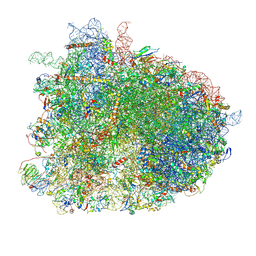 | | Cryo-EM reconstruction of yeast 80S ribosome in complex with mRNA, tRNA and eEF2 (GDP+AlF4/sordarin) | | 分子名称: | 18S ribosomal RNA, 40S ribosomal protein S0-A, 40S ribosomal protein S1-A, ... | | 著者 | Pellegrino, S, Yusupov, M, Yusupova, G, Hashem, Y. | | 登録日 | 2018-06-07 | | 公開日 | 2018-07-11 | | 最終更新日 | 2021-08-04 | | 実験手法 | ELECTRON MICROSCOPY (3.9 Å) | | 主引用文献 | Structural Insights into the Role of Diphthamide on Elongation Factor 2 in mRNA Reading-Frame Maintenance.
J. Mol. Biol., 430, 2018
|
|
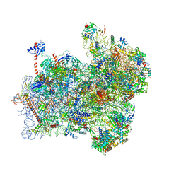
6GQ1
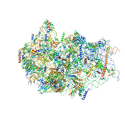
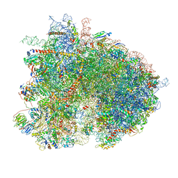 | | Cryo-EM reconstruction of yeast 80S ribosome in complex with mRNA, tRNA and eEF2 (GMPPCP/sordarin) | | 分子名称: | 18S ribosomal RNA, 40S ribosomal protein S0-A, 40S ribosomal protein S1-A, ... | | 著者 | Pellegrino, S, Demeshkina, N, Mancera-Martinez, E, Melnikov, S, Simonetti, A, Myasnikov, A, Yusupov, M, Yusupova, G, Hashem, Y. | | 登録日 | 2018-06-07 | | 公開日 | 2018-07-11 | | 最終更新日 | 2018-08-08 | | 実験手法 | ELECTRON MICROSCOPY (4.4 Å) | | 主引用文献 | Structural Insights into the Role of Diphthamide on Elongation Factor 2 in mRNA Reading-Frame Maintenance.
J. Mol. Biol., 430, 2018
|
|
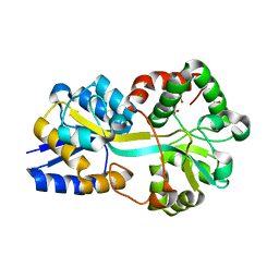
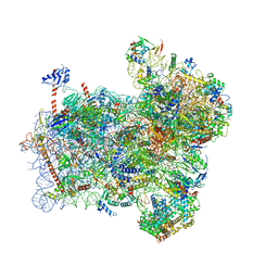
6GP1
 | | Structure of mEos4b in the red long-lived dark state | | 分子名称: | Green to red photoconvertible GFP-like protein EosFP | | 著者 | De Zitter, E, Adam, V, Byrdin, M, Van Meervelt, L, Dedecker, P, Bourgeois, D. | | 登録日 | 2018-06-04 | | 公開日 | 2019-05-22 | | 最終更新日 | 2024-01-17 | | 実験手法 | X-RAY DIFFRACTION (1.504 Å) | | 主引用文献 | Mechanistic investigation of mEos4b reveals a strategy to reduce track interruptions in sptPALM.
Nat.Methods, 16, 2019
|
|
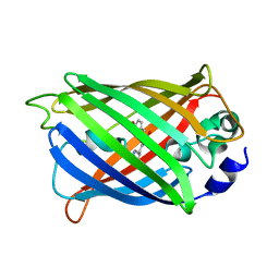
6GP0
 | | Structure of mEos4b in the red fluorescent state | | 分子名称: | Green to red photoconvertible GFP-like protein EosFP | | 著者 | De Zitter, E, Adam, V, Byrdin, M, Van Meervelt, L, Dedecker, P, Bourgeois, D. | | 登録日 | 2018-06-04 | | 公開日 | 2019-05-22 | | 最終更新日 | 2024-01-17 | | 実験手法 | X-RAY DIFFRACTION (1.5 Å) | | 主引用文献 | Mechanistic investigation of mEos4b reveals a strategy to reduce track interruptions in sptPALM.
Nat.Methods, 16, 2019
|
|
6GOZ
 | | Structure of mEos4b in the green long-lived dark state | | 分子名称: | 1,2-ETHANEDIOL, DI(HYDROXYETHYL)ETHER, GLYCEROL, ... | | 著者 | De Zitter, E, Adam, V, Byrdin, M, Van Meervelt, L, Dedecker, P, Bourgeois, D. | | 登録日 | 2018-06-04 | | 公開日 | 2019-11-13 | | 最終更新日 | 2023-11-15 | | 実験手法 | X-RAY DIFFRACTION (2.406 Å) | | 主引用文献 | Mechanistic Investigations of Green mEos4b Reveal a Dynamic Long-Lived Dark State.
J.Am.Chem.Soc., 2020
|
|
6GOY
 | | Structure of mEos4b in the green fluorescent state | | 分子名称: | 1,2-ETHANEDIOL, DI(HYDROXYETHYL)ETHER, GLYCEROL, ... | | 著者 | De Zitter, E, Adam, V, Byrdin, M, Van Meervelt, L, Dedecker, P, Bourgeois, D. | | 登録日 | 2018-06-04 | | 公開日 | 2019-05-22 | | 最終更新日 | 2024-01-17 | | 実験手法 | X-RAY DIFFRACTION (1.65 Å) | | 主引用文献 | Mechanistic investigation of mEos4b reveals a strategy to reduce track interruptions in sptPALM.
Nat.Methods, 16, 2019
|
|
6GO9
 | | Structure of GFPmut2 crystallized at pH 6 and transferred to pH 7 | | 分子名称: | (4R)-2-METHYLPENTANE-2,4-DIOL, (4S)-2-METHYL-2,4-PENTANEDIOL, Green fluorescent protein | | 著者 | Lolli, G, Raboni, S, Pasqualetto, E, Campanini, B, Mozzarelli, A, Bettati, S, Battistutta, R. | | 登録日 | 2018-06-01 | | 公開日 | 2018-12-19 | | 最終更新日 | 2024-01-17 | | 実験手法 | X-RAY DIFFRACTION (1.672 Å) | | 主引用文献 | Insight into GFPmut2 pH Dependence by Single Crystal Microspectrophotometry and X-ray Crystallography.
J.Phys.Chem.B, 122, 2018
|
|
6GO8
 | | Structure of GFPmut2 crystallized at pH 6 | | 分子名称: | (4R)-2-METHYLPENTANE-2,4-DIOL, (4S)-2-METHYL-2,4-PENTANEDIOL, Green fluorescent protein | | 著者 | Lolli, G, Raboni, S, Pasqualetto, E, Campanini, B, Mozzarelli, A, Bettati, S, Battistutta, R. | | 登録日 | 2018-06-01 | | 公開日 | 2018-12-19 | | 最終更新日 | 2024-01-17 | | 実験手法 | X-RAY DIFFRACTION (1.648 Å) | | 主引用文献 | Insight into GFPmut2 pH Dependence by Single Crystal Microspectrophotometry and X-ray Crystallography.
J.Phys.Chem.B, 122, 2018
|
|
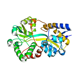
6GFP
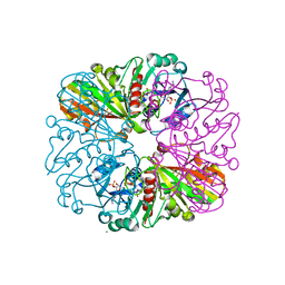 | | cyanobacterial GAPDH with NADP bound | | 分子名称: | FORMIC ACID, Glyceraldehyde-3-phosphate dehydrogenase, MAGNESIUM ION, ... | | 著者 | McFarlane, C.R, Briggs, L, Murray, J.W. | | 登録日 | 2018-05-01 | | 公開日 | 2019-05-08 | | 最終更新日 | 2024-01-17 | | 実験手法 | X-RAY DIFFRACTION (1.54 Å) | | 主引用文献 | Structural basis of light-induced redox regulation in the Calvin-Benson cycle in cyanobacteria.
Proc.Natl.Acad.Sci.USA, 116, 2019
|
|
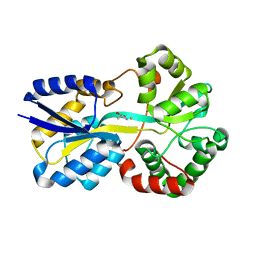
6GEZ
 | | THE STRUCTURE OF TWITCH-2B N532F | | 分子名称: | CALCIUM ION, FORMIC ACID, Green fluorescent protein,Optimized Ratiometric Calcium Sensor,Green fluorescent protein,Green fluorescent protein | | 著者 | Trigo Mourino, P, Paulat, M, Thestrup, T, Griesbeck, O, Griesinger, C, Becker, S. | | 登録日 | 2018-04-27 | | 公開日 | 2019-08-21 | | 最終更新日 | 2024-01-17 | | 実験手法 | X-RAY DIFFRACTION (2.47 Å) | | 主引用文献 | Dynamic tuning of FRET in a green fluorescent protein biosensor.
Sci Adv, 5, 2019
|
|
6GEL
 | | The structure of TWITCH-2B | | 分子名称: | CALCIUM ION, FORMIC ACID, GLYCEROL, ... | | 著者 | Trigo Mourino, P, Paulat, M, Thestrup, T, Griesbeck, O, Griesinger, C, Becker, S. | | 登録日 | 2018-04-26 | | 公開日 | 2019-08-21 | | 最終更新日 | 2019-09-11 | | 実験手法 | X-RAY DIFFRACTION (2.51 Å) | | 主引用文献 | Dynamic tuning of FRET in a green fluorescent protein biosensor.
Sci Adv, 5, 2019
|
|
6G7Q
 | |
6G7P
 | |
6G7N
 | |
6G5I
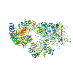
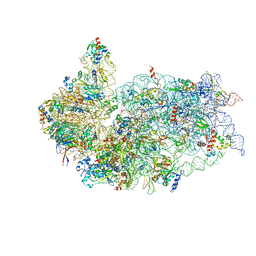 | | Cryo-EM structure of a late human pre-40S ribosomal subunit - State R | | 分子名称: | 18S ribosomal RNA, 40S ribosomal protein S10, 40S ribosomal protein S11, ... | | 著者 | Ameismeier, M, Cheng, J, Berninghausen, O, Beckmann, R. | | 登録日 | 2018-03-29 | | 公開日 | 2018-06-06 | | 最終更新日 | 2024-05-15 | | 実験手法 | ELECTRON MICROSCOPY (3.5 Å) | | 主引用文献 | Visualizing late states of human 40S ribosomal subunit maturation.
Nature, 558, 2018
|
|
6G5H
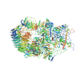
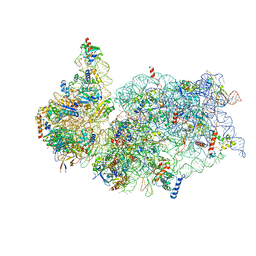 | | Cryo-EM structure of a late human pre-40S ribosomal subunit - Mature | | 分子名称: | 18S ribosomal RNA, 40S ribosomal protein S10, 40S ribosomal protein S11, ... | | 著者 | Ameismeier, M, Cheng, J, Berninghausen, O, Beckmann, R. | | 登録日 | 2018-03-29 | | 公開日 | 2018-06-06 | | 最終更新日 | 2024-05-15 | | 実験手法 | ELECTRON MICROSCOPY (3.6 Å) | | 主引用文献 | Visualizing late states of human 40S ribosomal subunit maturation.
Nature, 558, 2018
|
|
6G53
 | | Cryo-EM structure of a late human pre-40S ribosomal subunit - State E | | 分子名称: | 18S ribosomal RNA, 40S ribosomal protein S10, 40S ribosomal protein S11, ... | | 著者 | Ameismeier, M, Cheng, J, Berninghausen, O, Beckmann, R. | | 登録日 | 2018-03-28 | | 公開日 | 2018-06-06 | | 最終更新日 | 2024-05-15 | | 実験手法 | ELECTRON MICROSCOPY (4.5 Å) | | 主引用文献 | Visualizing late states of human 40S ribosomal subunit maturation.
Nature, 558, 2018
|
|
6G51
 | | Cryo-EM structure of a late human pre-40S ribosomal subunit - State D | | 分子名称: | 18S ribosomal RNA, 40S ribosomal protein S10, 40S ribosomal protein S11, ... | | 著者 | Ameismeier, M, Cheng, J, Berninghausen, O, Beckmann, R. | | 登録日 | 2018-03-28 | | 公開日 | 2018-06-06 | | 最終更新日 | 2024-05-15 | | 実験手法 | ELECTRON MICROSCOPY (4.1 Å) | | 主引用文献 | Visualizing late states of human 40S ribosomal subunit maturation.
Nature, 558, 2018
|
|
6G4W
 | | Cryo-EM structure of a late human pre-40S ribosomal subunit - State A | | 分子名称: | 18S ribosomal RNA, 40S ribosomal protein S11, 40S ribosomal protein S13, ... | | 著者 | Ameismeier, M, Cheng, J, Berninghausen, O, Beckmann, R. | | 登録日 | 2018-03-28 | | 公開日 | 2018-06-06 | | 最終更新日 | 2024-05-15 | | 実験手法 | ELECTRON MICROSCOPY (4.5 Å) | | 主引用文献 | Visualizing late states of human 40S ribosomal subunit maturation.
Nature, 558, 2018
|
|
6G4S
 | | Cryo-EM structure of a late human pre-40S ribosomal subunit - State B | | 分子名称: | 40S ribosomal protein S11, 40S ribosomal protein S13, 40S ribosomal protein S14, ... | | 著者 | Ameismeier, M, Cheng, J, Berninghausen, O, Beckmann, R. | | 登録日 | 2018-03-28 | | 公開日 | 2018-06-06 | | 最終更新日 | 2024-05-15 | | 実験手法 | ELECTRON MICROSCOPY (4 Å) | | 主引用文献 | Visualizing late states of human 40S ribosomal subunit maturation.
Nature, 558, 2018
|
|
6G18
 | | Cryo-EM structure of a late human pre-40S ribosomal subunit - State C | | 分子名称: | 40S ribosomal protein S11, 40S ribosomal protein S12, 40S ribosomal protein S13, ... | | 著者 | Ameismeier, M, Cheng, J, Berninghausen, O, Beckmann, R. | | 登録日 | 2018-03-20 | | 公開日 | 2018-06-06 | | 最終更新日 | 2024-05-15 | | 実験手法 | ELECTRON MICROSCOPY (3.6 Å) | | 主引用文献 | Visualizing late states of human 40S ribosomal subunit maturation.
Nature, 558, 2018
|
|
6FYY
 | | Structure of a partial yeast 48S preinitiation complex with eIF5 N-terminal domain (model C2) | | 分子名称: | 18S ribosomal RNA, 40S ribosomal protein S0, 40S ribosomal protein S1, ... | | 著者 | Llacer, J.L, Hussain, T, Gordiyenko, Y, Ramakrishnan, V. | | 登録日 | 2018-03-12 | | 公開日 | 2018-12-05 | | 最終更新日 | 2024-04-24 | | 実験手法 | ELECTRON MICROSCOPY (3.02 Å) | | 主引用文献 | Translational initiation factor eIF5 replaces eIF1 on the 40S ribosomal subunit to promote start-codon recognition.
Elife, 7, 2018
|
|
6FYX
 | | Structure of a partial yeast 48S preinitiation complex with eIF5 N-terminal domain (model C1) | | 分子名称: | 18S ribosomal RNA, 40S ribosomal protein S0, 40S ribosomal protein S1, ... | | 著者 | Llacer, J.L, Hussain, T, Gordiyenko, Y, Ramakrishnan, V. | | 登録日 | 2018-03-12 | | 公開日 | 2018-12-05 | | 最終更新日 | 2024-04-24 | | 実験手法 | ELECTRON MICROSCOPY (3.5 Å) | | 主引用文献 | Translational initiation factor eIF5 replaces eIF1 on the 40S ribosomal subunit to promote start-codon recognition.
Elife, 7, 2018
|
|
6FWW
 | | GFP/KKK. A redesigned GFP with improved solubility | | 分子名称: | Green fluorescent protein | | 著者 | Varejao, N, Lascorz, J, Gil-Garcia, M, Diaz-Caballero, M, Navarro, S, Ventura, S, Reverter, D. | | 登録日 | 2018-03-07 | | 公開日 | 2018-08-01 | | 最終更新日 | 2024-01-17 | | 実験手法 | X-RAY DIFFRACTION (1.131 Å) | | 主引用文献 | Combining Structural Aggregation Propensity and Stability Predictions To Redesign Protein Solubility.
Mol. Pharm., 15, 2018
|
|
6FSQ
 | |


