6VVU
 
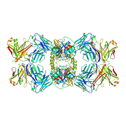 | | Anti-Tryptase fab E104.v1 bound to tryptase | | 分子名称: | CALCIUM ION, Fab E104.v1 heavy chain, Fab E104.v1 light chain, ... | | 著者 | Ultsch, M, Koerber, J.T. | | 登録日 | 2020-02-18 | | 公開日 | 2020-12-30 | | 最終更新日 | 2023-10-11 | | 実験手法 | X-RAY DIFFRACTION (3 Å) | | 主引用文献 | Bivalent antibody pliers inhibit beta-tryptase by an allosteric mechanism dependent on the IgG hinge.
Nat Commun, 11, 2020
|
|
5R42
 | | Crystal Structure of deuterated gamma-Chymotrypsin at pH 7.5, room temperature | | 分子名称: | IODIDE ION, gamma-Chymotrypsin, peptide SWPW, ... | | 著者 | Kreinbring, C.A, Wilson, M.A, Kovalevsky, A.Y, Blakeley, M.P, Fisher, S.Z, Lazar, L.M, Moulin, A.G, Novak, W.R, Petsko, G.A, Ringe, D. | | 登録日 | 2020-02-18 | | 公開日 | 2021-09-01 | | 実験手法 | X-RAY DIFFRACTION (1.05 Å) | | 主引用文献 | Effect of Temperature and pH on Ionizable Residues in gamma-Chymotrypsin: a X-ray and Neutron Crystallography Study
To be published
|
|
5R49
 | | Crystal Structure of gamma-Chymotrypsin at pH 5.6, cryo temperature | | 分子名称: | IODIDE ION, MALONATE ION, gamma-chymotrypsin, ... | | 著者 | Kreinbring, C.A, Wilson, M.A, Kovalevsky, A.Y, Blakeley, M.P, Fisher, S.Z, Lazar, L.M, Moulin, A.G, Novak, W.R, Petsko, G.A, Ringe, D. | | 登録日 | 2020-02-18 | | 公開日 | 2021-09-01 | | 実験手法 | X-RAY DIFFRACTION (1.05 Å) | | 主引用文献 | Effect of Temperature and pH on Ionizable Residues in gamma-Chymotrypsin: a X-ray and Neutron Crystallography Study
To be published
|
|
5R45
 | | Crystal Structure of gamma-Chymotrypsin at pH 7.5, cryo temperature | | 分子名称: | Chymotrypsinogen A, IODIDE ION, MALONATE ION, ... | | 著者 | Kreinbring, C.A, Wilson, M.A, Kovalevsky, A.Y, Blakeley, M.P, Fisher, S.Z, Lazar, L.M, Moulin, A.G, Novak, W.R, Petsko, G.A, Ringe, D. | | 登録日 | 2020-02-18 | | 公開日 | 2021-09-01 | | 実験手法 | X-RAY DIFFRACTION (1.05 Å) | | 主引用文献 | Effect of Temperature and pH on Ionizable Residues in gamma-Chymotrypsin: a X-ray and Neutron Crystallography Study
To be published
|
|
5R4C
 | | Crystal Structure of gamma-Chymotrypsin at pH 9, room temperature | | 分子名称: | IODIDE ION, SULFATE ION, gamma-chymotrypsin, ... | | 著者 | Kreinbring, C.A, Wilson, M.A, Kovalevsky, A.Y, Blakeley, M.P, Fisher, S.Z, Lazar, L.M, Moulin, A.G, Novak, W.R, Petsko, G.A, Ringe, D. | | 登録日 | 2020-02-18 | | 公開日 | 2021-09-01 | | 実験手法 | X-RAY DIFFRACTION (1.15 Å) | | 主引用文献 | Effect of Temperature and pH on Ionizable Residues in gamma-Chymotrypsin: a X-ray and Neutron Crystallography Study
To be published
|
|
5R48
 | | Crystal Structure of gamma-Chymotrypsin at pH 5.6, room temperature | | 分子名称: | IODIDE ION, SULFATE ION, gamma-chymotrypsin, ... | | 著者 | Kreinbring, C.A, Wilson, M.A, Kovalevsky, A.Y, Blakeley, M.P, Fisher, S.Z, Lazar, L.M, Moulin, A.G, Novak, W.R, Petsko, G.A, Ringe, D. | | 登録日 | 2020-02-18 | | 公開日 | 2021-09-01 | | 実験手法 | X-RAY DIFFRACTION (1.05 Å) | | 主引用文献 | Effect of Temperature and pH on Ionizable Residues in gamma-Chymotrypsin: a X-ray and Neutron Crystallography Study
To be published
|
|
5R4A
 | | Crystal Structure of deuterated gamma-Chymotrypsin at pH 9, room temperature | | 分子名称: | IODIDE ION, SULFATE ION, gamma-chymotrypsin, ... | | 著者 | Kreinbring, C.A, Wilson, M.A, Kovalevsky, A.Y, Blakeley, M.P, Fisher, S.Z, Lazar, L.M, Moulin, A.G, Novak, W.R, Petsko, G.A, Ringe, D. | | 登録日 | 2020-02-18 | | 公開日 | 2021-09-01 | | 実験手法 | X-RAY DIFFRACTION (1.2 Å) | | 主引用文献 | Effect of Temperature and pH on Ionizable Residues in gamma-Chymotrypsin: a X-ray and Neutron Crystallography Study
To be published
|
|
5R44
 | | Crystal Structure of gamma-Chymotrypsin at pH 7.5, room temperature | | 分子名称: | Chymotrypsinogen A, IODIDE ION, peptide SWPW, ... | | 著者 | Kreinbring, C.A, Wilson, M.A, Kovalevsky, A.Y, Blakeley, M.P, Fisher, S.Z, Lazar, L.M, Moulin, A.G, Novak, W.R, Petsko, G.A, Ringe, D. | | 登録日 | 2020-02-18 | | 公開日 | 2021-09-01 | | 実験手法 | X-RAY DIFFRACTION (1.05 Å) | | 主引用文献 | Effect of Temperature and pH on Ionizable Residues in gamma-Chymotrypsin: a X-ray and Neutron Crystallography Study
To be published
|
|
5R46
 | | Crystal Structure of deuterated gamma-Chymotrypsin at pH 5.6, room temperature | | 分子名称: | IODIDE ION, SULFATE ION, gamma-chymotrypsin, ... | | 著者 | Kreinbring, C.A, Wilson, M.A, Kovalevsky, A.Y, Blakeley, M.P, Fisher, S.Z, Lazar, L.M, Moulin, A.G, Novak, W.R, Petsko, G.A, Ringe, D. | | 登録日 | 2020-02-18 | | 公開日 | 2021-09-01 | | 実験手法 | X-RAY DIFFRACTION (1.05 Å) | | 主引用文献 | Effect of Temperature and pH on Ionizable Residues in gamma-Chymotrypsin: a X-ray and Neutron Crystallography Study
To be published
|
|
5R43
 | | Crystal Structure of deuterated gamma-Chymotrypsin at pH 7.5, cryo temperature | | 分子名称: | Chymotrypsinogen A, IODIDE ION, MALONIC ACID, ... | | 著者 | Kreinbring, C.A, Wilson, M.A, Kovalevsky, A.Y, Blakeley, M.P, Fisher, S.Z, Lazar, L.M, Moulin, A.G, Novak, W.R, Petsko, G.A, Ringe, D. | | 登録日 | 2020-02-18 | | 公開日 | 2021-09-01 | | 実験手法 | X-RAY DIFFRACTION (1 Å) | | 主引用文献 | Effect of Temperature and pH on Ionizable Residues in gamma-Chymotrypsin: a X-ray and Neutron Crystallography Study
To be published
|
|
5R4B
 | | Crystal Structure of deuterated gamma-Chymotrypsin at pH 9, cryo temperature | | 分子名称: | IODIDE ION, SULFATE ION, gamma-chymotrypsin, ... | | 著者 | Kreinbring, C.A, Wilson, M.A, Kovalevsky, A.Y, Blakeley, M.P, Fisher, S.Z, Lazar, L.M, Moulin, A.G, Novak, W.R, Petsko, G.A, Ringe, D. | | 登録日 | 2020-02-18 | | 公開日 | 2021-09-01 | | 実験手法 | X-RAY DIFFRACTION (1.05 Å) | | 主引用文献 | Effect of Temperature and pH on Ionizable Residues in gamma-Chymotrypsin: a X-ray and Neutron Crystallography Study
To be published
|
|
5R4D
 | | Crystal Structure of gamma-Chymotrypsin at pH 9, cryo temperature | | 分子名称: | IODIDE ION, SULFATE ION, gamma-chymotrypsin, ... | | 著者 | Kreinbring, C.A, Wilson, M.A, Kovalevsky, A.Y, Blakeley, M.P, Fisher, S.Z, Lazar, L.M, Moulin, A.G, Novak, W.R, Petsko, G.A, Ringe, D. | | 登録日 | 2020-02-18 | | 公開日 | 2021-09-01 | | 実験手法 | X-RAY DIFFRACTION (1.05 Å) | | 主引用文献 | Effect of Temperature and pH on Ionizable Residues in gamma-Chymotrypsin: a X-ray and Neutron Crystallography Study
To be published
|
|
5R47
 | | Crystal Structure of deuterated gamma-Chymotrypsin at pH 5.6, cryo temperature | | 分子名称: | IODIDE ION, MALONIC ACID, gamma-chymotrypsin, ... | | 著者 | Kreinbring, C.A, Wilson, M.A, Kovalevsky, A.Y, Blakeley, M.P, Fisher, S.Z, Lazar, L.M, Moulin, A.G, Novak, W.R, Petsko, G.A, Ringe, D. | | 登録日 | 2020-02-18 | | 公開日 | 2021-09-01 | | 実験手法 | X-RAY DIFFRACTION (1.1 Å) | | 主引用文献 | Effect of Temperature and pH on Ionizable Residues in gamma-Chymotrypsin: a X-ray and Neutron Crystallography Study
To be published
|
|
6Y2U
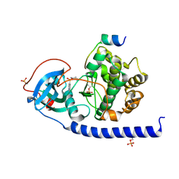 | | Crystal structure of the cAMP-dependent protein kinase A cocrystallized with aminofasudil and PKI (5-24) | | 分子名称: | 5-(1,4-diazepan-1-ylsulfonyl)isoquinolin-1-amine, cAMP-dependent protein kinase catalytic subunit alpha, cAMP-dependent protein kinase inhibitor alpha | | 著者 | Oebbeke, M, Gerber, H.-D, Heine, A, Klebe, G. | | 登録日 | 2020-02-17 | | 公開日 | 2021-03-31 | | 最終更新日 | 2024-01-24 | | 実験手法 | X-RAY DIFFRACTION (1.93 Å) | | 主引用文献 | Fragment based drug design - Small chemical changes of fragments effecting big changes in binding
To Be Published
|
|
6Y2O
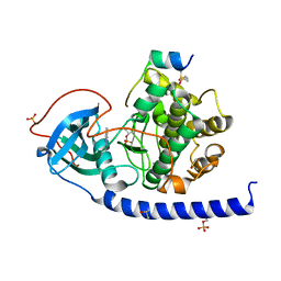 | | Crystal structure of the cAMP-dependent protein kinase A cocrystallized with 1,7-Naphthyridin-8-amine and PKI (5-24) | | 分子名称: | 1,7-naphthyridin-8-amine, DIMETHYL SULFOXIDE, cAMP-dependent protein kinase catalytic subunit alpha, ... | | 著者 | Oebbeke, M, Heine, A, Klebe, G. | | 登録日 | 2020-02-17 | | 公開日 | 2020-09-30 | | 最終更新日 | 2024-01-24 | | 実験手法 | X-RAY DIFFRACTION (2.01 Å) | | 主引用文献 | Fragment based drug design - Small chemical changes of fragments effecting big changes in binding
To Be Published
|
|
6VVG
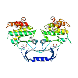 | | Structure of the Cydia pomonella Granulovirus kinase, PK-1 | | 分子名称: | ADENOSINE MONOPHOSPHATE, Arginine kinase | | 著者 | Oliver, M.R, Horne, C.R, Keown, J.R, Murphy, J.M, Metcalf, P. | | 登録日 | 2020-02-17 | | 公開日 | 2021-02-03 | | 最終更新日 | 2023-10-11 | | 実験手法 | X-RAY DIFFRACTION (2.01 Å) | | 主引用文献 | Granulovirus PK-1 kinase activity relies on a side-to-side dimerization mode centered on the regulatory alpha C helix.
Nat Commun, 12, 2021
|
|
6LYX
 | | Crystal structure of oxidized ACHT1 | | 分子名称: | GLYCEROL, SULFATE ION, Thioredoxin-like 2-1, ... | | 著者 | Wang, J.C, Pan, W.M, Cai, W.G, Wang, M.Z, Zhang, M. | | 登録日 | 2020-02-16 | | 公開日 | 2020-05-13 | | 最終更新日 | 2023-11-29 | | 実験手法 | X-RAY DIFFRACTION (1.696 Å) | | 主引用文献 | Structural insight into the biological functions of Arabidopsis thaliana ACHT1.
Int.J.Biol.Macromol., 158, 2020
|
|
6LYW
 | | Structural insight into the biological functions of Arabidopsis thaliana ACHT1 | | 分子名称: | GLYCEROL, SULFATE ION, Thioredoxin-like 2-1, ... | | 著者 | Wang, J.C, Pan, W.M, Wang, M.Z, Zhang, M. | | 登録日 | 2020-02-16 | | 公開日 | 2020-05-13 | | 最終更新日 | 2023-11-29 | | 実験手法 | X-RAY DIFFRACTION (1.7 Å) | | 主引用文献 | Structural insight into the biological functions of Arabidopsis thaliana ACHT1.
Int.J.Biol.Macromol., 158, 2020
|
|
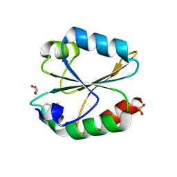
6Y2D
 | | Crystal structure of the second KH domain of FUBP1 | | 分子名称: | Far upstream element-binding protein 1, GLYCEROL, SULFATE ION | | 著者 | Ni, X, Chaikuad, A, Joerger, A.C, Arrowsmith, C.H, Edwards, A.M, Bountra, C, Knapp, S, Structural Genomics Consortium (SGC) | | 登録日 | 2020-02-15 | | 公開日 | 2020-03-25 | | 最終更新日 | 2024-01-24 | | 実験手法 | X-RAY DIFFRACTION (1.9 Å) | | 主引用文献 | Comparative structural analyses and nucleotide-binding characterization of the four KH domains of FUBP1.
Sci Rep, 10, 2020
|
|
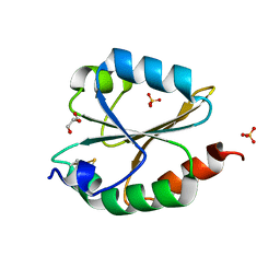
6Y2C
 | | Crystal structure of the third KH domain of FUBP1 | | 分子名称: | 1,2-ETHANEDIOL, Far upstream element-binding protein 1, ZINC ION | | 著者 | Ni, X, Joerger, A.C, Chaikuad, A, Arrowsmith, C.H, Edwards, A.M, Bountra, C, Knapp, S, Structural Genomics Consortium (SGC) | | 登録日 | 2020-02-15 | | 公開日 | 2020-03-25 | | 最終更新日 | 2024-01-24 | | 実験手法 | X-RAY DIFFRACTION (2 Å) | | 主引用文献 | Comparative structural analyses and nucleotide-binding characterization of the four KH domains of FUBP1.
Sci Rep, 10, 2020
|
|
6VUG
 | |
6Y24
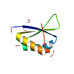 | | Crystal structure of fourth KH domain of FUBP1 | | 分子名称: | 1,2-ETHANEDIOL, Far upstream element-binding protein 1 | | 著者 | Ni, X, Joerger, A.C, Chaikuad, A, Arrowsmith, C.H, Edwards, A.M, Bountra, C, Knapp, S, Structural Genomics Consortium (SGC) | | 登録日 | 2020-02-14 | | 公開日 | 2020-03-25 | | 最終更新日 | 2024-01-24 | | 実験手法 | X-RAY DIFFRACTION (1.86 Å) | | 主引用文献 | Comparative structural analyses and nucleotide-binding characterization of the four KH domains of FUBP1.
Sci Rep, 10, 2020
|
|
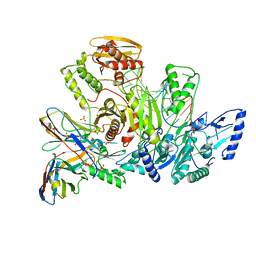
6LY4
 | | The crystal structure of the BM3 mutant LG-23 in complex with testosterone | | 分子名称: | 1,2-ETHANEDIOL, Bifunctional cytochrome P450/NADPH--P450 reductase, IMIDAZOLE, ... | | 著者 | Peng, Y, Chen, J, Zhou, J, Li, A, ReetZ, M.T. | | 登録日 | 2020-02-13 | | 公開日 | 2020-04-29 | | 最終更新日 | 2023-11-29 | | 実験手法 | X-RAY DIFFRACTION (1.68 Å) | | 主引用文献 | Regio- and Stereoselective Steroid Hydroxylation at C7 by Cytochrome P450 Monooxygenase Mutants.
Angew.Chem.Int.Ed.Engl., 59, 2020
|
|
6Y1U
 | | Mycobacterium tuberculosis FtsZ-GDP in complex with 4-hydroxycoumarin | | 分子名称: | 4-HYDROXY-2H-CHROMEN-2-ONE, Cell division protein FtsZ, DIMETHYL SULFOXIDE, ... | | 著者 | Alnami, A.T, Norton, R.S, Pena, H.P, Haider, M, kozielski, F. | | 登録日 | 2020-02-13 | | 公開日 | 2021-06-09 | | 最終更新日 | 2024-01-24 | | 実験手法 | X-RAY DIFFRACTION (1.68 Å) | | 主引用文献 | Conformational Flexibility of A Highly Conserved Helix Controls Cryptic Pocket Formation in FtsZ.
J.Mol.Biol., 433, 2021
|
|
6Y1V
 | | Mycobacterium tuberculosis FtsZ-GTP-gamma-S in complex with 4-hydroxycoumarin | | 分子名称: | 4-HYDROXY-2H-CHROMEN-2-ONE, 5'-GUANOSINE-DIPHOSPHATE-MONOTHIOPHOSPHATE, Cell division protein FtsZ, ... | | 著者 | Alnami, A.T, Norton, R.S, Pena, H.P, Haider, M, kozielski, F. | | 登録日 | 2020-02-13 | | 公開日 | 2021-06-09 | | 最終更新日 | 2024-01-24 | | 実験手法 | X-RAY DIFFRACTION (2.4 Å) | | 主引用文献 | Conformational Flexibility of A Highly Conserved Helix Controls Cryptic Pocket Formation in FtsZ.
J.Mol.Biol., 433, 2021
|
|


