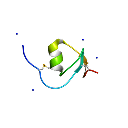
5XR0
 
 | | Crystal structure of Notched-fin eelpout type III antifreeze protein A20T mutant (NFE6, AFP), P21 form | | 分子名称: | Ice-structuring protein | | 著者 | Adachi, M, Shimizu, R, Shibazaki, C, Kondo, H, Tsuda, S. | | 登録日 | 2017-06-07 | | 公開日 | 2018-05-16 | | 最終更新日 | 2023-11-22 | | 実験手法 | X-RAY DIFFRACTION (0.98 Å) | | 主引用文献 | Polypentagonal ice-like water networks emerge solely in an activity-improved variant of ice-binding protein
Proc. Natl. Acad. Sci. U.S.A., 115, 2018
|
|
2G58
 | | Crystal structure of a complex of phospholipase A2 with a designed peptide inhibitor Dehydro-Ile-Ala-Arg-Ser at 0.98 A resolution | | 分子名称: | (PHQ)IARS, Phospholipase A2 VRV-PL-VIIIa, SULFATE ION | | 著者 | Prem Kumar, R, Singh, N, Somvanshi, R.K, Ethayathulla, A.S, Dey, S, Sharma, S, Kaur, P, Perbandt, M, Betzel, C, Singh, T.P. | | 登録日 | 2006-02-22 | | 公開日 | 2006-03-28 | | 最終更新日 | 2023-11-15 | | 実験手法 | X-RAY DIFFRACTION (0.98 Å) | | 主引用文献 | Crystal structure of a complex of phospholipase A2 with a designed peptide inhibitor Dehydro-Ile-Ala-Arg-Ser at 0.98 A resolution
To be Published
|
|
4YEO
 | | Triclinic HEWL co-crystallised with cisplatin, studied at a data collection temperature of 150K - new refinement | | 分子名称: | 1,2-ETHANEDIOL, ACETATE ION, Cisplatin, ... | | 著者 | Shabalin, I.G, Dauter, Z, Jaskolski, M, Minor, W, Wlodawer, A. | | 登録日 | 2015-02-24 | | 公開日 | 2015-03-04 | | 最終更新日 | 2023-09-27 | | 実験手法 | X-RAY DIFFRACTION (0.98 Å) | | 主引用文献 | Crystallography and chemistry should always go together: a cautionary tale of protein complexes with cisplatin and carboplatin.
Acta Crystallogr.,Sect.D, 71, 2015
|
|
7BCU
 | | The adduct of NAMI-A with Hen Egg White Lysozyme at 1.5 hours. | | 分子名称: | CHLORIDE ION, DIMETHYL SULFOXIDE, IMIDAZOLE, ... | | 著者 | Chiniadis, L, Giastas, P, Bratsos, I, Papakyriakou, A. | | 登録日 | 2020-12-21 | | 公開日 | 2021-07-28 | | 最終更新日 | 2024-01-31 | | 実験手法 | X-RAY DIFFRACTION (0.98 Å) | | 主引用文献 | Insights into the Protein Ruthenation Mechanism by Antimetastatic Metallodrugs: High-Resolution X-ray Structures of the Adduct Formed between Hen Egg-White Lysozyme and NAMI-A at Various Time Points.
Inorg.Chem., 60, 2021
|
|
5SAR
 | | Endothiapepsin in complex with compound FU290-1 | | 分子名称: | (1,4-phenylene)bis(methylene) dicarbamimidothioate, Endothiapepsin | | 著者 | Wollenhaupt, J, Metz, A, Messini, N, Barthel, T, Klebe, G, Weiss, M.S. | | 登録日 | 2021-05-28 | | 公開日 | 2021-09-01 | | 最終更新日 | 2021-09-29 | | 実験手法 | X-RAY DIFFRACTION (0.98 Å) | | 主引用文献 | Frag4Lead: growing crystallographic fragment hits by catalog using fragment-guided template docking.
Acta Crystallogr D Struct Biol, 77, 2021
|
|
7AVE
 | | Perdeuterated refolded hen egg-white lysozyme at 100 K | | 分子名称: | ACETATE ION, Lysozyme C, NITRATE ION | | 著者 | Ramos, J, Laux, V, Haertlein, M, Erba Boeri, E, Forsyth, V.T, Larsen, S, Mossou, E, Langkilde, A.E. | | 登録日 | 2020-11-05 | | 公開日 | 2021-05-12 | | 最終更新日 | 2024-01-31 | | 実験手法 | X-RAY DIFFRACTION (0.98 Å) | | 主引用文献 | Structural insights into protein folding, stability and activity using in vivo perdeuteration of hen egg-white lysozyme.
Iucrj, 8, 2021
|
|
1GHG
 | | CRYSTAL STRUCTURE OF VANCOMYCIN AGLYCON | | 分子名称: | ACETIC ACID, DIMETHYL SULFOXIDE, VANCOMYCIN AGLYCON | | 著者 | Kaplan, J, Korty, B.D, Axelsen, P.H, Loll, P.J. | | 登録日 | 2000-12-13 | | 公開日 | 2001-02-12 | | 最終更新日 | 2023-12-27 | | 実験手法 | X-RAY DIFFRACTION (0.98 Å) | | 主引用文献 | The Role of Sugar Residues in Molecular Recognition by Vancomycin
J.Med.Chem., 44, 2001
|
|
4HGU
 | | Crystal Structure of Galleria mellonella Silk Protease Inhibitor 2 | | 分子名称: | SODIUM ION, Silk protease inhibitor 2 | | 著者 | Krzywda, S, Jaskolski, M, Dvornyk, A, Kludkiewicz, B, Grzelak, K, Zagorski, W, Bal, W, Kopera, E. | | 登録日 | 2012-10-08 | | 公開日 | 2013-10-09 | | 最終更新日 | 2023-09-20 | | 実験手法 | X-RAY DIFFRACTION (0.98 Å) | | 主引用文献 | Atomic resolution structure of a protein prepared by non-enzymatic His-tag removal. Crystallographic and NMR study of GmSPI-2 inhibitor.
Plos One, 9, 2014
|
|
1TQG
 | | CheA phosphotransferase domain from Thermotoga maritima | | 分子名称: | Chemotaxis protein cheA | | 著者 | Quezada, C.M, Gradinaru, C, Simon, M.I, Bilwes, A.M, Crane, B.R. | | 登録日 | 2004-06-17 | | 公開日 | 2004-09-07 | | 最終更新日 | 2024-02-14 | | 実験手法 | X-RAY DIFFRACTION (0.98 Å) | | 主引用文献 | Helical Shifts Generate Two Distinct Conformers in the Atomic Resolution Structure of the CheA Phosphotransferase Domain from Thermotoga maritima.
J.Mol.Biol., 341, 2004
|
|
6SG6
 | |
2BZZ
 | | Crystal Structures of Eosinophil-derived Neurotoxin in Complex with the Inhibitors 5'-ATP, Ap3A, Ap4A and Ap5A | | 分子名称: | ACETIC ACID, BIS(ADENOSINE)-5'-PENTAPHOSPHATE, NONSECRETORY RIBONUCLEASE | | 著者 | Baker, M.D, Holloway, D.E, Swaminathan, G.J, Acharya, K.R. | | 登録日 | 2005-08-24 | | 公開日 | 2006-01-18 | | 最終更新日 | 2023-12-13 | | 実験手法 | X-RAY DIFFRACTION (0.98 Å) | | 主引用文献 | Crystal Structures of Eosinophil-Derived Neurotoxin (Edn) in Complex with the Inhibitors 5'- ATP, Ap(3)A, Ap(4)A, and Ap(5)A.
Biochemistry, 45, 2006
|
|
3I5E
 | |
4BCT
 | | Crystal structure of kiwi-fruit allergen Act d 2 | | 分子名称: | 4-(2-HYDROXYETHYL)-1-PIPERAZINE ETHANESULFONIC ACID, THAUMATIN-LIKE PROTEIN | | 著者 | Pavkov-Keller, T, Bublin, M, Jankovic, M, Breiteneder, H, Keller, W. | | 登録日 | 2012-10-03 | | 公開日 | 2013-10-16 | | 実験手法 | X-RAY DIFFRACTION (0.98 Å) | | 主引用文献 | Crystal Structure of Kiwi-Fruit Allergen Act D 2
To be Published
|
|
4QBX
 | |
5NFK
 | |
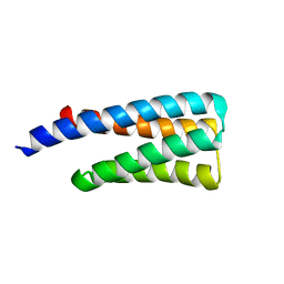
3V1A
 | | Crystal structure of de novo designed MID1-apo1 | | 分子名称: | Computational design, MID1-apo1 | | 著者 | Der, B.S, Machius, M, Miley, M.J, Kuhlman, B. | | 登録日 | 2011-12-09 | | 公開日 | 2012-01-11 | | 最終更新日 | 2023-09-13 | | 実験手法 | X-RAY DIFFRACTION (0.98 Å) | | 主引用文献 | Metal-mediated affinity and orientation specificity in a computationally designed protein homodimer.
J.Am.Chem.Soc., 134, 2012
|
|
5ZGI
 | |
5RC3
 | | PanDDA analysis group deposition -- Endothiapepsin changed state model for fragment F2X-Entry Library F03a | | 分子名称: | ACETATE ION, DIMETHYL SULFOXIDE, Endothiapepsin, ... | | 著者 | Weiss, M.S, Wollenhaupt, J, Metz, A, Barthel, T, Lima, G.M.A, Heine, A, Mueller, U, Klebe, G. | | 登録日 | 2020-03-24 | | 公開日 | 2020-06-03 | | 最終更新日 | 2020-06-17 | | 実験手法 | X-RAY DIFFRACTION (0.98 Å) | | 主引用文献 | F2X-Universal and F2X-Entry: Structurally Diverse Compound Libraries for Crystallographic Fragment Screening.
Structure, 28, 2020
|
|
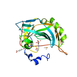
1K4I
 | | Crystal Structure of 3,4-dihydroxy-2-butanone 4-phosphate synthase in complex with two Magnesium ions | | 分子名称: | 3,4-Dihydroxy-2-Butanone 4-Phosphate Synthase, MAGNESIUM ION, SULFATE ION | | 著者 | Liao, D.-I, Zheng, Y.-J, Viitanen, P.V, Jordan, D.B. | | 登録日 | 2001-10-08 | | 公開日 | 2002-03-06 | | 最終更新日 | 2023-08-16 | | 実験手法 | X-RAY DIFFRACTION (0.98 Å) | | 主引用文献 | Structural definition of the active site and catalytic mechanism of 3,4-dihydroxy-2-butanone-4-phosphate synthase.
Biochemistry, 41, 2002
|
|
1V0L
 | | Xylanase Xyn10A from Streptomyces lividans in complex with xylobio-isofagomine at pH 5.8 | | 分子名称: | ENDO-1,4-BETA-XYLANASE A, PIPERIDINE-3,4-DIOL, beta-D-xylopyranose | | 著者 | Gloster, T.M, Williams, S.J, Roberts, S, Tarling, C.A, Wicki, J, Withers, S.G, Davies, G.J. | | 登録日 | 2004-03-31 | | 公開日 | 2004-08-16 | | 最終更新日 | 2023-12-13 | | 実験手法 | X-RAY DIFFRACTION (0.98 Å) | | 主引用文献 | Atomic Resolution Analyses of the Binding of Xylobiose-Derived Deoxynojirimycin and Isofagomine to Xylanase Xyn10A
Chem.Commun.(Camb.), 16, 2004
|
|
5RDN
 | | PanDDA analysis group deposition -- Endothiapepsin ground state model 47 | | 分子名称: | ACETATE ION, DI(HYDROXYETHYL)ETHER, Endothiapepsin, ... | | 著者 | Weiss, M.S, Wollenhaupt, J, Metz, A, Barthel, T, Lima, G.M.A, Heine, A, Mueller, U, Klebe, G. | | 登録日 | 2020-03-24 | | 公開日 | 2020-06-03 | | 最終更新日 | 2020-06-17 | | 実験手法 | X-RAY DIFFRACTION (0.98 Å) | | 主引用文献 | F2X-Universal and F2X-Entry: Structurally Diverse Compound Libraries for Crystallographic Fragment Screening.
Structure, 28, 2020
|
|
5CE4
 | | High Resolution X-Ray and Neutron diffraction structure of H-FABP | | 分子名称: | Fatty acid-binding protein, heart, OLEIC ACID | | 著者 | Podjarny, A.D, Howard, E.I, Blakeley, M.P, Guillot, B. | | 登録日 | 2015-07-06 | | 公開日 | 2016-03-09 | | 最終更新日 | 2024-05-08 | | 実験手法 | NEUTRON DIFFRACTION (0.98 Å), X-RAY DIFFRACTION | | 主引用文献 | High-resolution neutron and X-ray diffraction room-temperature studies of an H-FABP-oleic acid complex: study of the internal water cluster and ligand binding by a transferred multipolar electron-density distribution.
Iucrj, 3, 2016
|
|
5RVH
 | | PanDDA analysis group deposition -- Crystal structure of SARS-CoV-2 NSP3 macrodomain in complex with ZINC000000265642 | | 分子名称: | Non-structural protein 3, quinoline-3-carboxylic acid | | 著者 | Correy, G.J, Young, I.D, Thompson, M.C, Fraser, J.S. | | 登録日 | 2020-09-28 | | 公開日 | 2020-12-16 | | 最終更新日 | 2024-05-22 | | 実験手法 | X-RAY DIFFRACTION (0.98 Å) | | 主引用文献 | Fragment binding to the Nsp3 macrodomain of SARS-CoV-2 identified through crystallographic screening and computational docking.
Sci Adv, 7, 2021
|
|
3D1P
 | | Atomic resolution structure of uncharacterized protein from Saccharomyces cerevisiae | | 分子名称: | ACETATE ION, CHLORIDE ION, Putative thiosulfate sulfurtransferase YOR285W | | 著者 | Nocek, B, Evdokimova, E, Kudritska, M, Savchenko, A, Edwards, A.M, Joachimiak, A, Midwest Center for Structural Genomics (MCSG) | | 登録日 | 2008-05-06 | | 公開日 | 2008-07-08 | | 最終更新日 | 2011-07-13 | | 実験手法 | X-RAY DIFFRACTION (0.98 Å) | | 主引用文献 | Atomic resolution structure of uncharacterized protein from Saccharomyces cerevisiae.
To be Published
|
|
2NLS
 | |


