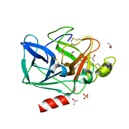
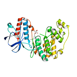
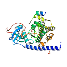
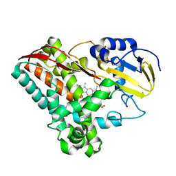
2FOF
 
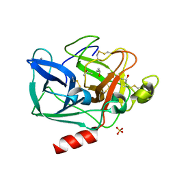 | | Structure of porcine pancreatic elastase in 80% isopropanol | | 分子名称: | CALCIUM ION, ISOPROPYL ALCOHOL, SULFATE ION, ... | | 著者 | Mattos, C, Bellamacina, C.R, Peisach, E, Pereira, A, Vitkup, D, Petsko, G.A, Ringe, D. | | 登録日 | 2006-01-13 | | 公開日 | 2006-04-18 | | 最終更新日 | 2023-08-30 | | 実験手法 | X-RAY DIFFRACTION (2.2 Å) | | 主引用文献 | Multiple solvent crystal structures: Probing binding sites, plasticity and hydration
J.Mol.Biol., 357, 2006
|
|
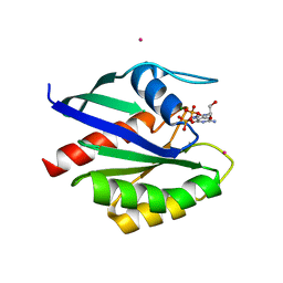
2FTB
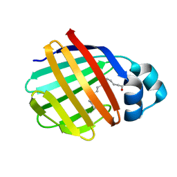 | | Crystal structure of axolotl (Ambystoma mexicanum) liver bile acid-binding protein bound to oleic acid | | 分子名称: | Fatty acid-binding protein 2, liver, OLEIC ACID | | 著者 | Capaldi, S, Guariento, M, Perduca, M, Di Pietro, S.M, Santome, J.A, Monaco, H.L. | | 登録日 | 2006-01-24 | | 公開日 | 2006-04-11 | | 最終更新日 | 2023-10-25 | | 実験手法 | X-RAY DIFFRACTION (2 Å) | | 主引用文献 | Crystal structure of axolotl (Ambystoma mexicanum) liver bile acid-binding protein bound to cholic and oleic acid
Proteins, 64, 2006
|
|
2FMX
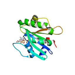 | | An open conformation of switch I revealed by Sar1-GDP crystal structure at low Mg(2+) | | 分子名称: | GTP-binding protein SAR1b, GUANOSINE-5'-DIPHOSPHATE, MAGNESIUM ION, ... | | 著者 | Rao, Y, Bian, C, Yuan, C, Li, Y, Huang, M. | | 登録日 | 2006-01-10 | | 公開日 | 2006-09-05 | | 最終更新日 | 2024-03-13 | | 実験手法 | X-RAY DIFFRACTION (1.82 Å) | | 主引用文献 | An open conformation of switch I revealed by Sar1-GDP crystal structure at low Mg(2+)
Biochem.Biophys.Res.Commun., 348, 2006
|
|
2LNX
 | |
2FX4
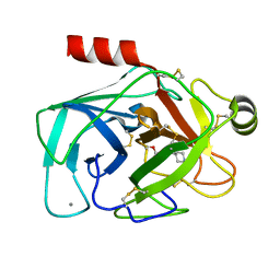 | |
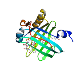
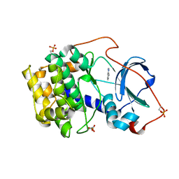
2FOB
 | | Structure of porcine pancreatic elastase in 40/50/10 cyclohexane | | 分子名称: | CALCIUM ION, ISOPROPYL ALCOHOL, SULFATE ION, ... | | 著者 | Mattos, C, Bellamacina, C.R, Peisach, E, Pereira, A, Vitkup, D, Petsko, G.A, Ringe, D. | | 登録日 | 2006-01-13 | | 公開日 | 2006-04-18 | | 最終更新日 | 2023-08-30 | | 実験手法 | X-RAY DIFFRACTION (1.9 Å) | | 主引用文献 | Multiple solvent crystal structures: Probing binding sites, plasticity and hydration
J.Mol.Biol., 357, 2006
|
|
2LBV
 | | Siderocalin Q83 reveals a dual ligand binding mode | | 分子名称: | ARACHIDONIC ACID, Extracellular fatty acid-binding protein, GALLIUM (III) ION, ... | | 著者 | Coudevylle, N, Hoetzinger, M, Geist, L, Kontaxis, G, Bister, K, Konrat, R. | | 登録日 | 2011-04-07 | | 公開日 | 2012-02-22 | | 最終更新日 | 2023-06-14 | | 実験手法 | SOLUTION NMR | | 主引用文献 | Lipocalin Q83 reveals a dual ligand binding mode with potential implications for the functions of siderocalins
Biochemistry, 50, 2011
|
|
2FRY
 | |
2FSM
 | |
2FST
 | |
6YOU
 | |
6YPP
 | |
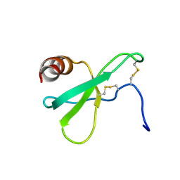
2FSP
 | | NMR SOLUTION STRUCTURE OF BACILLUS SUBTILIS SPO0F PROTEIN, MINIMIZED AVERAGE STRUCTURE | | 分子名称: | STAGE 0 SPORULATION PROTEIN F | | 著者 | Feher, V.A, Skelton, N.J, Dahlquist, F.W, Cavanagh, J. | | 登録日 | 1997-06-06 | | 公開日 | 1997-12-10 | | 最終更新日 | 2024-05-29 | | 実験手法 | SOLUTION NMR | | 主引用文献 | High-resolution NMR structure and backbone dynamics of the Bacillus subtilis response regulator, Spo0F: implications for phosphorylation and molecular recognition.
Biochemistry, 36, 1997
|
|
6YOT
 | |
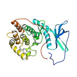
2FOL
 | | Crystal structure of human RAB1A in complex with GDP | | 分子名称: | GUANOSINE-5'-DIPHOSPHATE, MAGNESIUM ION, Ras-related protein Rab-1A, ... | | 著者 | Wang, J, Tempel, W, Shen, Y, Shen, L, Arrowsmith, C, Edwards, A, Sundstrom, M, Weigelt, J, Bochkarev, A, Park, H, Structural Genomics Consortium (SGC) | | 登録日 | 2006-01-13 | | 公開日 | 2006-01-31 | | 最終更新日 | 2023-08-30 | | 実験手法 | X-RAY DIFFRACTION (2.631 Å) | | 主引用文献 | Crystal structure of human RAB1A in complex with GDP
To be Published
|
|
2L7Z
 | | NMR Structure of A13 homedomain | | 分子名称: | Homeobox protein Hox-A13 | | 著者 | Ames, J. | | 登録日 | 2010-12-27 | | 公開日 | 2011-11-09 | | 最終更新日 | 2024-05-15 | | 実験手法 | SOLUTION NMR | | 主引用文献 | Structural basis for sequence specific DNA binding and protein dimerization of HOXA13.
Plos One, 6, 2011
|
|
2LMT
 | | NMR structure of Androcam | | 分子名称: | CALCIUM ION, Calmodulin-related protein 97A | | 著者 | Joshi, M.K, Moran, S.T, Beckingham, K.M, Mackenzie, K.R. | | 登録日 | 2011-12-12 | | 公開日 | 2012-08-22 | | 最終更新日 | 2024-05-15 | | 実験手法 | SOLUTION NMR | | 主引用文献 | Structure of androcam supports specialized interactions with myosin VI.
Proc.Natl.Acad.Sci.USA, 109, 2012
|
|
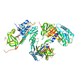
2FR7
 | | Crystal Structure of Cytochrome P450 CYP199A2 | | 分子名称: | PROTOPORPHYRIN IX CONTAINING FE, putative cytochrome P450 | | 著者 | Rao, Z, Wong, L.L, Xu, F, Bell, S.G. | | 登録日 | 2006-01-19 | | 公開日 | 2007-01-16 | | 最終更新日 | 2024-03-13 | | 実験手法 | X-RAY DIFFRACTION (2.01 Å) | | 主引用文献 | Crystal structure of CYP199A2, a para-substituted benzoic acid oxidizing cytochrome P450 from Rhodopseudomonas palustris
J.Mol.Biol., 383, 2008
|
|
2FRS
 | |
2LCW
 | |
2LAV
 | |
2GMT
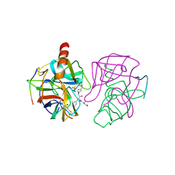 | | THREE-DIMENSIONAL STRUCTURE OF CHYMOTRYPSIN INACTIVATED WITH (2S) N-ACETYL-L-ALANYL-L-PHENYLALANYL-CHLOROETHYL KETONE: IMPLICATIONS FOR THE MECHANISM OF INACTIVATION OF SERINE PROTEASES BY CHLOROKETONES | | 分子名称: | (2S) N-ACETYL-L-ALANYL-ALPHAL-PHENYLALANYL-CHLOROETHYLKETONE, GAMMA-CHYMOTRYPSIN | | 著者 | Kreutter, K, Steinmetz, A.C.U, Liang, T.-C, Prorok, M, Abeles, R, Ringe, D. | | 登録日 | 1994-09-07 | | 公開日 | 1994-11-01 | | 最終更新日 | 2024-06-05 | | 実験手法 | X-RAY DIFFRACTION (1.8 Å) | | 主引用文献 | Three-dimensional structure of chymotrypsin inactivated with (2S)-N-acetyl-L-alanyl-L-phenylalanyl alpha-chloroethane: implications for the mechanism of inactivation of serine proteases by chloroketones.
Biochemistry, 33, 1994
|
|
2HBS
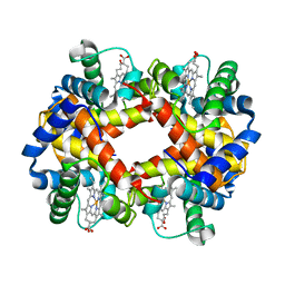 | | THE HIGH RESOLUTION CRYSTAL STRUCTURE OF DEOXYHEMOGLOBIN S | | 分子名称: | HEMOGLOBIN S (DEOXY), ALPHA CHAIN, BETA CHAIN, ... | | 著者 | Harrington, D.J, Adachi, K, Royer Junior, W.E. | | 登録日 | 1997-05-06 | | 公開日 | 1997-07-23 | | 最終更新日 | 2024-02-14 | | 実験手法 | X-RAY DIFFRACTION (2.05 Å) | | 主引用文献 | The high resolution crystal structure of deoxyhemoglobin S.
J.Mol.Biol., 272, 1997
|
|
2L4N
 | |
2HI9
 | |


