2O9J
 
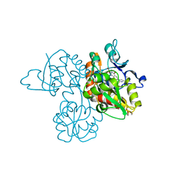
 | | Crystal structure of calcium atpase with bound magnesium fluoride and cyclopiazonic acid | | 分子名称: | (6AR,11AS,11BR)-10-ACETYL-9-HYDROXY-7,7-DIMETHYL-2,6,6A,7,11A,11B-HEXAHYDRO-11H-PYRROLO[1',2':2,3]ISOINDOLO[4,5,6-CD]INDOL-11-ONE, MAGNESIUM ION, SODIUM ION, ... | | 著者 | Young, H.S, Moncoq, K.A. | | 登録日 | 2006-12-13 | | 公開日 | 2007-02-06 | | 最終更新日 | 2023-08-30 | | 実験手法 | X-RAY DIFFRACTION (2.65 Å) | | 主引用文献 | The molecular basis for cyclopiazonic Acid inhibition of the sarcoplasmic reticulum calcium pump.
J.Biol.Chem., 282, 2007
|
|
3U7U
 | | Crystal structure of extracellular region of human epidermal growth factor receptor 4 in complex with neuregulin-1 beta | | 分子名称: | 2-acetamido-2-deoxy-beta-D-glucopyranose, Neuregulin 1, Receptor tyrosine-protein kinase erbB-4 | | 著者 | Liu, P, Cleveland IV, T.E, Bouyain, S, Longo, P.A, Leahy, D.J. | | 登録日 | 2011-10-14 | | 公開日 | 2012-08-29 | | 最終更新日 | 2023-09-13 | | 実験手法 | X-RAY DIFFRACTION (3.03 Å) | | 主引用文献 | A single ligand is sufficient to activate EGFR dimers.
Proc.Natl.Acad.Sci.USA, 109, 2012
|
|
4EKR
 | |
4H9F
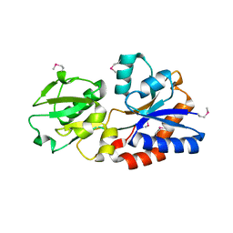
 | | Radiation damage study of lysozyme - 0.91 MGy | | 分子名称: | 1,2-ETHANEDIOL, CHLORIDE ION, Lysozyme C | | 著者 | Sutton, K.A. | | 登録日 | 2012-09-24 | | 公開日 | 2013-05-15 | | 最終更新日 | 2017-11-15 | | 実験手法 | X-RAY DIFFRACTION (1.2003 Å) | | 主引用文献 | Insights into the mechanism of X-ray-induced disulfide-bond cleavage in lysozyme crystals based on EPR, optical absorption and X-ray diffraction studies.
Acta Crystallogr.,Sect.D, 69, 2013
|
|
3F3T
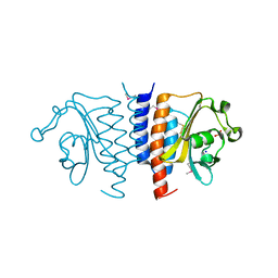
 | | Kinase domain of cSrc in complex with inhibitor RL38 (Type III) | | 分子名称: | 1-[1-(3-aminophenyl)-3-tert-butyl-1H-pyrazol-5-yl]-3-naphthalen-1-ylurea, Proto-oncogene tyrosine-protein kinase Src | | 著者 | Gruetter, C, Klueter, S, Getlik, M, Rauh, D. | | 登録日 | 2008-10-31 | | 公開日 | 2009-03-03 | | 最終更新日 | 2023-11-01 | | 実験手法 | X-RAY DIFFRACTION (2.5 Å) | | 主引用文献 | A new screening assay for allosteric inhibitors of cSrc
Nat.Chem.Biol., 5, 2009
|
|
4ELA
 | | Final Thaumatin Structure for Radiation Damage Experiment at 300 K | | 分子名称: | L(+)-TARTARIC ACID, Thaumatin-1 | | 著者 | Warkentin, M, Badeau, R, Hopkins, J.B, Thorne, R.E. | | 登録日 | 2012-04-10 | | 公開日 | 2012-08-29 | | 最終更新日 | 2013-01-23 | | 実験手法 | X-RAY DIFFRACTION (2 Å) | | 主引用文献 | Spatial distribution of radiation damage to crystalline proteins at 25-300 K.
Acta Crystallogr.,Sect.D, 68, 2012
|
|
3F10
 | |
3U9Z
 | | Crystal structure between actin and a protein construct containing the first beta-thymosin domain of drosophila ciboulot (residues 2-58) with the three mutations N26D/Q27K/D28S | | 分子名称: | ADENOSINE-5'-DIPHOSPHATE, Actin, alpha skeletal muscle, ... | | 著者 | Renault, L, Husson, C, Carlier, M.F, Didry, D. | | 登録日 | 2011-10-20 | | 公開日 | 2012-01-25 | | 最終更新日 | 2023-09-13 | | 実験手法 | X-RAY DIFFRACTION (2.09 Å) | | 主引用文献 | How a single residue in individual beta-thymosin/WH2 domains controls their functions in actin assembly
Embo J., 31, 2012
|
|
3EKP
 | | Crystal Structure of the inhibitor Amprenavir (APV) in complex with a multi-drug resistant HIV-1 protease variant (L10I/G48V/I54V/V64I/V82A)Refer: FLAP+ in citation | | 分子名称: | ACETATE ION, PHOSPHATE ION, Protease, ... | | 著者 | Prabu-Jayabalan, M, King, N.M, Bandaranayake, R.M. | | 登録日 | 2008-09-19 | | 公開日 | 2009-09-01 | | 最終更新日 | 2024-02-21 | | 実験手法 | X-RAY DIFFRACTION (2.15 Å) | | 主引用文献 | Extreme Entropy-Enthalpy Compensation in a Drug-Resistant Variant of HIV-1 Protease.
Acs Chem.Biol., 7, 2012
|
|
4HA0
 | | Structure of Geobacillus kaustophilus lactonase, mutant R230D with Zn2+ | | 分子名称: | FE (III) ION, HYDROXIDE ION, Phosphotriesterase, ... | | 著者 | Xue, B, Chow, J.Y, Yew, W.S, Robinson, R.C. | | 登録日 | 2012-09-25 | | 公開日 | 2012-11-07 | | 最終更新日 | 2013-05-22 | | 実験手法 | X-RAY DIFFRACTION (1.902 Å) | | 主引用文献 | Structural evidence of a productive active site architecture for an evolved quorum-quenching GKL lactonase.
Biochemistry, 52, 2013
|
|
3UA8
 | |
3TQW
 | | Structure of a ABC transporter, periplasmic substrate-binding protein from Coxiella burnetii | | 分子名称: | METHIONINE, Methionine-binding protein, SULFATE ION | | 著者 | Cheung, J, Franklin, M.C, Rudolph, M, Cassidy, M, Gary, E, Burshteyn, F, Love, J. | | 登録日 | 2011-09-09 | | 公開日 | 2011-09-21 | | 最終更新日 | 2017-11-08 | | 実験手法 | X-RAY DIFFRACTION (2 Å) | | 主引用文献 | Structural genomics for drug design against the pathogen Coxiella burnetii.
Proteins, 83, 2015
|
|
3E9Z
 | | Crystal structure of purine nucleoside phosphorylase from Schistosoma mansoni in complex with 6-chloroguanine | | 分子名称: | 6-chloroguanine, ACETATE ION, DIMETHYL SULFOXIDE, ... | | 著者 | Pereira, H.M, Rezende, M.M, Oliva, G, Garratt, R.C. | | 登録日 | 2008-08-24 | | 公開日 | 2009-09-01 | | 最終更新日 | 2023-08-30 | | 実験手法 | X-RAY DIFFRACTION (2.31 Å) | | 主引用文献 | Crystal structure of Schistosoma mansoni purine nucleoside phosphorylase (SmPNP) in complex with adenine, 8-aminoguanine, 8-azaguanine and 6-chloroguanine.
To be Published
|
|
3TRC
 | | Structure of the GAF domain from a phosphoenolpyruvate-protein phosphotransferase (ptsP) from Coxiella burnetii | | 分子名称: | PHOSPHATE ION, Phosphoenolpyruvate-protein phosphotransferase, SODIUM ION | | 著者 | Cheung, J, Franklin, M.C, Rudolph, M, Cassidy, M, Gary, E, Burshteyn, F, Love, J. | | 登録日 | 2011-09-09 | | 公開日 | 2011-09-28 | | 最終更新日 | 2017-11-08 | | 実験手法 | X-RAY DIFFRACTION (1.65 Å) | | 主引用文献 | Structural genomics for drug design against the pathogen Coxiella burnetii.
Proteins, 83, 2015
|
|
3TRK
 | | Structure of the Chikungunya virus nsP2 protease | | 分子名称: | Nonstructural polyprotein, SODIUM ION | | 著者 | Cheung, J, Franklin, M, Mancia, F, Rudolph, M, Cassidy, M, Gary, E, Burshteyn, F, Love, J. | | 登録日 | 2011-09-09 | | 公開日 | 2011-09-28 | | 最終更新日 | 2023-12-06 | | 実験手法 | X-RAY DIFFRACTION (2.397 Å) | | 主引用文献 | Structure of the Chikungunya virus nsP2 protease
To be Published
|
|
4ENP
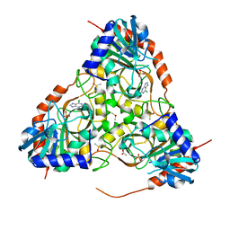
 | | Structure of E530A variant E. coli KatE | | 分子名称: | Catalase HPII, PROTOPORPHYRIN IX CONTAINING FE | | 著者 | Loewen, P.C, Jha, V. | | 登録日 | 2012-04-13 | | 公開日 | 2012-05-09 | | 最終更新日 | 2024-02-28 | | 実験手法 | X-RAY DIFFRACTION (1.5 Å) | | 主引用文献 | Influence of main channel structure on H(2)O(2) access to the heme cavity of catalase KatE of Escherichia coli.
Arch.Biochem.Biophys., 526, 2012
|
|
3EMK
 | |
4HBO
 | |
4EPD
 | | Initial Urease Structure for Radiation Damage Experiment at 300 K | | 分子名称: | NICKEL (II) ION, Urease subunit alpha, Urease subunit beta, ... | | 著者 | Warkentin, M, Badeau, R, Hopkins, J.B, Thorne, R.E. | | 登録日 | 2012-04-17 | | 公開日 | 2012-08-29 | | 最終更新日 | 2013-01-23 | | 実験手法 | X-RAY DIFFRACTION (1.7 Å) | | 主引用文献 | Spatial distribution of radiation damage to crystalline proteins at 25-300 K.
Acta Crystallogr.,Sect.D, 68, 2012
|
|
3TK8
 | | Structure of a 2,3,4,5-tetrahydropyridine-2,6-dicarboxylate N-succinyltransferase from Burkholderia pseudomallei | | 分子名称: | 1,2-ETHANEDIOL, 2,3,4,5-tetrahydropyridine-2,6-dicarboxylate N-succinyltransferase, SULFATE ION, ... | | 著者 | Seattle Structural Genomics Center for Infectious Disease (SSGCID) | | 登録日 | 2011-08-25 | | 公開日 | 2011-10-12 | | 最終更新日 | 2024-02-28 | | 実験手法 | X-RAY DIFFRACTION (1.8 Å) | | 主引用文献 | Structure of a 2,3,4,5-tetrahydropyridine-2,6-dicarboxylate N-succinyltransferase from Burkholderia pseudomallei
TO BE PUBLISHED
|
|
3EEI
 | | Crystal structure of 5'-methylthioadenosine/S-adenosylhomocysteine nucleosidase from neisseria meningitidis in complex with methylthio-immucillin-A | | 分子名称: | (3S,4R)-2-(4-AMINO-5H-PYRROLO[3,2-D]PYRIMIDIN-7-YL)-5-[(METHYLSULFANYL)METHYL]PYRROLIDINE-3,4-DIOL, 5-methylthioadenosine nucleosidase/S-adenosylhomocysteine nucleosidase | | 著者 | Ho, M, Rinaldo-matthis, A, Brown, R.L, Norris, G.E, Tyler, P.C, Furneaux, R.H, Almo, S.C, Schramm, V.L. | | 登録日 | 2008-09-04 | | 公開日 | 2009-09-08 | | 最終更新日 | 2024-02-21 | | 実験手法 | X-RAY DIFFRACTION (1.78 Å) | | 主引用文献 | Crystal structure of 5'-methylthioadenosine/S-adenosylhomocysteine nucleosidase from neisseria meningitidis in complex with methylthio-immucillin-A
To be Published
|
|
4EDK
 | | The structure of the S. aureus DnaG RNA Polymerase Domain bound to GTP and Manganese | | 分子名称: | BENZAMIDINE, DNA primase, GUANOSINE-5'-TRIPHOSPHATE, ... | | 著者 | Rymer, R.U, Solorio, F.A, Chu, C, Corn, J.E, Wang, J.D, Berger, J.M. | | 登録日 | 2012-03-27 | | 公開日 | 2012-07-25 | | 最終更新日 | 2024-02-28 | | 実験手法 | X-RAY DIFFRACTION (2 Å) | | 主引用文献 | Binding Mechanism of Metal-NTP Substrates and Stringent-Response Alarmones to Bacterial DnaG-Type Primases.
Structure, 20, 2012
|
|
2OIL
 | | Crystal structure of human RAB25 in complex with GDP | | 分子名称: | GUANOSINE-5'-DIPHOSPHATE, MAGNESIUM ION, Ras-related protein Rab-25, ... | | 著者 | Zhu, H, Wang, J, Shen, Y, Tempel, W, Landry, R, Arrowsmith, C.H, Edwards, A.M, Sundstrom, M, Weigelt, J, Bochkarev, A, Park, H, Structural Genomics Consortium (SGC) | | 登録日 | 2007-01-11 | | 公開日 | 2007-01-23 | | 最終更新日 | 2023-08-30 | | 実験手法 | X-RAY DIFFRACTION (2.3 Å) | | 主引用文献 | Crystal structure of human RAB25 in complex with GDP
To be Published
|
|
4EQZ
 | | Crystal structure of human DOT1L in complex with inhibitor FED2 | | 分子名称: | 5'-deoxy-5'-[(3-{[(4-methylphenyl)carbamoyl]amino}propyl)(propan-2-yl)amino]adenosine, Histone-lysine N-methyltransferase, H3 lysine-79 specific, ... | | 著者 | Wernimont, A.K, Tempel, W, Yu, W, Li, Y, Nguyen, K.T, Federation, A, Marineau, J, Qi, J, Vedadi, M, Bradner, J.E, Schapira, M, Arrowsmith, C.H, Edwards, A.M, Bountra, C, Brown, P.J, Structural Genomics Consortium (SGC) | | 登録日 | 2012-04-19 | | 公開日 | 2012-05-02 | | 最終更新日 | 2024-02-28 | | 実験手法 | X-RAY DIFFRACTION (2.15 Å) | | 主引用文献 | Catalytic site remodelling of the DOT1L methyltransferase by selective inhibitors.
Nat Commun, 3, 2012
|
|
4HF5
 | |


