3UR2
 
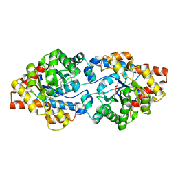
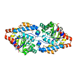 | | Crystal Structure of PTE mutant H254G/H257W/L303T/K185R/I274N/A80V | | 分子名称: | 1,2-ETHANEDIOL, COBALT (II) ION, IMIDAZOLE, ... | | 著者 | Tsai, P, Fox, N.G, Li, Y, Barondeau, D.P, Raushel, F.M. | | 登録日 | 2011-11-21 | | 公開日 | 2012-08-01 | | 最終更新日 | 2023-12-06 | | 実験手法 | X-RAY DIFFRACTION (2 Å) | | 主引用文献 | Enzymes for the homeland defense: optimizing phosphotriesterase for the hydrolysis of organophosphate nerve agents.
Biochemistry, 51, 2012
|
|
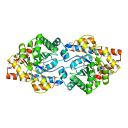
3UR5
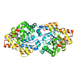 | | Crystal Structure of PTE mutant K185R/I274N | | 分子名称: | COBALT (II) ION, DIETHYL HYDROGEN PHOSPHATE, Parathion hydrolase | | 著者 | Tsai, P, Fox, N.G, Li, Y, Barondeau, D.P, Raushel, F.M. | | 登録日 | 2011-11-21 | | 公開日 | 2012-08-01 | | 最終更新日 | 2023-12-06 | | 実験手法 | X-RAY DIFFRACTION (1.6 Å) | | 主引用文献 | Enzymes for the homeland defense: optimizing phosphotriesterase for the hydrolysis of organophosphate nerve agents.
Biochemistry, 51, 2012
|
|
3URQ
 | | Crystal Structure of PTE mutant H254G/H257W/L303T/M317L/I106C/F132I/L271I/K185R/I274N/A80V/R67H with cyclohexyl methylphosphonate inhibitor | | 分子名称: | COBALT (II) ION, IMIDAZOLE, Parathion hydrolase, ... | | 著者 | Tsai, P, Fox, N.G, Li, Y, Barondeau, D.P, Raushel, F.M. | | 登録日 | 2011-11-22 | | 公開日 | 2012-08-01 | | 最終更新日 | 2023-12-06 | | 実験手法 | X-RAY DIFFRACTION (2.1 Å) | | 主引用文献 | Enzymes for the homeland defense: optimizing phosphotriesterase for the hydrolysis of organophosphate nerve agents.
Biochemistry, 51, 2012
|
|
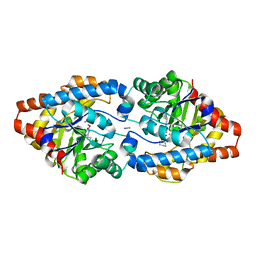
3URB
 | | Crystal Structure of PTE mutant H254G/H257W/L303T/M317L/I106C/F132I/L271I/K185R/I274N/A80V/R67H | | 分子名称: | COBALT (II) ION, DIETHYL HYDROGEN PHOSPHATE, IMIDAZOLE, ... | | 著者 | Tsai, P, Fox, N.G, Li, Y, Barondeau, D.P, Raushel, F.M. | | 登録日 | 2011-11-21 | | 公開日 | 2012-08-01 | | 最終更新日 | 2023-12-06 | | 実験手法 | X-RAY DIFFRACTION (1.77 Å) | | 主引用文献 | Enzymes for the homeland defense: optimizing phosphotriesterase for the hydrolysis of organophosphate nerve agents.
Biochemistry, 51, 2012
|
|
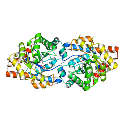
3URA
 | | Crystal Structure of PTE mutant H254G/H257W/L303T/K185R/I274N/A80V/S61T | | 分子名称: | COBALT (II) ION, IMIDAZOLE, Parathion hydrolase | | 著者 | Tsai, P, Fox, N.G, Li, Y, Barondeau, D.P, Raushel, F.M. | | 登録日 | 2011-11-21 | | 公開日 | 2012-08-01 | | 最終更新日 | 2023-12-06 | | 実験手法 | X-RAY DIFFRACTION (1.88 Å) | | 主引用文献 | Enzymes for the homeland defense: optimizing phosphotriesterase for the hydrolysis of organophosphate nerve agents.
Biochemistry, 51, 2012
|
|
3URN
 | | Crystal Structure of PTE mutant H254G/H257W/L303T/K185R/I274N/A80V/S61T with cyclohexyl methylphosphonate inhibitor | | 分子名称: | COBALT (II) ION, IMIDAZOLE, Parathion hydrolase, ... | | 著者 | Tsai, P, Fox, N.G, Li, Y, Barondeau, D.P, Raushel, F.M. | | 登録日 | 2011-11-22 | | 公開日 | 2012-08-01 | | 最終更新日 | 2023-12-06 | | 実験手法 | X-RAY DIFFRACTION (1.95 Å) | | 主引用文献 | Enzymes for the homeland defense: optimizing phosphotriesterase for the hydrolysis of organophosphate nerve agents.
Biochemistry, 51, 2012
|
|
3UPM
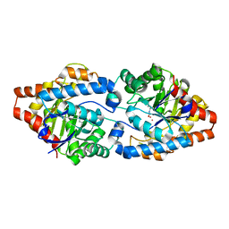 | | Crystal Structure of PTE mutant H254Q/H257F/K185R/I274N | | 分子名称: | COBALT (II) ION, Parathion hydrolase | | 著者 | Tsai, P, Fox, N.G, Li, Y, Barondeau, D.P, Raushel, F.M. | | 登録日 | 2011-11-18 | | 公開日 | 2012-08-01 | | 最終更新日 | 2023-12-06 | | 実験手法 | X-RAY DIFFRACTION (1.95 Å) | | 主引用文献 | Enzymes for the homeland defense: optimizing phosphotriesterase for the hydrolysis of organophosphate nerve agents.
Biochemistry, 51, 2012
|
|
6U95
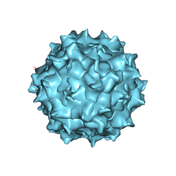 | | Adeno-associated virus strain AAVhu.37 capsid icosahedral structure | | 分子名称: | Capsid protein VP1 | | 著者 | Kaelber, J.T, Yost, S.A, Firlar, E, Mercer, A.C. | | 登録日 | 2019-09-06 | | 公開日 | 2019-09-18 | | 最終更新日 | 2024-03-20 | | 実験手法 | ELECTRON MICROSCOPY (2.56 Å) | | 主引用文献 | Structure of the AAVhu.37 capsid by cryoelectron microscopy.
Acta Crystallogr.,Sect.F, 76, 2020
|
|
6WV5
 | | Human VKOR C43S mutant with vitamin K1 epoxide | | 分子名称: | (2R,3R)-2-hydroxy-3-methyl-2-[(2E,7S)-3,7,11,15-tetramethylhexadec-2-en-1-yl]-2,3-dihydronaphthalene-1,4-dione, Vitamin K epoxide reductase Cys43Ser mutant, termini restrained by green fluorescent protein | | 著者 | Liu, S, Sukumar, N, Li, W. | | 登録日 | 2020-05-05 | | 公開日 | 2020-11-11 | | 最終更新日 | 2023-11-15 | | 実験手法 | X-RAY DIFFRACTION (2.8 Å) | | 主引用文献 | Structural basis of antagonizing the vitamin K catalytic cycle for anticoagulation.
Science, 371, 2021
|
|
6WV6
 | | Human VKOR with phenindione | | 分子名称: | (2R)-2,3-dihydroxypropyl (9Z)-octadec-9-enoate, Phenindione, Vitamin K epoxide reductase, ... | | 著者 | Liu, S, Sukumar, N, Li, W. | | 登録日 | 2020-05-05 | | 公開日 | 2020-11-11 | | 最終更新日 | 2023-11-15 | | 実験手法 | X-RAY DIFFRACTION (2.7 Å) | | 主引用文献 | Structural basis of antagonizing the vitamin K catalytic cycle for anticoagulation.
Science, 371, 2021
|
|
6WV3
 | | Human VKOR with warfarin | | 分子名称: | (2R)-2,3-dihydroxypropyl (9Z)-octadec-9-enoate, S-WARFARIN, Vitamin K epoxide reductase, ... | | 著者 | Liu, S, Sukumar, N, Li, W. | | 登録日 | 2020-05-05 | | 公開日 | 2020-11-11 | | 最終更新日 | 2023-11-15 | | 実験手法 | X-RAY DIFFRACTION (2.197 Å) | | 主引用文献 | Structural basis of antagonizing the vitamin K catalytic cycle for anticoagulation.
Science, 371, 2021
|
|
6WVH
 | | Human VKOR with Brodifacoum | | 分子名称: | (2R)-2,3-dihydroxypropyl (9Z)-octadec-9-enoate, Brodifacoum, Vitamin K epoxide reductase, ... | | 著者 | Liu, S, Sukumar, N, Li, W. | | 登録日 | 2020-05-06 | | 公開日 | 2020-11-11 | | 最終更新日 | 2023-11-15 | | 実験手法 | X-RAY DIFFRACTION (1.99 Å) | | 主引用文献 | Structural basis of antagonizing the vitamin K catalytic cycle for anticoagulation.
Science, 371, 2021
|
|
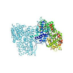
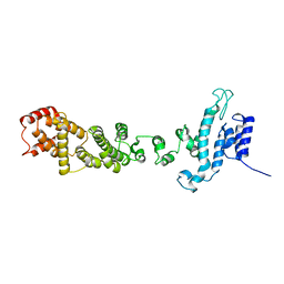
4WFC
 | | Structure of the Rrp6-Rrp47 interaction | | 分子名称: | Exosome complex exonuclease RRP6, Exosome complex protein LRP1, SULFATE ION | | 著者 | Schuch, B, Conti, E. | | 登録日 | 2014-09-14 | | 公開日 | 2014-10-29 | | 最終更新日 | 2024-05-01 | | 実験手法 | X-RAY DIFFRACTION (2.35 Å) | | 主引用文献 | The exosome-binding factors Rrp6 and Rrp47 form a composite surface for recruiting the Mtr4 helicase.
Embo J., 33, 2014
|
|
4GPB
 | |
6ZC5
 | | Human Adenovirus serotype D10 FiberKnob protein | | 分子名称: | Fiber | | 著者 | Baker, A.T, Mundy, R.M, Rizkallah, P.J, Parker, A.L. | | 登録日 | 2020-06-09 | | 公開日 | 2021-06-30 | | 最終更新日 | 2024-01-24 | | 実験手法 | X-RAY DIFFRACTION (2.5 Å) | | 主引用文献 | Development of a low-seroprevalence, alpha v beta 6 integrin-selective virotherapy based on human adenovirus type 10.
Mol Ther Oncolytics, 25, 2022
|
|
1TWB
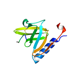 | | SspB disulfide crosslinked to an ssrA degradation tag | | 分子名称: | Stringent starvation protein B homolog, ssrA peptide | | 著者 | Bolon, D.N, Grant, R.A, Baker, T.A, Sauer, R.T. | | 登録日 | 2004-06-30 | | 公開日 | 2004-11-16 | | 最終更新日 | 2023-08-23 | | 実験手法 | X-RAY DIFFRACTION (1.9 Å) | | 主引用文献 | Nucleotide-Dependent Substrate Handoff from the SspB Adaptor to the AAA+ ClpXP Protease.
Mol.Cell, 16, 2004
|
|
7EYF
 | |
1DV0
 | |
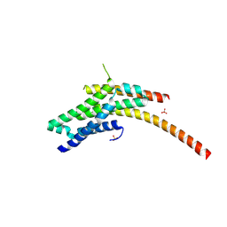
6TA7
 | | CRYSTAL STRUCTURE OF HUMAN G3BP1-NTF2 IN COMPLEX WITH HUMAN CAPRIN1-DERIVED SOLOMON MOTIF | | 分子名称: | CHLORIDE ION, Caprin-1, Ras GTPase-activating protein-binding protein 1, ... | | 著者 | Schulte, T, Achour, A, Panas, M.D, McInerney, G.M. | | 登録日 | 2019-10-29 | | 公開日 | 2021-05-12 | | 最終更新日 | 2024-01-24 | | 実験手法 | X-RAY DIFFRACTION (1.93 Å) | | 主引用文献 | Caprin-1 binding to the critical stress granule protein G3BP1 is regulated by pH
Biorxiv, 2021
|
|
3MMI
 | |
6NHT
 | |
3VHS
 | |
5WTS
 | |
5XG8
 | |
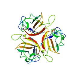
5XG7
 | | Galectin-13/Placental Protein 13 crystal structure | | 分子名称: | Galactoside-binding soluble lectin 13 | | 著者 | Su, J.Y, Wang, Y. | | 登録日 | 2017-04-12 | | 公開日 | 2018-01-31 | | 最終更新日 | 2024-04-17 | | 実験手法 | X-RAY DIFFRACTION (1.55 Å) | | 主引用文献 | Galectin-13, a different prototype galectin, does not bind beta-galacto-sides and forms dimers via intermolecular disulfide bridges between Cys-136 and Cys-138
Sci Rep, 8, 2018
|
|


