7YF7
 
 | |
3USJ
 | |
4F8K
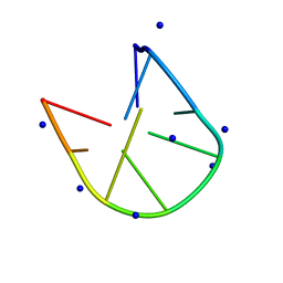
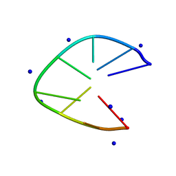
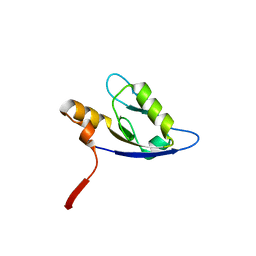 | | Molecular analysis of the interaction between the prostacyclin receptor and the first PDZ domain of PDZK1 | | 分子名称: | Na(+)/H(+) exchange regulatory cofactor NHE-RF3, Prostacyclin receptor | | 著者 | Kocher, O, Birrane, G, Kinsella, B.T, Mulvaney, E.P. | | 登録日 | 2012-05-17 | | 公開日 | 2013-02-27 | | 最終更新日 | 2023-09-13 | | 実験手法 | X-RAY DIFFRACTION (1.7 Å) | | 主引用文献 | Molecular Analysis of the Prostacyclin Receptor's Interaction with the PDZ1 Domain of Its Adaptor Protein PDZK1.
Plos One, 8, 2013
|
|
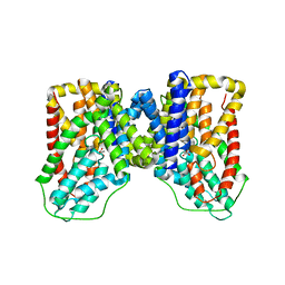
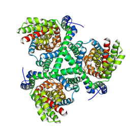
3UQ4
 | | X-ray structure of a pentameric ligand gated ion channel from Erwinia chrysanthemi (ELIC) mutant F247L (F16L) | | 分子名称: | Gamma-aminobutyric-acid receptor subunit beta-1, SODIUM ION | | 著者 | Gonzalez-Gutierrez, G, Lukk, T, Agarwal, V, Papke, D, Nair, S.K, Grosman, C. | | 登録日 | 2011-11-19 | | 公開日 | 2012-04-04 | | 最終更新日 | 2023-09-13 | | 実験手法 | X-RAY DIFFRACTION (3.5 Å) | | 主引用文献 | Mutations that stabilize the open state of the Erwinia chrisanthemi ligand-gated ion channel fail to change the conformation of the pore domain in crystals.
Proc.Natl.Acad.Sci.USA, 109, 2012
|
|
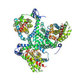
6DFT
 | | Trypanosoma brucei deoxyhypusine synthase | | 分子名称: | Deoxyhypusine synthase, Deoxyhypusine synthase regulatory subunit, NICOTINAMIDE-ADENINE-DINUCLEOTIDE, ... | | 著者 | Tomchick, D.R, Phillips, M.A, Afanador, G.A. | | 登録日 | 2018-05-15 | | 公開日 | 2018-08-08 | | 最終更新日 | 2023-10-11 | | 実験手法 | X-RAY DIFFRACTION (3.5 Å) | | 主引用文献 | Trypanosomatid Deoxyhypusine Synthase Activity Is Dependent on Shared Active-Site Complementation between Pseudoenzyme Paralogs.
Structure, 26, 2018
|
|
6M6K
 | |
6M6J
 | |
6OL1
 | |
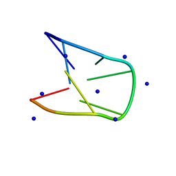
3UQ5
 | | X-ray structure of a pentameric ligand gated ion channel from Erwinia chrysanthemi (ELIC) mutant L240A F247L (L9A F16L) in the presence of 10 mM cysteamine | | 分子名称: | Gamma-aminobutyric-acid receptor subunit beta-1, SODIUM ION | | 著者 | Gonzalez-Gutierrez, G, Lukk, T, Agarwal, V, Papke, D, Nair, S.K, Grosman, C. | | 登録日 | 2011-11-19 | | 公開日 | 2012-04-04 | | 最終更新日 | 2023-09-13 | | 実験手法 | X-RAY DIFFRACTION (4.2 Å) | | 主引用文献 | Mutations that stabilize the open state of the Erwinia chrisanthemi ligand-gated ion channel fail to change the conformation of the pore domain in crystals.
Proc.Natl.Acad.Sci.USA, 109, 2012
|
|
6M0B
 | |
6WTX
 | |
6M0C
 | |
4X2S
 | |
6V8G
 | | GltPh mutant - Y204L A345V V366A | | 分子名称: | ASPARTIC ACID, Glutamate transporter homolog, SODIUM ION | | 著者 | Boudker, O, Huysmans, G.H.M. | | 登録日 | 2019-12-11 | | 公開日 | 2020-11-18 | | 最終更新日 | 2023-10-11 | | 実験手法 | X-RAY DIFFRACTION (3.38 Å) | | 主引用文献 | The high-energy transition state of the glutamate transporter homologue GltPh.
Embo J., 40, 2021
|
|
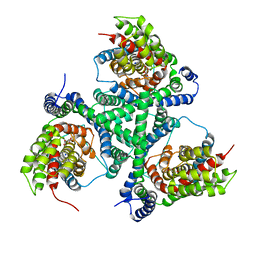
4LD7
 | | Crystal structure of AnaPT from Neosartorya fischeri | | 分子名称: | Dimethylallyl tryptophan synthase, SODIUM ION, TRIHYDROGEN THIODIPHOSPHATE | | 著者 | Zocher, G, Stehle, T. | | 登録日 | 2013-06-24 | | 公開日 | 2013-12-11 | | 最終更新日 | 2024-02-28 | | 実験手法 | X-RAY DIFFRACTION (2.83 Å) | | 主引用文献 | Catalytic Mechanism of Stereospecific Formation of cis-Configured Prenylated Pyrroloindoline Diketopiperazines by Indole Prenyltransferases.
Chem.Biol., 20, 2013
|
|
6IY5
 | | NMR solution structures of 5'-ATTCTATTCT-3 | | 分子名称: | DNA (5'-D(*AP*TP*TP*CP*TP*AP*TP*TP*CP*T)-3'), SODIUM ION | | 著者 | Lam, S.L, Guo, P. | | 登録日 | 2018-12-13 | | 公開日 | 2020-06-10 | | 最終更新日 | 2024-05-15 | | 実験手法 | SOLUTION NMR | | 主引用文献 | Minidumbbell structures formed by ATTCT pentanucleotide repeats in spinocerebellar ataxia type 10.
Nucleic Acids Res., 48, 2020
|
|
6BAU
 | | Crystal Structure of GltPh R397C in complex with L-Cysteine | | 分子名称: | CYSTEINE, Glutamate transporter homolog, SODIUM ION | | 著者 | Font, J, Scopelliti, A.J, Vandenberg, R.J, Boudker, O, Ryan, R.M. | | 登録日 | 2017-10-15 | | 公開日 | 2018-01-17 | | 最終更新日 | 2023-10-04 | | 実験手法 | X-RAY DIFFRACTION (3.8 Å) | | 主引用文献 | Structural characterisation reveals insights into substrate recognition by the glutamine transporter ASCT2/SLC1A5.
Nat Commun, 9, 2018
|
|
6BAT
 | | Crystal Structure of Wild-Type GltPh in complex with L-aspartate | | 分子名称: | ASPARTIC ACID, Glutamate transporter homolog, SODIUM ION | | 著者 | Font, J, Scopelliti, A.J, Vandenberg, R.J, Boudker, O, Ryan, R.M. | | 登録日 | 2017-10-15 | | 公開日 | 2018-01-17 | | 最終更新日 | 2023-10-04 | | 実験手法 | X-RAY DIFFRACTION (3.4 Å) | | 主引用文献 | Structural characterisation reveals insights into substrate recognition by the glutamine transporter ASCT2/SLC1A5.
Nat Commun, 9, 2018
|
|
5GWL
 | | Structure of two CCTG repeats | | 分子名称: | DNA (5'-D(*CP*CP*TP*GP*CP*CP*TP*G)-3'), SODIUM ION | | 著者 | Guo, P, Lam, S.L. | | 登録日 | 2016-09-12 | | 公開日 | 2016-10-12 | | 最終更新日 | 2024-05-01 | | 実験手法 | SOLUTION NMR | | 主引用文献 | Minidumbbell: A New Form of Native DNA Structure
J.Am.Chem.Soc., 138, 2016
|
|
5GWQ
 | | Structure of two TTTA repeats | | 分子名称: | DNA (5'-D(*TP*TP*TP*AP*TP*TP*TP*A)-3'), SODIUM ION | | 著者 | Guo, P, Lam, S.L. | | 登録日 | 2016-09-13 | | 公開日 | 2016-10-12 | | 最終更新日 | 2024-05-01 | | 実験手法 | SOLUTION NMR | | 主引用文献 | Minidumbbell: A New Form of Native DNA Structure
J.Am.Chem.Soc., 138, 2016
|
|
6J37
 | | DNA minidumbbell structure of two CTTG repeats | | 分子名称: | DNA (5'-D(*CP*TP*TP*GP*CP*TP*TP*G)-3'), SODIUM ION | | 著者 | Lam, S.L, Guo, P. | | 登録日 | 2019-01-04 | | 公開日 | 2019-05-29 | | 最終更新日 | 2024-05-01 | | 実験手法 | SOLUTION NMR | | 主引用文献 | Unprecedented hydrophobic stabilizations from a reverse wobble T·T mispair in DNA minidumbbell.
J.Biomol.Struct.Dyn., 38, 2020
|
|
6CTF
 | |
3V8G
 | |
6X2Z
 | | hEAAT3-OFS-Asp | | 分子名称: | ASPARTIC ACID, Excitatory amino acid transporter 3, SODIUM ION | | 著者 | Qiu, B, Matthies, D, Boudker, O. | | 登録日 | 2020-05-21 | | 公開日 | 2021-03-17 | | 最終更新日 | 2024-03-06 | | 実験手法 | ELECTRON MICROSCOPY (3.03 Å) | | 主引用文献 | Cryo-EM structures of excitatory amino acid transporter 3 visualize coupled substrate, sodium, and proton binding and transport.
Sci Adv, 7, 2021
|
|
6X3E
 | | hEAAT3-Asymmetric-1o2i | | 分子名称: | ASPARTIC ACID, Excitatory amino acid transporter 3, SODIUM ION | | 著者 | Qiu, B, Matthies, D, Boudker, O. | | 登録日 | 2020-05-21 | | 公開日 | 2021-03-17 | | 最終更新日 | 2024-03-06 | | 実験手法 | ELECTRON MICROSCOPY (3.42 Å) | | 主引用文献 | Cryo-EM structures of excitatory amino acid transporter 3 visualize coupled substrate, sodium, and proton binding and transport.
Sci Adv, 7, 2021
|
|


