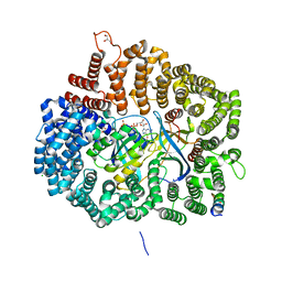
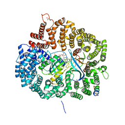
6I7S
 
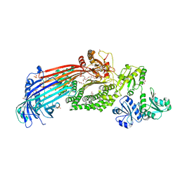 | | Microsomal triglyceride transfer protein | | 分子名称: | 1,2-ETHANEDIOL, 2-AMINO-2-HYDROXYMETHYL-PROPANE-1,3-DIOL, 2-{2-[2-(2-{2-[2-(2-ETHOXY-ETHOXY)-ETHOXY]-ETHOXY}-ETHOXY)-ETHOXY]-ETHOXY}-ETHANOL, ... | | 著者 | Biterova, E, Isupov, M.N, Keegan, R.M, Lebedev, A.A, Ruddock, L.W. | | 登録日 | 2018-11-17 | | 公開日 | 2019-08-21 | | 最終更新日 | 2024-01-24 | | 実験手法 | X-RAY DIFFRACTION (2.5 Å) | | 主引用文献 | The crystal structure of human microsomal triglyceride transfer protein.
Proc.Natl.Acad.Sci.USA, 116, 2019
|
|
6A3E
 | | MVM NES mutant Nm15 in complex with CRM1-Ran-RanBP1 | | 分子名称: | Exportin-1, GTP-binding nuclear protein Ran, GUANOSINE-5'-TRIPHOSPHATE, ... | | 著者 | Sun, Q, Li, Y. | | 登録日 | 2018-06-15 | | 公開日 | 2019-06-19 | | 最終更新日 | 2023-11-22 | | 実験手法 | X-RAY DIFFRACTION (2.7 Å) | | 主引用文献 | Cancer Therapy with Nanoparticle-Medicated Intracellular Expression of Peptide CRM1-Inhibitor.
Int J Nanomedicine, 16, 2021
|
|
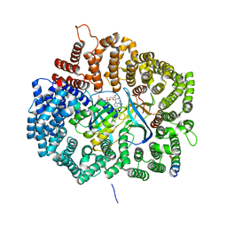
3ELO
 | | Crystal Structure of Human Pancreatic Prophospholipase A2 | | 分子名称: | Phospholipase A2, SULFATE ION | | 著者 | Xu, W, Yi, L, Feng, Y, Chen, L, Liu, J. | | 登録日 | 2008-09-22 | | 公開日 | 2009-04-14 | | 最終更新日 | 2014-02-05 | | 実験手法 | X-RAY DIFFRACTION (1.55 Å) | | 主引用文献 | Structural insight into the activation mechanism of human pancreatic prophospholipase A2
J.Biol.Chem., 284, 2009
|
|
6IPY
 | |
1MSH
 | |
6A3C
 | | MVM NES mutant Nm12 in complex with CRM1-Ran-RanBP1 | | 分子名称: | 1,2-ETHANEDIOL, CHLORIDE ION, Exportin-1, ... | | 著者 | Sun, Q, Li, Y. | | 登録日 | 2018-06-15 | | 公開日 | 2019-06-19 | | 最終更新日 | 2023-11-22 | | 実験手法 | X-RAY DIFFRACTION (2.35 Å) | | 主引用文献 | Cancer Therapy with Nanoparticle-Medicated Intracellular Expression of Peptide CRM1-Inhibitor.
Int J Nanomedicine, 16, 2021
|
|
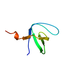
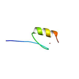
3E2B
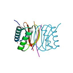 | | Crystal structure of Dynein Light chain LC8 in complex with a peptide derived from Swallow | | 分子名称: | ACETATE ION, Dynein light chain 1, cytoplasmic, ... | | 著者 | Benison, G, Barbar, E, Karplus, P.A. | | 登録日 | 2008-08-05 | | 公開日 | 2008-08-12 | | 最終更新日 | 2023-08-30 | | 実験手法 | X-RAY DIFFRACTION (2 Å) | | 主引用文献 | The interplay of ligand binding and quaternary structure in the diverse interactions of dynein light chain LC8.
J.Mol.Biol., 384, 2008
|
|
6A38
 | | MVM NS2 NES in complex with CRM1-Ran-RanBP1 | | 分子名称: | 1,2-ETHANEDIOL, Exportin-1, GLYCEROL, ... | | 著者 | Sun, Q, Li, Y. | | 登録日 | 2018-06-15 | | 公開日 | 2019-06-19 | | 最終更新日 | 2023-11-22 | | 実験手法 | X-RAY DIFFRACTION (2.69 Å) | | 主引用文献 | Cancer Therapy with Nanoparticle-Medicated Intracellular Expression of Peptide CRM1-Inhibitor.
Int J Nanomedicine, 16, 2021
|
|
1OOT
 | |
5AAZ
 | | TBK1 recruitment to cytosol-invading Salmonella induces anti- bacterial autophagy | | 分子名称: | OPTINEURIN, ZINC ION | | 著者 | Thurston, T.l, Allen, M.D, Ravenhill, B, Karpiyevitch, M, Bloor, S, Kaul, A, Matthews, S, Komander, D, Holden, D, Bycroft, M, Randow, F. | | 登録日 | 2015-07-31 | | 公開日 | 2016-07-13 | | 最終更新日 | 2024-05-15 | | 実験手法 | SOLUTION NMR | | 主引用文献 | Recruitment of Tbk1 to Cytosol-Invading Salmonella Induces Wipi2-Dependent Antibacterial Autophagy.
Embo J., 35, 2016
|
|
6AGV
 | |
4Q6F
 | | Crystal structure of human BAZ2A PHD zinc finger in complex with unmodified H3K4 histone peptide | | 分子名称: | 1,2-ETHANEDIOL, Bromodomain adjacent to zinc finger domain protein 2A, ZINC ION, ... | | 著者 | Tallant, C, Overvoorde, L, Krojer, T, Filippakopoulos, P, von Delft, F, Arrowsmith, C.H, Edwards, A.M, Bountra, C, Ciulli, A, Knapp, S, Structural Genomics Consortium (SGC) | | 登録日 | 2014-04-22 | | 公開日 | 2014-05-21 | | 最終更新日 | 2024-02-28 | | 実験手法 | X-RAY DIFFRACTION (1.91 Å) | | 主引用文献 | Molecular basis of histone tail recognition by human TIP5 PHD finger and bromodomain of the chromatin remodeling complex NoRC.
Structure, 23, 2015
|
|
7A36
 | |
7A39
 | |
7A3E
 | |
7A3C
 | |
7A2T
 | |
7A2Y
 | |
7A2Z
 | |
7A31
 | |
7A32
 | |
7A35
 | |
7A38
 | |
5KK5
 | | AsCpf1(E993A)-crRNA-DNA ternary complex | | 分子名称: | CRISPR-associated endonuclease Cpf1, DNA (28-MER), DNA (8-mer), ... | | 著者 | Gao, P, Yang, H, Rajashankar, K.R, Huang, Z, Patel, D.J. | | 登録日 | 2016-06-21 | | 公開日 | 2016-08-10 | | 最終更新日 | 2024-03-06 | | 実験手法 | X-RAY DIFFRACTION (3.289 Å) | | 主引用文献 | Type V CRISPR-Cas Cpf1 endonuclease employs a unique mechanism for crRNA-mediated target DNA recognition.
Cell Res., 26, 2016
|
|
7A2X
 | |


