Loading
PDBj
メニューPDBj@FacebookPDBj@TwitterPDBj@YouTubewwPDB FoundationwwPDB
RCSB PDBPDBeBMRBAdv. SearchSearch help

2HGY
DownloadVisualize
BU of 2hgy by Molmil
Structure of S65A Y66F E222A GFP variant after cyclization and carbon-carbon bond cleavage
分子名称: Green fluorescent protein
著者Barondeau, D.P, Kassmann, C.J, Tainer, J.A, Getzoff, E.D.
登録日2006-06-27
公開日2007-03-27
最終更新日2023-11-15
実験手法X-RAY DIFFRACTION (2.05 Å)
主引用文献The Case of the Missing Ring: Radical Cleavage of a Carbon-Carbon Bond and Implications for GFP Chromophore Biosynthesis
J.Am.Chem.Soc., 129, 2007
2HGD
DownloadVisualize
BU of 2hgd by Molmil
Structure of S65A Y66F GFP variant with an oxidized chromophore
分子名称: Green fluorescent protein
著者Barondeau, D.P, Kassmann, C.J, Tainer, J.A, Getzoff, E.D.
登録日2006-06-26
公開日2007-03-27
最終更新日2023-11-15
実験手法X-RAY DIFFRACTION (1.6 Å)
主引用文献The Case of the Missing Ring: Radical Cleavage of a Carbon-Carbon Bond and Implications for GFP Chromophore Biosynthesis
J.Am.Chem.Soc., 129, 2007
2HFC
DownloadVisualize
BU of 2hfc by Molmil
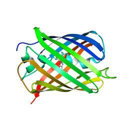
Structure of S65T Y66F R96A GFP variant in precursor state
分子名称: Green fluorescent protein, MAGNESIUM ION
著者Barondeau, D.P, Kassmann, C.J, Tainer, J.A, Getzoff, E.D.
登録日2006-06-23
公開日2007-03-27
最終更新日2023-08-30
実験手法X-RAY DIFFRACTION (1.2 Å)
主引用文献The Case of the Missing Ring: Radical Cleavage of a Carbon-Carbon Bond and Implications for GFP Chromophore Biosynthesis
J.Am.Chem.Soc., 129, 2007
2HCG
DownloadVisualize
BU of 2hcg by Molmil
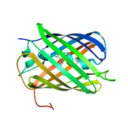
Structure of S65T Y66F GFP variant after cyclization, carbon-carbon bond cleavage, and oxygen incorporation reactions
分子名称: Green fluorescent protein, MAGNESIUM ION
著者Barondeau, D.P, Kassmann, C.J, Tainer, J.A, Getzoff, E.D.
登録日2006-06-16
公開日2007-03-27
最終更新日2023-11-15
実験手法X-RAY DIFFRACTION (1.35 Å)
主引用文献The Case of the Missing Ring: Radical Cleavage of a Carbon-Carbon Bond and Implications for GFP Chromophore Biosynthesis
J.Am.Chem.Soc., 129, 2007
2H9W
DownloadVisualize
BU of 2h9w by Molmil
Green fluorescent protein ground states: the influence of a second protonation site near the chromophore
分子名称: Green fluorescent protein, SULFATE ION
著者Garau, G.
登録日2006-06-12
公開日2007-04-24
最終更新日2023-11-15
実験手法X-RAY DIFFRACTION (1.82 Å)
主引用文献Green Fluorescent Protein Ground States: The Influence of a Second Protonation Site near the Chromophore.
Biochemistry, 46, 2007
2H8Q
DownloadVisualize
BU of 2h8q by Molmil
Crystal Structure of a Redshifted Mutant (K83M) of the Red Fluorescent Protein dRFP583/dsRed
分子名称: Red fluorescent protein drFP583
著者Yarbrough, C.A, Remington, S.J.
登録日2006-06-07
公開日2006-08-22
最終更新日2023-11-15
実験手法X-RAY DIFFRACTION (2 Å)
主引用文献Novel Chromophores and Buried Charges Control Color in mFruits(,).
Biochemistry, 45, 2006
2H6V
DownloadVisualize
BU of 2h6v by Molmil
Spectroscopic and structural study of the heterotropic linkage between halide and proton ion binding to GFP proteins- E2(GFP) APO FORM
分子名称: Green fluorescent protein, SODIUM ION
著者Garau, G.
登録日2006-06-01
公開日2007-05-15
最終更新日2021-10-20
実験手法X-RAY DIFFRACTION (1.47 Å)
主引用文献Spectroscopic and Structural Study of Proton and Halide Ion Cooperative Binding to GFP
Biophys.J., 93, 2007
2H5O
DownloadVisualize
BU of 2h5o by Molmil
Crystal structure of mOrange
分子名称: MAGNESIUM ION, mOrange
著者Shu, X, Remington, S.J.
登録日2006-05-26
公開日2006-08-22
最終更新日2017-10-18
実験手法X-RAY DIFFRACTION (1.08 Å)
主引用文献Novel Chromophores and Buried Charges Control Color in mFruits
Biochemistry, 45, 2006
2H5Q
DownloadVisualize
BU of 2h5q by Molmil
Crystal structure of mCherry
分子名称: mCherry
著者Shu, X, Remington, S.J.
登録日2006-05-26
公開日2006-08-22
最終更新日2017-10-18
実験手法X-RAY DIFFRACTION (1.36 Å)
主引用文献Novel Chromophores and Buried Charges Control Color in mFruits
Biochemistry, 45, 2006
2H5R
DownloadVisualize
BU of 2h5r by Molmil
Crystal structure of mStrawberry at pH 10.5
分子名称: mStrawberry
著者Shu, X, Remington, S.J.
登録日2006-05-26
公開日2006-08-22
最終更新日2011-07-13
実験手法X-RAY DIFFRACTION (1.6 Å)
主引用文献Novel Chromophores and Buried Charges Control Color in mFruits
Biochemistry, 45, 2006
2H5P
DownloadVisualize
BU of 2h5p by Molmil
Crystal structure of mStrawberry at pH 9.5
分子名称: mStrawberry
著者Shu, X, Remington, S.J.
登録日2006-05-26
公開日2006-08-22
最終更新日2017-10-18
実験手法X-RAY DIFFRACTION (1.21 Å)
主引用文献Novel Chromophores and Buried Charges Control Color in mFruits
Biochemistry, 45, 2006
2GX0
DownloadVisualize
BU of 2gx0 by Molmil
Crystal structural and functional analysis of GFP-like fluorescent protein
分子名称: fluorescent protein Dronpa
著者Hwang, K.Y, Nam, K.-H, Park, S.-Y, Sugiyama, K.
登録日2006-05-08
公開日2007-05-08
最終更新日2023-11-15
実験手法X-RAY DIFFRACTION (1.9 Å)
主引用文献Structural characterization of the photoswitchable fluorescent protein Dronpa-C62S
Biochem.Biophys.Res.Commun., 354, 2007
2GX2
DownloadVisualize
BU of 2gx2 by Molmil
Crystal structural and functional analysis of GFP-like fluorescent protein Dronpa
分子名称: MAGNESIUM ION, fluorescent protein Dronpa
著者Hwang, K.Y, Nam, K.-H, Park, S.-Y, Sugiyama, K.
登録日2006-05-08
公開日2007-05-08
最終更新日2023-11-15
実験手法X-RAY DIFFRACTION (1.8 Å)
主引用文献Structural characterization of the photoswitchable fluorescent protein Dronpa-C62S
Biochem.Biophys.Res.Commun., 354, 2007
2GW4
DownloadVisualize
BU of 2gw4 by Molmil
Crystal structure of stony coral fluorescent protein Kaede, red form
分子名称: Kaede, NICKEL (II) ION
著者Hayashi, I, Mizuno, H, Miyawako, A, Ikura, M.
登録日2006-05-03
公開日2007-05-08
最終更新日2023-11-15
実験手法X-RAY DIFFRACTION (1.6 Å)
主引用文献Crystallographic evidence for water-assisted photo-induced peptide cleavage in the stony coral fluorescent protein Kaede.
J.Mol.Biol., 372, 2007
2GW3
DownloadVisualize
BU of 2gw3 by Molmil
Crystal structure of stony coral fluorescent protein Kaede, green form
分子名称: Kaede, NICKEL (II) ION
著者Hayashi, I, Mizuno, H, Miyawaki, A, Ikura, M.
登録日2006-05-03
公開日2007-05-08
最終更新日2023-11-15
実験手法X-RAY DIFFRACTION (1.4 Å)
主引用文献Crystallographic evidence for water-assisted photo-induced peptide cleavage in the stony coral fluorescent protein Kaede.
J.Mol.Biol., 372, 2007
2G6Y
DownloadVisualize
BU of 2g6y by Molmil
Crystal structure of the novel green fluorescent protein from marine copepod Pontellina plumata
分子名称: green fluorescent protein 2
著者Evdokimov, A.G, Pokross, M.E, Chudakov, D.M.
登録日2006-02-26
公開日2006-03-28
最終更新日2021-10-20
実験手法X-RAY DIFFRACTION (1.6 Å)
主引用文献Structural basis for the fast maturation of Arthropoda green fluorescent protein.
Embo Rep., 7, 2006
2G6X
DownloadVisualize
BU of 2g6x by Molmil
Crystal structure of a novel green fluorescent protein from marine copepod Pontellina plumata
分子名称: green fluorescent protein 2
著者Evdokimov, A.G, Pokross, M.E, Chudakov, D.M.
登録日2006-02-26
公開日2006-03-28
最終更新日2021-10-20
実験手法X-RAY DIFFRACTION (2 Å)
主引用文献Structural basis for the fast maturation of Arthropoda green fluorescent protein.
Embo Rep., 7, 2006
2G6E
DownloadVisualize
BU of 2g6e by Molmil
Structure of cyclized F64L S65A Y66S GFP variant
分子名称: Green fluorescent protein, MAGNESIUM ION
著者Barondeau, D.P.
登録日2006-02-24
公開日2006-04-18
最終更新日2023-11-15
実験手法X-RAY DIFFRACTION (1.3 Å)
主引用文献Understanding GFP Posttranslational Chemistry: Structures of Designed Variants that Achieve Backbone Fragmentation, Hydrolysis, and Decarboxylation.
J.Am.Chem.Soc., 128, 2006
2G5Z
DownloadVisualize
BU of 2g5z by Molmil
Structure of S65G Y66S GFP variant after spontaneous peptide hydrolysis and decarboxylation
分子名称: Green fluorescent protein, MAGNESIUM ION
著者Barondeau, D.P, Kassmann, C.J, Tainer, J.A, Getzoff, E.D.
登録日2006-02-23
公開日2006-04-18
最終更新日2023-08-30
実験手法X-RAY DIFFRACTION (1.8 Å)
主引用文献Understanding GFP Posttranslational Chemistry: Structures of Designed Variants that Achieve Backbone Fragmentation, Hydrolysis, and Decarboxylation.
J.Am.Chem.Soc., 128, 2006
2G3O
DownloadVisualize
BU of 2g3o by Molmil
The 2.1A crystal structure of copGFP
分子名称: green fluorescent protein 2
著者Wilmann, P.G.
登録日2006-02-20
公開日2006-08-15
最終更新日2017-10-18
実験手法X-RAY DIFFRACTION (2.1 Å)
主引用文献The 2.1A crystal structure of copGFP, a representative member of the copepod clade within the green fluorescent protein superfamily
J.Mol.Biol., 359, 2006
2G3D
DownloadVisualize
BU of 2g3d by Molmil
Structure of S65G Y66A GFP variant after spontaneous peptide hydrolysis
分子名称: Green fluorescent protein, MAGNESIUM ION
著者Barondeau, D.P.
登録日2006-02-17
公開日2006-04-18
最終更新日2023-08-30
実験手法X-RAY DIFFRACTION (1.35 Å)
主引用文献Understanding GFP Posttranslational Chemistry: Structures of Designed Variants that Achieve Backbone Fragmentation, Hydrolysis, and Decarboxylation.
J.Am.Chem.Soc., 128, 2006
2G2S
DownloadVisualize
BU of 2g2s by Molmil
Structure of S65G Y66S GFP variant after spontaneous peptide hydrolysis
分子名称: Green fluorescent protein, MAGNESIUM ION
著者Barondeau, D.P.
登録日2006-02-16
公開日2006-04-18
最終更新日2023-08-30
実験手法X-RAY DIFFRACTION (1.2 Å)
主引用文献Understanding GFP Posttranslational Chemistry: Structures of Designed Variants that Achieve Backbone Fragmentation, Hydrolysis, and Decarboxylation.
J.Am.Chem.Soc., 128, 2006
2G16
DownloadVisualize
BU of 2g16 by Molmil
Structure of S65A Y66S GFP variant after backbone fragmentation
分子名称: Green fluorescent protein
著者Barondeau, D.P.
登録日2006-02-13
公開日2006-04-18
最終更新日2023-11-15
実験手法X-RAY DIFFRACTION (2 Å)
主引用文献Understanding GFP Posttranslational Chemistry: Structures of Designed Variants that Achieve Backbone Fragmentation, Hydrolysis, and Decarboxylation.
J.Am.Chem.Soc., 128, 2006
2FZU
DownloadVisualize
BU of 2fzu by Molmil
Reduced enolate chromophore intermediate for GFP variant
分子名称: 1,2-ETHANEDIOL, Green fluorescent protein, MAGNESIUM ION
著者Barondeau, D.P, Tainer, J.A, Getzoff, E.D.
登録日2006-02-10
公開日2006-03-14
最終更新日2024-07-10
実験手法X-RAY DIFFRACTION (1.25 Å)
主引用文献Structural evidence for an enolate intermediate in GFP fluorophore biosynthesis.
J.Am.Chem.Soc., 128, 2006
2FWQ
DownloadVisualize
BU of 2fwq by Molmil
Reduced enolate chromophore intermediate for Y66H GFP variant
分子名称: Green fluorescent protein, MAGNESIUM ION
著者Barondeau, D.P, Tainer, J.A, Getzoff, E.D.
登録日2006-02-02
公開日2006-03-14
最終更新日2023-11-15
実験手法X-RAY DIFFRACTION (1.4 Å)
主引用文献Structural evidence for an enolate intermediate in GFP fluorophore biosynthesis.
J.Am.Chem.Soc., 128, 2006

223532

件を2024-08-07に公開中

PDB statisticsPDBj update infoContact PDBjnumon