2PCO
 
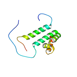
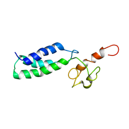
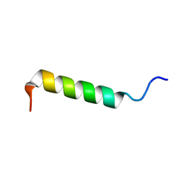 | | Spatial Structure and Membrane Permeabilization for Latarcin-1, a Spider Antimicrobial Peptide | | 分子名称: | Latarcin-1 | | 著者 | Dubovskii, P.V, Volynsky, P.E, Polyansky, A.A, Chupin, V.V, Efremov, R.G, Arseniev, A.S. | | 登録日 | 2007-03-30 | | 公開日 | 2008-03-18 | | 最終更新日 | 2024-05-22 | | 実験手法 | SOLUTION NMR | | 主引用文献 | Three-dimensional structure/hydrophobicity of latarcins specifies their mode of membrane activity.
Biochemistry, 47, 2008
|
|
2N8W
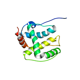
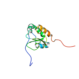
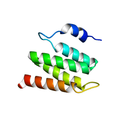 | | Solution NMR Structure of Designed Protein DA05R1, Northeast Structural Genomics Consortium (NESG) Target OR690 | | 分子名称: | Designed Protein DA05R1 | | 著者 | Eletsky, A, Federizon, J.F, Xu, X, Pulavarti, S, Jacobs, T.M, Kuhlman, B, Szyperski, T, Northeast Structural Genomics Consortium (NESG) | | 登録日 | 2015-10-27 | | 公開日 | 2015-11-25 | | 最終更新日 | 2024-05-15 | | 実験手法 | SOLUTION NMR | | 主引用文献 | Design of structurally distinct proteins using strategies inspired by evolution.
Science, 352, 2016
|
|
2N7A
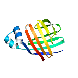
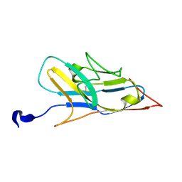 | | Solution structure of the human Siglec-8 lectin domain | | 分子名称: | Sialic acid-binding Ig-like lectin 8 | | 著者 | Proepster, J.M, Yang, F, Rabbani, S, Ernst, B, Allain, F.H.-T, Schubert, M. | | 登録日 | 2015-09-07 | | 公開日 | 2016-07-06 | | 最終更新日 | 2023-06-14 | | 実験手法 | SOLUTION NMR | | 主引用文献 | Structural basis for sulfation-dependent self-glycan recognition by the human immune-inhibitory receptor Siglec-8.
Proc.Natl.Acad.Sci.USA, 113, 2016
|
|
2N9E
 | |
2NOU
 | |
2N8V
 | | An NMR/SAXS structure of the PKI domain of the honeybee dicistrovirus, Israeli acute paralysis virus (IAPV) IRES | | 分子名称: | RNA (70-MER) | | 著者 | Au, H.H, Cornilescu, G, Mouzakis, K.D, Burke, J.E, Ren, Q, Lee, S, Butcher, S.E, Jan, E. | | 登録日 | 2015-10-27 | | 公開日 | 2015-11-11 | | 最終更新日 | 2024-05-01 | | 実験手法 | SOLUTION NMR | | 主引用文献 | Global shape mimicry of tRNA within a viral internal ribosome entry site mediates translational reading frame selection.
Proc.Natl.Acad.Sci.USA, 112, 2015
|
|
2N6N
 | | Structure Determination for spider toxin, U4-agatoxin-Ao1a | | 分子名称: | U4-agatoxin-Ao1a | | 著者 | Pineda, S.S, Chin, Y.K.-Y, Mobli, M.S, King, G.F. | | 登録日 | 2015-08-27 | | 公開日 | 2016-08-31 | | 最終更新日 | 2023-03-08 | | 実験手法 | SOLUTION NMR | | 主引用文献 | Structural venomics reveals evolution of a complex venom by duplication and diversification of an ancient peptide-encoding gene.
Proc.Natl.Acad.Sci.USA, 117, 2020
|
|
2N8I
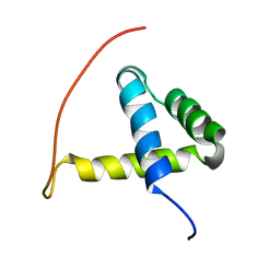
 | | Solution NMR Structure of Designed Protein DA05, Northeast Structural Genomics Consortium (NESG) Target OR626 | | 分子名称: | Designed Protein DA05 | | 著者 | Xu, X, Eletsky, A, Federizon, J.F, Jacobs, T.M, Kuhlman, B, Szyperski, T, Northeast Structural Genomics Consortium (NESG) | | 登録日 | 2015-10-15 | | 公開日 | 2016-01-20 | | 最終更新日 | 2024-05-15 | | 実験手法 | SOLUTION NMR | | 主引用文献 | Design of structurally distinct proteins using strategies inspired by evolution.
Science, 352, 2016
|
|
2N8K
 | | Chemical Shift Assignments and Structure Determination for spider toxin, U33-theraphotoxin-Cg1c | | 分子名称: | U33-theraphotoxin-Cg1b | | 著者 | Chin, Y.K.-Y, Pineda, S.S, Mobli, M, King, G.F. | | 登録日 | 2015-10-20 | | 公開日 | 2016-08-31 | | 最終更新日 | 2023-06-14 | | 実験手法 | SOLUTION NMR | | 主引用文献 | Structural venomics reveals evolution of a complex venom by duplication and diversification of an ancient peptide-encoding gene.
Proc.Natl.Acad.Sci.USA, 117, 2020
|
|
2NCN
 | |
2MZX
 | |
1W4E
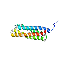
 | | Peripheral-subunit binding domains from mesophilic, thermophilic, and hyperthermophilic bacteria fold by ultrafast, apparently two-state transitions | | 分子名称: | DIHYDROLIPOYLLYSINE-RESIDUE ACETYLTRANSFERASE | | 著者 | Ferguson, N, Sharpe, T.D, Schartau, P.J, Allen, M.D, Johnson, C.M, Sato, S, Fersht, A.R. | | 登録日 | 2004-07-23 | | 公開日 | 2005-07-20 | | 最終更新日 | 2024-05-15 | | 実験手法 | SOLUTION NMR | | 主引用文献 | Ultra-Fast Barrier-Limited Folding in the Peripheral Subunit-Binding Domain Family.
J.Mol.Biol., 353, 2005
|
|
2DT7
 | | Solution structure of the second SURP domain of human splicing factor SF3a120 in complex with a fragment of human splicing factor SF3a60 | | 分子名称: | Splicing factor 3 subunit 1, Splicing factor 3A subunit 3 | | 著者 | He, F, Kuwasako, K, Inoue, M, Guntert, P, Muto, Y, Yokoyama, S, RIKEN Structural Genomics/Proteomics Initiative (RSGI) | | 登録日 | 2006-07-11 | | 公開日 | 2006-12-26 | | 最終更新日 | 2024-05-29 | | 実験手法 | SOLUTION NMR | | 主引用文献 | Solution structures of the SURP domains and the subunit-assembly mechanism within the splicing factor SF3a complex in 17S U2 snRNP
Structure, 14, 2006
|
|
2K36
 | | Structure ensemble Backbone and side-chain 1H, 13C, and 15N Chemical Shift Assignments, 1H-15N RDCs and 1H-1H nOe restraints for protein K7 from the Vaccinia virus | | 分子名称: | Protein K7 | | 著者 | Kalverda, A.P, Thompson, G.S, Vogel, A, Schr der, M, Bowie, A.G, Khan, A.R, Homans, S.W. | | 登録日 | 2008-04-22 | | 公開日 | 2008-10-28 | | 最終更新日 | 2024-05-01 | | 実験手法 | SOLUTION NMR | | 主引用文献 | Poxvirus K7 protein adopts a Bcl-2 fold: biochemical mapping of its interactions with human DEAD box RNA helicase DDX3.
J.Mol.Biol., 385, 2009
|
|
2JU3
 | | Solution-state NMR structures of apo-LFABP (Liver Fatty Acid-Binding Protein) | | 分子名称: | Fatty acid-binding protein, liver | | 著者 | He, Y, Yang, X, Wang, H, Estephan, R, Francis, F, Kodukula, S, Storch, J, Stark, R.E. | | 登録日 | 2007-08-14 | | 公開日 | 2007-11-20 | | 最終更新日 | 2024-05-29 | | 実験手法 | SOLUTION NMR | | 主引用文献 | Solution-State Molecular Structure of Apo and Oleate-Liganded Liver Fatty Acid-Binding Protein
Biochemistry, 46, 2007
|
|
2I8F
 | |
2JNK
 | |
2K8V
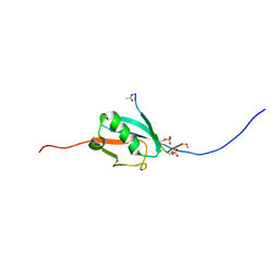
 | | Solution structure of Oxidised ERp18 | | 分子名称: | Thioredoxin domain-containing protein 12 | | 著者 | Rowe, M.L, Alanen, H.I, Ruddock, L.W, Kelly, G, Schmidt, J.M, Williamson, R.A, Howard, M.J. | | 登録日 | 2008-09-25 | | 公開日 | 2009-06-02 | | 最終更新日 | 2022-03-16 | | 実験手法 | SOLUTION NMR | | 主引用文献 | Solution structure and dynamics of ERp18, a small endoplasmic reticulum resident oxidoreductase .
Biochemistry, 48, 2009
|
|
2E61
 | | Solution structure of the zf-CW domain in zinc finger CW-type PWWP domain protein 1 | | 分子名称: | ZINC ION, Zinc finger CW-type PWWP domain protein 1 | | 著者 | He, F, Muto, Y, Inoue, M, Kigawa, T, Shirouzu, M, Terada, T, Yokoyama, S, RIKEN Structural Genomics/Proteomics Initiative (RSGI) | | 登録日 | 2006-12-25 | | 公開日 | 2007-06-26 | | 最終更新日 | 2024-05-01 | | 実験手法 | SOLUTION NMR | | 主引用文献 | Structural insight into the zinc finger CW domain as a histone modification reader
Structure, 18, 2010
|
|
2EBI
 | | Arabidopsis GT-1 DNA-binding domain with T133D phosphomimetic mutation | | 分子名称: | DNA binding protein GT-1 | | 著者 | Nagata, T, Noto, K, Niyada, E, Ikeda, Y, Yamamoto, Y, Uesugi, S, Murata, J, Hiratsuka, K, Katahira, M. | | 登録日 | 2007-02-08 | | 公開日 | 2008-02-19 | | 最終更新日 | 2024-05-22 | | 実験手法 | SOLUTION NMR | | 主引用文献 | Solution structures of the trihelix DNA-binding domains of the wild-type and a phosphomimetic mutant of Arabidopsis GT-1: mechanism for an increase in DNA-binding affinity through phosphorylation.
Proteins, 78, 2010
|
|
2B5X
 | |
2B0H
 | | Solution structure of VBS3 fragment of talin | | 分子名称: | Talin-1 | | 著者 | Gingras, A.R, Vogel, K.P, Steinhoff, H.J, Ziegler, W.H, Patel, B, Emsley, J, Critchley, D.R, Roberts, G.C, Barsukov, I.L. | | 登録日 | 2005-09-14 | | 公開日 | 2006-01-17 | | 最終更新日 | 2024-05-22 | | 実験手法 | SOLUTION NMR | | 主引用文献 | Structural and Dynamic Characterization of a Vinculin Binding Site in the Talin Rod
Biochemistry, 45, 2006
|
|
1ZR9
 | |
1Y5C
 | |
2KPL
 | | MAGI-1 PDZ1 / E6CT | | 分子名称: | Membrane-associated guanylate kinase, WW and PDZ domain-containing protein 1, Protein E6 | | 著者 | Charbonnier, S, Nomine, Y, Ramirez, J, Luck, K, Stote, R.H, Trave, G, Kieffer, B, Atkinson, R.A. | | 登録日 | 2009-10-16 | | 公開日 | 2010-10-27 | | 最終更新日 | 2024-05-01 | | 実験手法 | SOLUTION NMR | | 主引用文献 | The structural and dynamic response of MAGI-1 PDZ1 with non-canonical domain boundaries to binding of human papillomavirus (HPV) E6
J.Mol.Biol., 2011
|
|


