4UBV
 
 | |
5TVJ
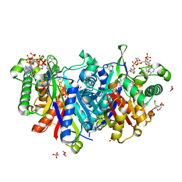
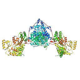
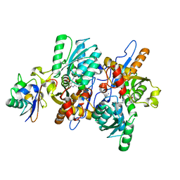
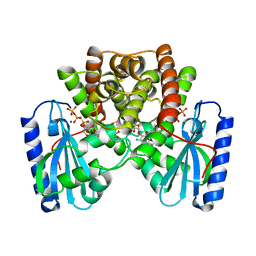
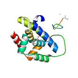
 | | Crystal structure of acetyltransferase Eis from Mycobacterium tuberculosis in complex with CoA and inhibitor 2k*: 1-(4-fluorophenyl)-2-[2-(4-methylphenyl)-2-oxoethyl]pyrrolo[1,2-a]pyrazin-2-ium | | 分子名称: | 1-(4-fluorophenyl)-2-[2-(4-methylphenyl)-2-oxoethyl]pyrrolo[1,2-a]pyrazin-2-ium, CHLORIDE ION, COENZYME A, ... | | 著者 | Gajadeera, C.S, Garzan, A, Hou, C, Garneau-Tsodikova, S, Tsodikov, O.V. | | 登録日 | 2016-11-09 | | 公開日 | 2017-03-01 | | 最終更新日 | 2023-10-04 | | 実験手法 | X-RAY DIFFRACTION (2.3 Å) | | 主引用文献 | Combating Enhanced Intracellular Survival (Eis)-Mediated Kanamycin Resistance of Mycobacterium tuberculosis by Novel Pyrrolo[1,5-a]pyrazine-Based Eis Inhibitors.
ACS Infect Dis, 3, 2017
|
|
7XAM
 | |
4LN9
 | |
9F48
 | |
7KIF
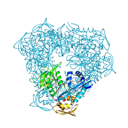
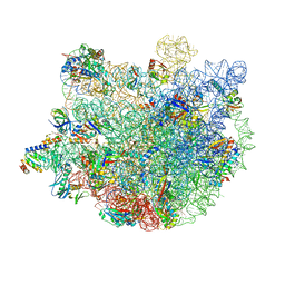
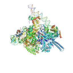 | | Mycobacterium tuberculosis WT RNAP transcription open promoter complex with WhiB7 transcription factor | | 分子名称: | DNA (55-MER), DNA (63-MER), DNA-directed RNA polymerase subunit alpha, ... | | 著者 | Lilic, M, Darst, S.A, Campbell, E.A. | | 登録日 | 2020-10-23 | | 公開日 | 2021-04-21 | | 最終更新日 | 2025-05-14 | | 実験手法 | ELECTRON MICROSCOPY (2.94 Å) | | 主引用文献 | Structural basis of transcriptional activation by the Mycobacterium tuberculosis intrinsic antibiotic-resistance transcription factor WhiB7.
Mol.Cell, 81, 2021
|
|
7KIM
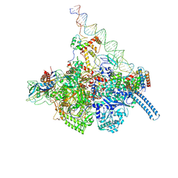 | | Mycobacterium tuberculosis WT RNAP transcription closed promoter complex with WhiB7 transcription factor | | 分子名称: | DNA (45-MER), DNA-directed RNA polymerase subunit alpha, DNA-directed RNA polymerase subunit beta, ... | | 著者 | Lilic, M, Darst, S.A, Campbell, E.A. | | 登録日 | 2020-10-23 | | 公開日 | 2021-04-21 | | 最終更新日 | 2025-05-28 | | 実験手法 | ELECTRON MICROSCOPY (3.38 Å) | | 主引用文献 | Structural basis of transcriptional activation by the Mycobacterium tuberculosis intrinsic antibiotic-resistance transcription factor WhiB7.
Mol.Cell, 81, 2021
|
|
7KIN
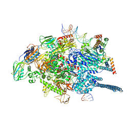 | | Mycobacterium tuberculosis WT RNAP transcription open promoter complex with WhiB7 promoter | | 分子名称: | DNA (49-MER), DNA (54-MER), DNA-directed RNA polymerase subunit alpha, ... | | 著者 | Lilic, M, Darst, S.A, Campbell, E.A. | | 登録日 | 2020-10-23 | | 公開日 | 2021-04-21 | | 最終更新日 | 2025-05-21 | | 実験手法 | ELECTRON MICROSCOPY (2.74 Å) | | 主引用文献 | Structural basis of transcriptional activation by the Mycobacterium tuberculosis intrinsic antibiotic-resistance transcription factor WhiB7.
Mol.Cell, 81, 2021
|
|
6S0U
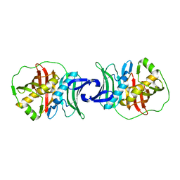
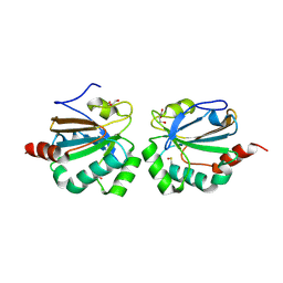
 | | The crystal structure of kanamycin B dioxygenase (KanJ) from Streptomyces kanamyceticus in complex with nickel and 2-oxoglutarate | | 分子名称: | 2-OXOGLUTARIC ACID, CHLORIDE ION, DI(HYDROXYETHYL)ETHER, ... | | 著者 | Mrugala, B, Porebski, P.J, Niedzialkowska, E, Minor, W, Borowski, T. | | 登録日 | 2019-06-18 | | 公開日 | 2020-07-08 | | 最終更新日 | 2024-01-24 | | 実験手法 | X-RAY DIFFRACTION (2.15 Å) | | 主引用文献 | A study on the structure, mechanism, and biochemistry of kanamycin B dioxygenase (KanJ)-an enzyme with a broad range of substrates.
Febs J., 288, 2021
|
|
3DWG
 | | Crystal structure of a sulfur carrier protein complex found in the cysteine biosynthetic pathway of Mycobacterium tuberculosis | | 分子名称: | 9.5 kDa culture filtrate antigen cfp10A, Cysteine synthase B, PYRIDOXAL-5'-PHOSPHATE | | 著者 | Jurgenson, C.T, Burns, K.E, Begley, T.P, Ealick, S.E. | | 登録日 | 2008-07-22 | | 公開日 | 2008-09-23 | | 最終更新日 | 2025-03-26 | | 実験手法 | X-RAY DIFFRACTION (1.53 Å) | | 主引用文献 | Crystal structure of a sulfur carrier protein complex found in the cysteine biosynthetic pathway of Mycobacterium tuberculosis.
Biochemistry, 47, 2008
|
|
6S0T
 | | The crystal structure of kanamycin B dioxygenase (KanJ) from Streptomyces kanamyceticus in complex with nickel, sulfate, soaked with iodide | | 分子名称: | IODIDE ION, Kanamycin B dioxygenase, NICKEL (II) ION, ... | | 著者 | Mrugala, B, Porebski, P.J, Niedzialkowska, E, Cymborowski, M.T, Minor, W, Borowski, T. | | 登録日 | 2019-06-18 | | 公開日 | 2020-07-08 | | 最終更新日 | 2024-01-24 | | 実験手法 | X-RAY DIFFRACTION (2.1 Å) | | 主引用文献 | A study on the structure, mechanism, and biochemistry of kanamycin B dioxygenase (KanJ)-an enzyme with a broad range of substrates.
Febs J., 288, 2021
|
|
5ID2
 | |
6S0R
 | | The crystal structure of kanamycin B dioxygenase (KanJ) from Streptomyces kanamyceticus complex with nickel, sulfate and chloride | | 分子名称: | CHLORIDE ION, Kanamycin B dioxygenase, NICKEL (II) ION, ... | | 著者 | Mrugala, B, Porebski, P.J, Niedzialkowska, E, Cymborowski, M.T, Minor, W, Borowski, T. | | 登録日 | 2019-06-18 | | 公開日 | 2020-07-08 | | 最終更新日 | 2024-06-19 | | 実験手法 | X-RAY DIFFRACTION (2.5 Å) | | 主引用文献 | A study on the structure, mechanism, and biochemistry of kanamycin B dioxygenase (KanJ)-an enzyme with a broad range of substrates.
Febs J., 288, 2021
|
|
1LQT
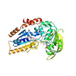 | | A covalent modification of NADP+ revealed by the atomic resolution structure of FprA, a Mycobacterium tuberculosis oxidoreductase | | 分子名称: | 4-OXO-NICOTINAMIDE-ADENINE DINUCLEOTIDE PHOSPHATE, ACETATE ION, FLAVIN-ADENINE DINUCLEOTIDE, ... | | 著者 | Bossi, R.T, Aliverti, A, Raimondi, D, Fischer, F, Zanetti, G, Ferrari, D, Tahallah, N, Maier, C.S, Heck, A.J.R, Rizzi, M, Mattevi, A, TB Structural Genomics Consortium (TBSGC) | | 登録日 | 2002-05-13 | | 公開日 | 2002-07-31 | | 最終更新日 | 2024-12-25 | | 実験手法 | X-RAY DIFFRACTION (1.05 Å) | | 主引用文献 | A covalent modification of NADP+ revealed by the atomic resolution structure of FprA, a Mycobacterium tuberculosis oxidoreductase.
Biochemistry, 41, 2002
|
|
7XKZ
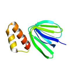 | | Solution structure of subunit epsilon of the Mycobacterium abscessus F-ATP synthase | | 分子名称: | ATP synthase epsilon chain | | 著者 | Shin, J, Grueber, G, Harikishore, A, Wong, C.F, Prya, R, Dick, T. | | 登録日 | 2022-04-20 | | 公開日 | 2023-03-08 | | 最終更新日 | 2024-05-15 | | 実験手法 | SOLUTION NMR | | 主引用文献 | Atomic solution structure of Mycobacterium abscessus F-ATP synthase subunit epsilon and identification of Ep1MabF1 as a targeted inhibitor.
Febs J., 289, 2022
|
|
1KNY
 | | KANAMYCIN NUCLEOTIDYLTRANSFERASE | | 分子名称: | DIPHOSPHOMETHYLPHOSPHONIC ACID ADENOSYL ESTER, KANAMYCIN A, KANAMYCIN NUCLEOTIDYLTRANSFERASE, ... | | 著者 | Pedersen, L.C, Benning, M.M, Holden, H.M. | | 登録日 | 1995-07-07 | | 公開日 | 1996-08-17 | | 最終更新日 | 2024-02-14 | | 実験手法 | X-RAY DIFFRACTION (2.5 Å) | | 主引用文献 | Structural investigation of the antibiotic and ATP-binding sites in kanamycin nucleotidyltransferase.
Biochemistry, 34, 1995
|
|
9HQ7
 | |
9HQC
 | |
9GOT
 | |
9BIN
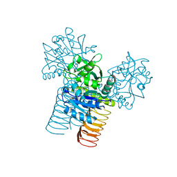
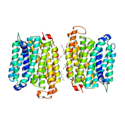 | | BRD-8000.3 bound EFPA transporter of Mycobacterium tuberculosis | | 分子名称: | (1S,3S)-N-[6-bromo-5-(pyrimidin-2-yl)pyridin-2-yl]-2,2-dimethyl-3-(2-methylprop-1-en-1-yl)cyclopropane-1-carboxamide, CHOLESTEROL HEMISUCCINATE, Uncharacterized MFS-type transporter EfpA, ... | | 著者 | Khandelwal, N.K, Gupta, M, Stroud, R.M. | | 登録日 | 2024-04-23 | | 公開日 | 2025-02-05 | | 最終更新日 | 2025-06-04 | | 実験手法 | ELECTRON MICROSCOPY (3.45 Å) | | 主引用文献 | Inhibitor BRD-8000.3 bound EfpA transporter of Mycobacterium tuberculosis
To Be Published
|
|
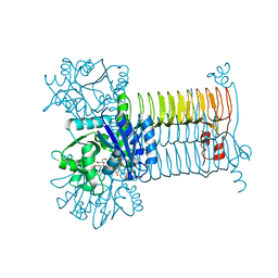
9BIQ
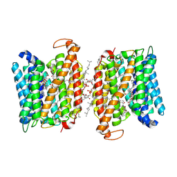 | | BRD-9327 bound EFPA transporter of Mycobacterium tuberculosis | | 分子名称: | CHOLESTEROL HEMISUCCINATE, N-(7-benzoyl-2,3-dihydro-1,4-benzodioxin-6-yl)-2-bromobenzamide, Uncharacterized MFS-type transporter EfpA, ... | | 著者 | Khandelwal, N.K, Gupta, M, Stroud, R.M. | | 登録日 | 2024-04-24 | | 公開日 | 2025-02-05 | | 最終更新日 | 2025-05-21 | | 実験手法 | ELECTRON MICROSCOPY (3 Å) | | 主引用文献 | BRD-8000.3 bound EFPA transporter of Mycobacterium tuberculosis
To Be Published
|
|
8IBO
 | |
8IBP
 | |
3D98
 | |
3D8V
 | |


